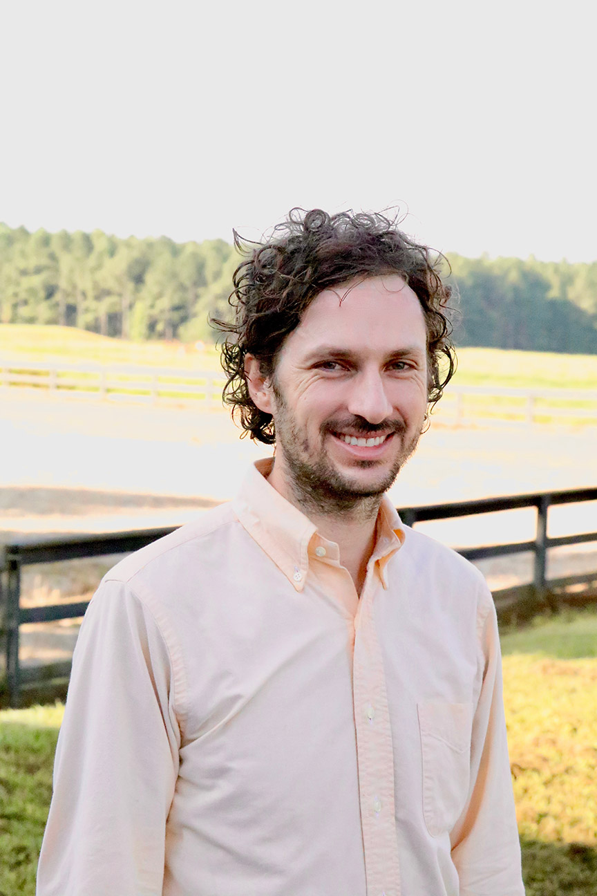
Christine R. Rose serves as Head of the Institute of Neurobiology at Heinrich-Heine-University Düsseldorf, Faculty of Mathematics and Natural Sciences. Her research focuses on intracellular ion signaling in the vertebrate brain, particularly astrocyte-neuron interactions at glutamatergic synapses in hippocampus, cortex, and cerebellum. Research Themes: Sodium signaling as ionic excitability, ion gradient maintenance under pathophysiological conditions, TRPV4 channel roles in ischemia, ATP dynamics in energy-deprived states Techniques: Multi-photon laser scanning microscopy, electrophysiology, fluorescence lifetime imaging, mathematical modeling Collaborations: International neuroscience network partnerships Article trends show consistent exploration of ion homeostasis in both neurons and astrocytes, with recent work extending to organoid slice protocols and pH regulation mechanisms . Most publications address synaptic dysfunction in ischemic and metabolic compromise scenarios.
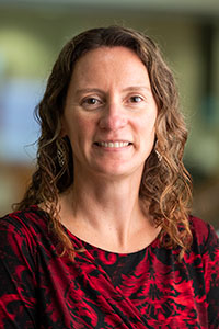
Karissa Tilbury is an Associate Professor at the Graduate School of Biomedical Science and Engineering at the University of Maine. She leads the Tilbury Biophotonics Group, focusing on advanced optical imaging techniques to study biological systems, particularly in the context of disease pathology and tissue engineering. Her research interests include: Biomedical Engineering Cancer and Extracellular Matrix Remodeling Cellular Metabolism Non-linear Microscopy, especially Second Harmonic Generation (SHG) and Multiphoton Microscopy Spatial Frequency Domain Imaging STEM Education The recent publications highlight a consistent focus on SHG microscopy for label-free, high-resolution imaging of collagen in various diseases, especially cancer and fibrosis. Her work spans pancreatic, ovarian, and breast cancers, pulmonary fibrosis, adipose tissue, and cartilage, demonstrating a multiscale approach from molecular to tissue-level analysis. The integration of polarization, 3D texture analysis, and open-source instrumentation (e.g., OpenSFDI) reflects a strong emphasis on methodological innovation and accessibility. Scientific contributions and collaborations are evident through publications in high-impact journals such as Frontiers in Oncology , eLife , Journal of Biomedical Optics , and Scientific Reports . She advises students through her research lab and has been involved in numerous collaborative grants and projects, particularly in developing imaging platforms for cancer and tissue analysis. While specific grants are not listed, her work involves instrumentation development, computational image analysis, and translational applications. Karissa leads the Tilbury Biophotonics Group, which develops and applies cutting-edge optical tools to understand biological interactions at spatio-temporal scales relevant to disease progression and therapeutic response.
Dr. Armin Hochreiner is an Assistant Professor at Linz University of Applied Sciences, specializing in Medical Engineering and Thermography. He leads research projects at the Research Center Linz and TIMed Center, focusing on advanced optical setups, bioprinting, and non-destructive testing. Projects: GBOLF (Greiner Bio One Leichtfried), BF-ABSAOS (Adaptive Beam Shaping), TC-LOEM (Laser-Induced Surface Modification). Research Areas: Optical coherence tomography, two-photon lithography, fiber lasers, and data acquisition systems. His recent work includes a 2025 publication on low-cost pulse generation for 3D-printed microfluidics and a 2024 study on lipid properties in high-density lipoproteins. Grants from Land OÖ Basisfinanzierung and FTI-Strukturförderung support his research on adaptive optics and biocompatible microstructures. Collaborations span biomedical imaging, machine learning integration in optical systems, and interdisciplinary projects with institutions like Greiner Bio One and Springer.
Jaroslaw Jacak is a Professor at the University of Applied Sciences Upper Austria (FH Linz), where he leads research in nanotechnology and biomedical engineering. He is affiliated with the Center of Excellence Medical Engineering/TIMed Center and directs NASAN - Nano Structuring and Bio-Analytics. His work bridges advanced imaging, nanofabrication, and biomedical applications with significant research output spanning over two decades. Dr. Jacak's research focuses on cutting-edge technologies in biomedical engineering: Nanoscale imaging and manipulation using STED lithography 3D bioprinting and microfabrication techniques Extracellular vesicle research and therapeutic applications Stem cell differentiation on engineered scaffolds Single-molecule analysis of cellular interactions Microfluidic systems for cell analysis Photochemical processes for biomaterial development His recent publications (2023-2025) demonstrate a clear trajectory toward precise nanoscale tools for biological investigation, with emphasis on multiphoton lithography, protein photoresists, and extracellular vesicle purification. The research shows strong integration of physics, engineering, and biology to solve complex biomedical challenges. Dr. Jacak has secured substantial research funding through multiple competitive projects: BF-SigSim (2024-2025): As Co-PI, developing signaling pathway simulation for gene expression prediction LiSSCeD (2018-2022): As PI, creating 3D lithographical scaffolds for stem cell differentiation Vascular MicroLab (2018-2021): As PI, characterizing thrombocyte aggregation under flow conditions Bioceta (2018-2023): As PI, biophysical characterization of extracellular bioparticles for therapeutic application He leads the NASAN research group which has developed groundbreaking techniques using STED lithography for single-protein capacity nano-anchors and advanced imaging applications. His team maintains strong collaborations across European research institutions and has produced over 116 research outputs with significant citation impact (h-index: 22).
Luke J Mortensen is an Associate Professor of Regenerative Medicine and Engineering in the Department of Animal and Dairy Science at the University of Georgia, with a joint appointment in the School of Chemical, Materials, and Biomedical Engineering. His research bridges engineering, regenerative medicine, and imaging technologies with a focus on bone and muscle regeneration. Dr. Mortensen's educational background includes: Ph.D. in Biomedical Engineering from the University of Rochester (2011) M.S. in Biomedical Engineering from the University of Rochester (2006) B.S. in Bioengineering from the University of Toledo (2003) Dr. Mortensen's research primarily focuses on regenerative medicine in bone and muscle. His long-term goals include creating next-generation super-resolution 2 and 3 photon microscopes for imaging cell organelles through opaque bone, and leveraging key imaging and lipid metabolite features associated with mesenchymal stem cell (MSC) therapeutic potency to biomanufacture efficacious cell therapies. His work integrates advanced imaging techniques with lipidomics to understand and enhance stem cell therapies for musculoskeletal conditions. Analysis of Dr. Mortensen's recent publications reveals a strong focus on MSC potency metrics, advanced imaging through bone tissue, and lipid-mediated regenerative processes. His work spans multiple disciplines including biomedical engineering, optical physics, and regenerative medicine, with particular emphasis on developing novel imaging approaches for studying cellular processes in challenging environments like bone tissue. Dr. Mortensen has secured significant research funding from diverse sources including the National Institutes of Health, National Science Foundation, Department of Defense, and private foundations. His sponsored projects cover areas such as bone microfracture repair, MSC potency assessment, and advanced imaging technologies. Dr. Mortensen leads a research team focused on regenerative medicine and engineering, working at the intersection of imaging technology development and therapeutic applications for musculoskeletal disorders. His lab combines expertise in microscopy, lipidomics, and stem cell biology to address challenges in regenerative medicine.
Antoine Anfray serves as an Instructor in Neuroscience at the Brain and Mind Research Institute, Weill Cornell Medical College (Cornell University) since 2023, focusing on neurovascular mechanisms in stroke, dementia, and hypertension. His work bridges vascular biology and neural function to address cerebrovascular disorders. His academic foundation includes: Ph.D. in Neuroscience, University of Caen (France), 2017 M.S. in Neuroscience, University of Caen (France), 2014 B.S. in Neuroscience, University of Caen (France), 2012 Dr. Anfray investigates immune-vascular interactions in neurodegenerative diseases, particularly how border-associated macrophages drive ApoE4-related neurovascular dysfunction and white matter injury. His research explores thrombotic stroke models in awake mice, circadian influences on cerebrovascular events, and tPA's dual roles in neuroprotection and vascular damage. Key themes include neurovascular coupling impairment in Alzheimer's pathology, hypertension-induced cognitive decline via T-cell mediated inflammation, and atherosclerosis progression post-stroke. Analysis of his 15 most recent publications (2016-2025) reveals dominant trends in cerebrovascular disease mechanisms, with 70% focusing on stroke models and neuroimmune interactions. His work consistently employs advanced in vivo imaging in awake rodents to dissect cellular mechanisms, emphasizing translational pathways for vascular cognitive impairment. He currently leads as Principal Investigator a BrightFocus Foundation grant (2022-2025) titled Role of perivascular macrophages in ApoE4-induced neurovascular dysfunction , examining macrophage-mediated vascular oxidative stress in Alzheimer's models. His mentorship includes doctoral candidates in neuroscience and vascular biology, though specific advisees are not listed in source materials. Based at the Brain and Mind Research Institute, Dr. Anfray collaborates with the Feil Family Brain and Mind Research Institute's neurovascular team, utilizing transgenic mouse models and multiphoton microscopy to study real-time neurovascular dynamics in awake animals.
Brian J. Bacskai is a Professor of Neurobiology at Harvard Medical School and Principal Investigator at the Alzheimer's Disease Research Unit of Massachusetts General Hospital . His work combines advanced optical imaging techniques with neurodegenerative disease research. Professor of Neurobiology, Harvard Medical School Principal Investigator, Alzheimer's Disease Research Unit, Mass General Lab Member, MassGeneral Institute for Neurodegenerative Disease Research Focus : Dr. Bacskai's laboratory specializes in multiphoton microscopy to study Alzheimer's disease pathology in live transgenic mouse models. Key areas include: Chronic in vivo imaging of senile plaques Development of fluorescence lifetime imaging microscopy (FLIM) Characterization of anti-amyloid therapeutics Neurovascular unit dynamics Non-invasive near-infrared (NIR) imaging approaches Scientific Contributions : Recent publications demonstrate expertise in: Optical imaging of neurovascular interactions Quantitative analysis of amyloid pathology Translational research from mice to human applications Development of novel neuroimaging technologies Collaborative Environment : The lab maintains multidisciplinary collaborations with: Chemists Physicists Clinical neurologists Medical imaging specialists
Dr. FANG Xiaofeng is a Research Professor in the Department of Biomedical Engineering at Southern University of Science and Technology (SUSTech). He leads a research group focused on developing advanced materials and imaging techniques for biomedical applications. His career includes postdoctoral training at SUSTech (2017-2019) following doctoral studies at Jilin University. Education: Ph.D. in Physical Electronics, Jilin University (2014-2017) M.S. in Organic Chemistry, Jilin University (2012-2014) B.S. in Chemistry, Jilin University (2008-2012) Research Focus: Dr. Fang's work spans three interconnected domains: 1) Design and synthesis of organic functional materials with tailored optical properties; 2) Super-resolution imaging techniques for biological systems; and 3) Integrated biosensing and phototherapy platforms. His research bridges materials chemistry, optical engineering, and translational medicine. Publication Trends: His recent articles (2021-2025) demonstrate a strong emphasis on polymer-based nanoplatforms for advanced bioimaging and therapy. Key innovations include NIR-II probes for deep-tissue imaging, photoswitchable materials for super-resolution microscopy, and targeted nanoconjugates for cancer theranostics. The work consistently integrates materials innovation with biomedical problem-solving. He currently leads a research group developing molecular probes and bio-optical imaging technologies. The laboratory focuses on four key areas: molecular probe design, bioanalysis and biosensors, optoelectronic functional materials, and biomedical applications. The team actively recruits postdoctoral researchers for cross-disciplinary projects.
Karsten Siller is an Associate Professor at the University of Virginia's School of Data Science. He previously served as an Assistant Professor in the Department of Biology (College of Arts & Sciences) and held the role of Associate Director for Research Computing User Services. His work bridges data science with biology, focusing on scalable, reproducible data workflows and computational methods. Ph.D., Biology , University of Tübingen, Germany Dipl. Biol., Biology , University of Tübingen, Germany His research interests include: Building reproducible and resilient, scalable data analysis workflows for diverse computing environments. Developing user-friendly, extensible interfaces for modular data pipelines with integrated visualization. Advancing open-source software and computational tools for biology and image analysis. Key trends in his publications show expertise in: Fluorescence Lifetime Imaging (FLIM) for cellular metabolism studies. High-performance computing (HPC) for data pipeline management. Mechanistic insights into cell division and neurodegenerative processes via imaging and computational modeling.
Melissa Skala, Ph.D., is the Carol Skornicka Chair of Biomedical Imaging at the Morgridge Institute for Research and a full Professor of Biomedical Engineering & Medical Physics at the University of Wisconsin–Madison. Her laboratory pioneers label-free optical imaging technologies—most notably fluorescence-lifetime microscopy and optical coherence tomography—to quantify metabolic heterogeneity in cancer, immune cells and engineered tissues, with direct translation to cell-therapy manufacturing and personalized cancer medicine. Education Ph.D. Biomedical Engineering, 2007, Duke University M.S. Biomedical Engineering, 2004, University of Wisconsin–Madison B.S. Physics, 2002, Washington State University Research Focus The Skala lab develops and applies cutting-edge photonics platforms—including autofluorescence lifetime imaging, optical redox ratio mapping, second-harmonic generation, and light-sheet microscopy—to interrogate metabolic states of single cells and organoids in vitro and in vivo . Major thrusts include: Cancer metabolism and immunotherapy response Immune cell activation and exhaustion Stem-cell and CAR-T manufacturing quality control Micro-physiological disease models Machine-learning–driven image analysis Publication Trends Across >250 peer-reviewed papers since 2004, she has progressed from foundational studies on NAD(P)H/FAD redox imaging in epithelial tissues to recent landmark reports defining metabolic biomarkers for T-cell activation, immune-cell subtyping, and patient-derived cancer organoid drug response. A 2025–2024 cluster emphasizes label-free metabolic monitoring of stem-cell-derived cardiomyocytes, neutrophil functional states, and collagen remodeling during immunotherapy. Scientific Honors Fellow, OSA, SPIE, AIMBE (2019) Carol Skornicka Chair, Morgridge Institute (2022) Daniel M. Albert Chair, Retina Research Foundation (2021) Stand Up To Cancer – Sharp Collaboration Award (2017) NSF CAREER Award (2016) NIH/NCI Pathway to Independence Award (K99/R00) (2010) Grants & Team Mentorship Dr. Skala directs multiple active NIH grants (R01, R35, P30) totaling several million dollars, focusing on metabolic imaging for cancer immunotherapy, stem-cell manufacturing, and infectious disease models. The Skala Laboratory actively recruits and mentors graduate students and post-doctoral researchers, fostering interdisciplinary collaborations across engineering, oncology and immunology. Laboratory & Infrastructure The Skala Lab is equipped with custom-built multimodal optical systems, high-throughput microfluidic platforms, and dedicated animal-imaging suites. Core capabilities include time-correlated single-photon counting, hyperspectral imaging, and AI-driven single-cell analytics.
Dr. Valdas Šablinskas is a Professor and Head of the Molecular Spectroscopy Group at Vilnius University's Institute of Chemical Physics . His work focuses on Vibrational Spectroscopy , Raman Spectroscopy , SERS , and Spectroscopic Imaging for biomedical and chemical applications. Current affiliations: Vilnius University, Institute of Chemical Physics Academic role: Professor (since 2010) Key research themes: Spectrochemical analysis, disease diagnostics, molecular structure studies His recent publications highlight advancements in Raman spectroscopy for urinary sediment analysis, conformational studies of organosilicon molecules via DFT/MP2 methods , and MXene-based SERS substrates for salicylic acid detection. These works span biomedical diagnostics , molecular physics , and computational chemistry . Scientific leadership includes: Lithuanian National Science Award (2012) Project leadership in FP7, Horizon 2020, and bilateral collaborations Expertise in FTIR/SERS instrumentation for disease studies He has supervised 7 completed doctoral theses and 2 ongoing projects , contributed to educational programs on wave optics and molecular spectroscopy, and participated in international committees for research evaluation.
Prof. Marc van Zandvoort is a Professor at the Institute for Molecular Cardiovascular Research (IMCAR), specializing in advanced microscopy applications for cardiovascular and tissue engineering research. His work bridges molecular imaging, cellular dynamics, and clinical pathology through interdisciplinary collaborations. His primary research focuses on cardiovascular microvasculature, tissue engineering maturation, and cellular death mechanisms. He employs multiphoton microscopy, two-photon endoscopy, and super-resolution techniques to investigate placental pathologies in preeclampsia, atherosclerotic plaque microvasculature, mitochondrial networks, and cerebrovascular glycocalyx integrity. Key methodologies include quantitative imaging of nuclear morphology, 3D structural analysis, and biomaterial characterization for drug delivery systems. Analysis of his 2023-2025 publications reveals dominant themes in cardiovascular imaging (68%), placental/obstetric research (20%), and neural applications (12%). Technical innovations center on multiphoton imaging for tissue engineering maturation (32%), hydrogel-based biomaterials (24%), and mitochondrial dynamics in cell death pathways (16%), with strong cross-disciplinary links to obstetrics, cardiology, and nanomedicine. Scientific Awards: None mentioned in the provided text. Information on student advising, research grants, and educational background was not provided in the available text. Prof. van Zandvoort leads microscopy-focused research within IMCAR, utilizing two-photon endoscopy systems and multiphoton imaging platforms. His collaborative network spans 17 institutions across Europe, with frequent co-authorship on projects involving tissue-engineered heart valves, placental microvasculature quantification, and super-resolution pathology profiling.
PD Dr. Steffen Dietzel is an Associate Professor affiliated with the Biomedical Center Munich (BMC) at Ludwig-Maximilians-Universität München. As Head of the Core Facility Bioimaging , he manages advanced light microscopy systems including confocal, multi-photon, and STED microscopes, providing technical support and training for researchers. Core Facility Bioimaging (European Reference Site for Leica Microsystems) Biomedical Center Munich (BMC) His research focuses on label-free imaging techniques for cellular and tissue analysis, including third harmonic generation (THG) and multiphoton microscopy. These methods enable non-invasive 3D visualization of biological structures and hemodynamic parameter quantification. His work also explores chromatin compaction, nuclear organization, and retroviral integration effects on genome architecture. The articles listed demonstrate expertise in microscopy innovation (2019: microscope performance workflow; 2010: mirror-based signal enhancement), 3D imaging (2011: muscle tissue visualization; 2008: nuclear gene positioning), and chromatin dynamics (2004: transgene array compaction; 2012: retroviral integration effects). Subfields span optical physics, image processing, and cellular biophysics. As facility head, he oversees instrumentation maintenance, user training, and advanced imaging methodology development. His group collaborates on projects requiring live-cell microscopy , deconvolution, and superresolution techniques.
Dr. Robert Knight is a Reader in Developmental Genetics at King’s College London, affiliated with the Centre for Craniofacial & Regenerative Biology and Faculty of Dentistry, Oral & Craniofacial Sciences. He obtained his PhD in Molecular Evolution from the University of Reading in 2000, followed by postdoctoral work at UC Irvine and University of Sheffield, focusing on AP-2 genes, neural crest, and cranial muscle development in zebrafish. His research explores molecular regulation of muscle regeneration and stem cell behavior, combining live imaging, transcriptomics, and zebrafish models. Key findings include the role of Ret tyrosine kinase in facial muscle development and FSHD therapy, RhoA coupling migration/differentiation, and NF-κB in macrophage function during repair. Collaborations span systems biology (Dresden), physiology (Amsterdam), and macrophage signaling (Lyon). Recent publications highlight trends in muscle stem cell dynamics , signaling pathways (Wnt, Notch, Ret), and age-related regeneration . His work aligns with SDGs via health (musculoskeletal diseases) and innovation (AI in biosciences, multiphoton microscopy). Scientific Awards: BBSRC Project Award MDUK Studentship NC3Rs Studentship Royal Society Partnership Award Carl Zeiss Collaboration Current projects address DUX4-activated Ret in FSHD, chromatin organization in aging, and macrophage-stem cell interactions . He leads grants from BBSRC, Dunhill Medical Trust, and Leverhulme Trust.
Michael Dailey is an Associate Professor at the University of Iowa, affiliated with the Department of Biology and Carver College of Medicine. He serves as Director of the Carver Center for Imaging (CCI) and leads a research laboratory focused on glial cell structure and function in mammalian brain development and pathology. PhD, Washington University (St. Louis) Research Interests Dr. Dailey’s work investigates microglial biology in brain development and injury response, with specific projects on: Purinergic signaling pathways regulating microglial activation Environmental toxicants (alcohol, pesticides) in neurodevelopmental disorders Microglial survival mechanisms in stroke models Neuroimmune interactions during developmental brain injury Advanced imaging techniques for live tissue analysis Recent Research Trends His 2025-2024 studies examine PCB exposure impacts on brain gene expression and maternal pesticide effects on fetal neurodevelopment. Earlier work from 2019-2013 explores alcohol-induced neuroinflammation, microglial dynamics in hippocampal injury, and imaging methodologies. Collaborations & Funding Dr. Dailey collaborates with Pediatrics and Neurology Departments (Carver College of Medicine) and Pharmacy/Public Health Colleges. His research on pesticide exposure receives funding from the National Institute for Environmental Health Sciences (NIEHS/NIH P30 ES005605). Laboratory The Dailey Lab (established 1996) specializes in time-lapse fluorescence imaging of rodent brain tissues to study neuronal-glial interactions, with strengths in confocal and multiphoton microscopy. The lab website provides further details on ongoing projects.