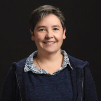
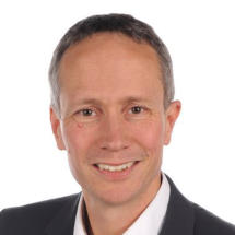
Paul Klenerman is a Professor at the Nuffield Department of Medicine within the Medical Sciences Division , University of Oxford. His research focuses on immune responses to infectious diseases including HIV, hepatitis B/C, and SARS-CoV-2, with emphasis on T-cell biology, vaccine development, and host-pathogen dynamics. Email: paul.klenerman@medawar.ox.ac.uk Collaborators: Ellie Barnes (Oxford), Adrian Hill (Oxford), Georg Lauer (Harvard), Robert Thimme (Freiburg), and others across 12 institutions. Key research themes include: CD161++/MAIT cell biology (bacterial/viral defense, liver immunology), HCV immune defense (vaccine trials using adenoviral vectors), and memory inflation (persistent immune responses post-viral infections). Recent publications (2024-2025) span vaccine immunology (hybrid immunity, bivalent boosters), liver immunobiology (MAIT cell repair mechanisms), infectious disease dynamics (HCV persistence, dengue progression), and single-cell analysis of gut/lung pathologies.
James C. Gee is a Professor of Radiologic Science in Radiology at the University of Pennsylvania's Perelman School of Medicine. He serves as Director of the Penn Image Computing and Science Laboratory and Co-Director of the Translational Biomedical Imaging Center , with affiliations in Bioengineering and Applied Mathematics graduate groups. His research focuses on biomedical image analysis, specialization in segmentation, registration, and morphometry applied to neurodegenerative diseases and multi-organ systems. Education : B.S. in Computer Science/Electrical Engineering (University of Washington, 1987), Ph.D. in Computer and Information Science (University of Pennsylvania, 1996) Research : Quantitative medical imaging methods, brain connectomics, neurodegeneration mapping, and translational imaging technologies Publications : 15+ recent works on AI-driven image analysis for Alzheimer's disease, cardiac amyloidosis, and radiomics applications Leadership : Directs MSE-DS Online Degree Program, co-chairs Radiology DCOAP Committee, and founded RISE (Radiology Initiative to Support Inclusive Excellence) His laboratory develops advanced computational tools like ITK-SNAP for biomedical imaging, with applications in both in vivo clinical imaging and ex vivo histology . The work spans cross-disciplinary collaborations in computer science, neuroscience, and clinical medicine.
Spencer L. Bowen, Ph.D., is an Assistant Professor in the Department of Radiology at UT Southwestern Medical Center, where he is a member of the Radiology Research section and serves as a PET research scientist. His work is centered on advancing nuclear imaging technologies for clinical and research applications in oncology, neurology, and cardiology. Education: Bachelor's in Biomedical Engineering – University of Washington, Seattle Ph.D. in Biomedical Engineering – University of California, Davis Research Fellow – Massachusetts General Hospital, Charlestown, MA Dr. Bowen's research focuses on the development of advanced PET imaging systems, including dedicated breast PET/CT scanners and hybrid PET-MR technologies. He investigates image acquisition techniques, reconstruction algorithms, attenuation and scatter correction methods, and partial volume correction to improve quantitative accuracy. His work spans hardware design, software development (e.g., the Masamune processing tool), and clinical translation. His recent publications highlight innovations in cardiac and neurological PET quantification, breast imaging, and hybrid PET/MR systems. Themes include attenuation correction in PET/MR, dynamic PET modeling, and the impact of image processing on clinical interpretation. Scientific Recognition: Research featured on the cover of the Journal of Nuclear Medicine Work covered by press outlets Dr. Bowen actively contributes to the scientific community as a reviewer for leading journals including Journal of Nuclear Medicine , Medical Physics , Physics in Medicine and Biology , and IEEE Transactions on Nuclear Science and Transactions on Medical Imaging . His lab, the Bowen Lab, is engaged in ongoing research and is currently recruiting PhD graduate students, indicating active grant support and research momentum. He leads a research team focused on developing tomographic tools for precision medicine. The Bowen Lab is dedicated to creating and refining nuclear imaging technologies to enhance both clinical care and scientific discovery, with a strong emphasis on quantitative, high-resolution imaging across multiple disease domains.
Dr. Oluwabunmi (Bunmi) Olaloye is an Assistant Professor of Pediatrics in the Division of Neonatology at Yale School of Medicine. She holds appointments in Neonatal-Perinatal Medicine and is affiliated with the Janeway Society. Her academic background includes an MD from Rutgers New Jersey Medical School, pediatrics residency at University of Texas Medical Branch, and neonatology fellowship at University of Pittsburgh Medical Center. Dr. Olaloye's research focuses on immune dysfunction underlying neonatal intestinal diseases such as necrotizing enterocolitis (NEC) and spontaneous intestinal perforation (SIP). Using cutting-edge techniques like single-cell RNA sequencing and mass cytometry, her work identifies biomarkers and therapeutic targets to improve outcomes for premature infants. Key research areas include fetal immune system maturation, placental immune interactions, and gestational age-specific inflammatory responses. Her publication record spans 12 peer-reviewed articles between 2019-2025, emphasizing translational immunology and neonatal pathophysiology. Notable contributions include defining immune cell trajectories in preterm infants and developing gating guidelines for high-dimensional cytometry data. Current projects involve constructing immune cell atlases across human lifespans and investigating nutritional interventions for intestinal disorders. Laboratory affiliations include the Laboratory for Surgery, Obstetrics & Gynecology where she explores neonatal mucosal immunity. Her work integrates clinical observations with systems immunology approaches to address critical gaps in understanding prematurity-associated gastrointestinal pathologies.
Shyni Varghese is the Laszlo Ormandy Distinguished Professor of Orthopaedic Surgery at Duke University, with joint appointments in Mechanical Engineering & Materials Science and Biomedical Engineering. She directs the Varghese Lab, an interdisciplinary team focused on smart biomaterials, organ-on-chip models, rejuvenation therapies, and translational medical technologies. Her research bridges tissue engineering, regenerative medicine, and disease modeling to address bone healing, osteoarthritis, and age-related tissue degeneration. Education: Ph.D. in Chemistry/Materials Science, National Chemical Laboratory (India), 2002 Research spans four pillars: Smart Biomaterials : Engineered ECM mimetics, self-healing hydrogels, and stimuli-responsive systems for tissue regeneration. Miniature Organs : Organoid and organ-on-chip platforms (e.g., tumor-on-chip, lung alveolus models) to study disease mechanisms. Rejuvenation : Targeting cellular senescence, adenosine signaling, and inflammation to enhance aged tissue repair. Bench to Bedside : Translating technologies like 'bone bandages' and nanocarriers for fracture healing and osteoporosis. Recent publications emphasize orthopaedic repair (fracture healing, osteoarthritis), immunomodulation (macrophage reprogramming, immunotherapy), and advanced biomaterials (self-healing lubricants, cartilage-penetrating carriers). Studies frequently employ mouse models and microengineered platforms to dissect pain mechanisms, senescence, and tissue regeneration pathways. Dr. Varghese advises 10+ doctoral students and postdoctoral researchers. Her lab has pioneered innovations like 'DraBot' (environment-responsive soft robot) and 'cell pouch' xenotransplantation devices. Collaborative projects include NIH-funded work on bone radioprotection and NSF-supported biomaterial design. The Varghese Lab occupies the Duke Medical Science Research Building, fostering collaborations with clinicians and engineers. Current projects explore: Senolysis for neuroinflammation mitigation Adenosine-based therapies for bone loss 3D tumor models for immunotherapy screening
Kaye Morgan is an Associate Professor in the School of Physics and Astronomy at Monash University, specializing in X-ray imaging technologies with applications in medical and respiratory research. She holds an Australian Research Council Future Fellowship and has held prestigious positions including a Hans Fischer Fellowship at Technische Universität München. Her research focuses on advancing X-ray optics methodologies, particularly phase contrast X-ray imaging (PCXI) and dark-field imaging, to enhance resolution, speed, and sensitivity. These techniques are applied to study airway health in cystic fibrosis and other respiratory diseases, using synchrotron facilities like SPring-8 and the Munich Compact Light Source. She has pioneered single-grid imaging and propagation-based dark-field approaches, enabling real-time visualization of lung dynamics and treatment efficacy. Morgan leads multiple high-impact projects funded by ARC and international collaborations, with over 85 publications in journals like Optics Express and Scientific Reports. Her work contributes to UN Sustainable Development Goals related to health and innovation. Key achievements include developing lab-based X-ray sources for clinical translation and quantifying lung microstructure through advanced imaging algorithms.
James Lacefield is a Professor in both the Department of Electrical and Computer Engineering and the Department of Medical Biophysics at Western University. He serves as Director of the School of Biomedical Engineering and maintains his research laboratory in the Amit Chakma Engineering Building. His academic appointments span multiple disciplines, reflecting the interdisciplinary nature of his work in biomedical ultrasound imaging. Dr. Lacefield earned his Ph.D. and B.S.E. in Biomedical Engineering from Duke University. His educational background established the foundation for his current research program that bridges engineering principles with medical applications. His research focuses on the physical acoustics and signal processing aspects of ultrasound imaging, with particular emphasis on quantitative vascular imaging applications. Dr. Lacefield's laboratory develops novel methods for color Doppler, power Doppler, and contrast-enhanced ultrasound imaging, with primary applications in cancer research. Current projects include optimization of high-resolution ultrasound systems for tumor vascular characterization and development of methods to quantify spatial blood flow distribution in tumors. Analysis of his recent publications reveals a strong focus on quantitative ultrasound techniques for cancer applications, with particular attention to tumor perfusion assessment using contrast-enhanced ultrasound. His work demonstrates increasing sophistication in speckle analysis methods to improve the reliability of perfusion measurements in preclinical tumor models. Associate Editor, IEEE Transactions on Ultrasonics, Ferroelectrics, and Frequency Control Member, Council of Chairs of Bioengineering and Biomedical Engineering Member, College of Reviewers, Canadian Institutes of Health Research Dr. Lacefield maintains active collaborations with multiple research groups including the Imaging Research Laboratories at Robarts Research Institute. His professional activities include editorial work for major ultrasound journals and participation in national review panels for biomedical research funding.
Toni M. Antalis, PhD, is a Professor in the Department of Pharmacology & Physiology at the University of Maryland School of Medicine. She serves as Associate Director of Training and Education for the Marlene and Stewart Greenebaum Comprehensive Cancer Center and Director of the Program in Molecular Medicine. Her research bridges vascular biology and cancer, focusing on membrane-anchored serine proteases and their role in tumor metastasis, inflammation, and coagulation. Doctorate in Biochemistry from Rice University Postdoctoral training in Cell Biology at Baylor College of Medicine Her laboratory investigates how protease-activated receptors (PARs) and the plasminogen activation system influence vascular disease and ovarian cancer progression. Current projects include studying fibrinolysis in thrombus resolution and developing protease-targeted therapies for metastatic ovarian cancer. Recent publications highlight roles of matriptase, testisin, and PAI-2 in tumor dissemination and vascular permeability. Dr. Antalis' research is funded by the National Institutes of Health (NIH), the Department of Defense, and a VA Merit Award. She has previously received support from the Lance Armstrong Foundation and Rivkin Center. She co-directs NIH-funded T32 and PREP programs for cancer training. Mentored numerous PhD students and postdoctoral fellows Developed engineered anthrax toxin prodrugs for ovarian cancer therapy (patents 10,568,929 and 11,013,784)
Sophie Lanone is a researcher and team leader of the Genetic-environment Interactions in COPD, Cystic Fibrosis, and Respiratory Pathologies (GEIC2O) team at the Mondor Institute of Biomedical Research (IMRB), affiliated with Université Paris-Est Créteil. Her work focuses on understanding the interplay between genetic and environmental factors in respiratory diseases, particularly COPD, cystic fibrosis, and surfactant-related pathologies. She leads a multidisciplinary team of clinicians and scientists investigating molecular mechanisms, inflammation resolution, and environmental impacts on pulmonary health. Key research themes include the molecular basis of cigarette smoke-induced COPD, genetic and cellular aspects of cystic fibrosis, and the role of specialized pro-resolving mediators in disease. Funding sources include EU programs (e.g., H2020 REMEDIA), ANR, and patient associations like Vaincre la Mucoviscidose. Recent advances include identifying lipid mediator defects in CF patients and demonstrating resolvin E1’s efficacy in correcting ciliary dysfunction. Team members have received awards, such as Khadeeja Adam Sy’s 2024 prize for active participation in CF research. Collaborations span in vitro/ex vivo models, patient cohort studies, and translational approaches toward personalized therapies. The team also explores environmental exposures (e.g., asbestos, nanoparticles) and their long-term respiratory health impacts.
Rainer Böckmann is a Professor of Computational Biology in the Department of Biology at Friedrich-Alexander-University Erlangen-Nürnberg (FAU), Germany, where he leads the Group for Theoretical and Computational Membrane Biophysics. His research integrates molecular dynamics simulations with biophysical analysis to study membrane structure, dynamics, and function. Research Interests: His work focuses on computational biophysics, particularly lipid bilayers, membrane proteins, molecular dynamics, and structural bioinformatics. He investigates how lipid composition, cholesterol, and embedded peptides influence membrane organization, curvature, and permeability, with applications in antimicrobial strategies and mRNA vaccine delivery systems. Recent Research Trends: His recent publications reflect a strong emphasis on lipid nanoparticles (LNPs), particularly their phase behavior, pH-dependent protonation, and structural transitions relevant to mRNA vaccines. He also explores antimicrobial peptides, membrane domain formation, and the role of cholesterol in modulating membrane properties. His group develops and applies advanced simulation techniques, including constant-pH MD and coarse-grained modeling. Member of Editorial Board, Biophysical Journal (2024–present) Elected Member, DFG Review Board for Biophysics (2020–present) Chairman, Molecular Biophysics Section, German Biophysical Society (2011–2012) Leadership and Service: Böckmann is actively involved in academic governance, serving on editorial boards, DFG committees, and as a guest editor for special issues in Frontiers journals. He contributes to graduate education and high-performance computing initiatives at FAU, including the NHR@FAU and Life@FAU Graduate School. He has organized major conferences and workshops in biophysics and membrane modeling. Laboratory and Collaboration: He leads a research group focused on biomembrane physics, collaborating with experimentalists and theorists. His lab develops and applies simulation tools to study membrane systems, bridging computational insights with biological function.
Alejandro Benjamin Balazs is an Assistant Professor of Medicine at the Ragon Institute of MGH, MIT, and Harvard. His laboratory specializes in synthetic immunology, gene transfer technologies, and immune system engineering to combat HIV and emerging viral threats like SARS-CoV-2. Current research focuses include: Understanding sterilizing immunity mechanisms against HIV. Studying pathogen escape from immunological pressure. Optimizing AAV vector delivery for broadly neutralizing antibodies. Investigating innate immune responses in viral neutralization. His lab has published extensively on: Antibody-mediated prophylaxis against HIV and SARS-CoV-2. Viral evolution and immune escape dynamics. AAV-based gene delivery systems. Humoral immunity in vulnerable populations. Immune signatures in humanized mouse models. Scientific accolades include: NIDA Avenir New Innovator (DP2) Grant (2015) Gilead Sciences Research Scholars Award (2016) MGH Transformative Research Scholars Award (2016) Doctoral students mentored in his lab include: Jackie Brady (Harvard BBS Program, 2015-2020) Allen Lin (Harvard Systems Biology, 2015-2020) Meredith Phelps (Harvard Virology, 2018-2022)
Professor Daniel Davis MBE FMedSci is the Head of the Department of Life Sciences and Professor of Immunology at Imperial College London. He holds affiliations with the Institute of Chemical Biology, the CDT in Chemical Biology: Innovation in Life Sciences (as a supervisor), and research groups in Immunology and Molecular Mechanisms of Disease. His academic journey includes a doctorate in Physics from Harvard University and prior roles as Director of Research at the Manchester Collaborative Centre for Inflammation Research (University of Manchester). His research focuses on nanoscale biology of immune cell interactions, employing advanced microscopy techniques to study immune synapse formation, cytotoxicity mechanisms, and immunological regulation. Notable contributions include elucidating how immune cells use adhesion, signaling, and structural reorganization to target pathogens and cancer cells. Professor Davis has authored four popular science books, including Self Defence: A Myth-Busting Guide to Immune Health (2025), The Beautiful Cure (2018), and The Compatibility Gene (2014), which bridge public understanding of immunology and biology. His work has been recognized with prestigious awards such as the Royal Society Science Book Prize and the Prose Award. His articles span topics like NK cell heterogeneity, gene therapy for neurological disorders, and super-resolution microscopy applications. Grants and collaborations include work on AAV-based gene therapies and immunomodulatory drug development. Davis actively engages in public science communication through festivals, media outlets (e.g., BBC, Guardian), and international speaking engagements. His research labs at Imperial College focus on interdisciplinary approaches, combining biophysics, genetics, and clinical applications to advance immunology and translational medicine.
Associate Professor Freda Passam is a Clinical Academic Haematologist at Royal Prince Alfred Hospital, University of Sydney, specializing in thrombosis and haemostasis. She leads the Haematology Research Group at the Charles Perkins Centre, focusing on platelet biology, endothelial cell dysfunction, and translational research in cardiovascular diseases. Her work integrates basic science with clinical applications, including developing microfluidic technologies like the Endo-chip for thrombosis diagnosis and therapy screening. Education: MD and PhD in Greece (angiogenesis in Hodgkin’s lymphoma), postdoctoral training at UNSW and Harvard University. Research spans platelet hyperactivity in diabetes, immune thrombosis (e.g., HIT), and thiol isomerase inhibitors as antithrombotics. Collaborates globally with institutions like Harvard and the University of Utah. Research Interests: 1) Platelet biomarkers in diabetes-related cardiovascular risk; 2) Immune thrombosis diagnostics/therapies (e.g., Endo-chip); 3) Bone marrow-on-chip models for thrombopoiesis. Current projects include SEC61B regulation of calcium flux, endothelial thromboinflammation, and ERp5/ERp57 roles in platelet production. Grants: Over $6M in funding from NHMRC, MRFF, NSW Health, and industry partnerships. Notable awards include Sydney Nano Grand Challenge Award (2022) and SLHD VTE Stewardship Award (2020). Advising: Mentors over 6 PhD/master’s students in thrombosis research, emphasizing clinical/research integration. Teaching includes Sydney Medicine curriculum, bedside tutorials, and student engagement in lab activities. Labs/Teams: Haematology Research Group (Charles Perkins Centre), collaborations with Sydney Health Partners, Harvard, and international haematology groups. Develops technologies like the Endo-chip and bone marrow-on-chip models.
Dr. Athma A Pai is an Associate Professor at UMass Chan Medical School, holding primary appointments in the RNA Therapeutics Institute and the T.H. Chan School of Medicine. She maintains extensive secondary appointments across multiple departments including Genomics and Computational Biology, Systems Biology, and several graduate programs at the Morningside Graduate School of Biomedical Sciences, reflecting the highly interdisciplinary nature of her work. Education: BS in Biochemistry/Anthropology from University of Pennsylvania PhD in Human Genetics from University of Chicago Postdoctoral training in RNA Genomics from MIT Dr. Pai's research program centers on RNA biology with particular emphasis on RNA processing, splicing mechanisms, and the regulation of gene expression. Her work investigates how environmental factors influence RNA processing through biochemical, molecular, and genetic mechanisms. She employs cutting-edge genomic and transcriptomic approaches to study alternative polyadenylation, mRNA transcript initiation and termination, and the spatial organization of RNA processing events within cells. Her research has significant implications for understanding fundamental gene regulation mechanisms and their roles in disease processes. Analysis of Dr. Pai's recent publications reveals a strong focus on developing high-resolution profiling methods for understanding transcriptional and translational regulation. Her work increasingly integrates computational approaches with experimental biology to investigate how RNA processing events are coordinated across the transcriptome. A notable trend is her exploration of how RNA processing contributes to inflammatory responses and cellular defense mechanisms, with implications for therapeutic development. Dr. Pai actively mentors students through multiple graduate programs at UMass Chan Medical School, including Biochemistry and Molecular Biotechnology, Biophysical Chemical and Computational Biology, Interdisciplinary Graduate Program, MD/PhD Program, RNA Therapeutics and Biology Program, and Systems Computational and Quantitative Biology. She maintains an active laboratory (Pai Lab) that welcomes postdoctoral researchers interested in RNA biology. Her laboratory website provides additional information about ongoing research projects and opportunities for collaboration and training, and she maintains a professional presence through her Twitter account (@athmapai).
Cornelius Faber is a University Professor in the Department of Radiology at the University of Münster, Germany, where he leads the Experimental Nuclear Magnetic Resonance research group. His work focuses on developing and implementing novel MRI techniques that extend the boundaries of magnetic resonance imaging in terms of spatial and temporal resolution, sensitivity, and specificity for physiological, structural, and molecular changes. He actively participates in the "Cells in Motion" interdisciplinary research initiative at the university. Professor Faber's research spans multiple critical areas in medical imaging and biomedical science. His primary expertise lies in MRI cell tracking , enabling visualization of cellular dynamics in vivo. He has made significant contributions to infection imaging , developing methods to detect and characterize microbial infections using MRI. His work on MR methodology development has advanced quantitative imaging techniques, while his research on multimodal integration in MR and MRI contrast mechanisms has provided deeper insights into molecular and cellular processes. His research bridges physics, engineering, and biomedical applications, with particular relevance to inflammation, cancer, neurological disorders, and cardiovascular disease. Analysis of Professor Faber's extensive publication record reveals a clear evolution from fundamental MRI technique development toward increasingly sophisticated applications in disease models. His recent work demonstrates a strong trend toward multimodal imaging approaches that combine MRI with complementary techniques such as mass spectrometry, optical imaging, and PET. This integration creates comprehensive diagnostic platforms that provide both anatomical and molecular information. A notable pattern is the focus on cellular dynamics, particularly immune cell behavior in inflammatory conditions and tumor microenvironments, with applications spanning neuroscience, oncology, and cardiology. Professor Faber leads a multidisciplinary research team of approximately 15 members, including scientists, doctoral students, technicians, and medical students. His laboratory is deeply integrated with the University of Münster's research infrastructure, particularly the Multiscale Imaging Centre. The group's work contributes significantly to advancing preclinical MRI methodologies while maintaining strong clinical relevance, with numerous publications in high-impact journals across medical imaging, neuroscience, and biomedical engineering disciplines.