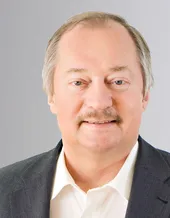
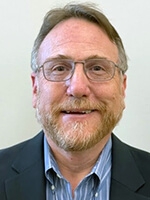
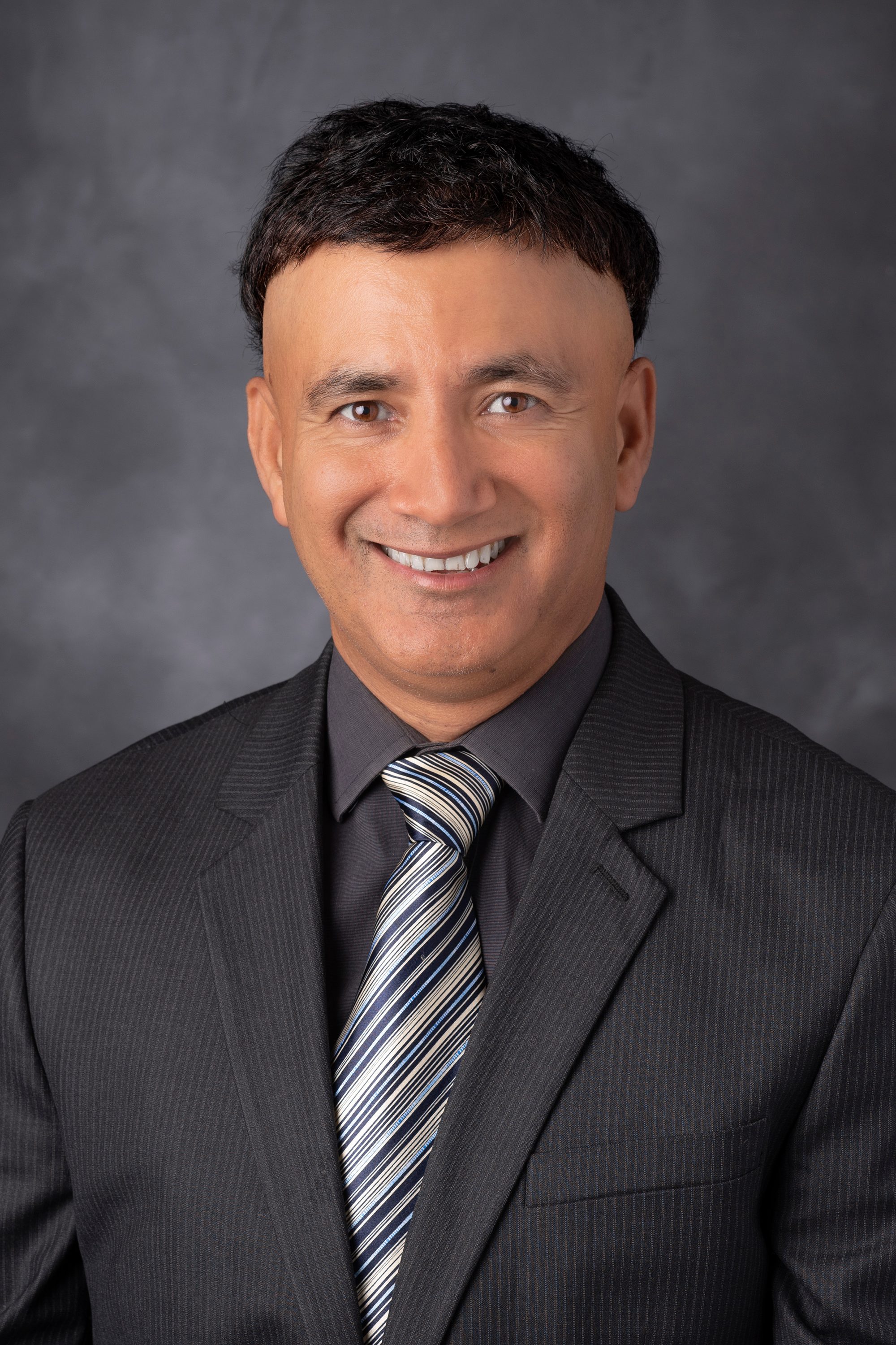
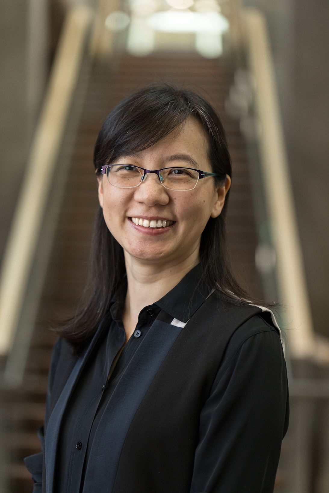
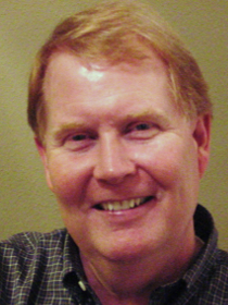
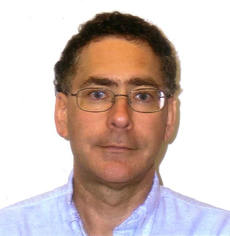
Craig H. Meyer is a Professor in Biomedical Engineering and Radiology & Medical Imaging at the University of Virginia. He holds a Ph.D. from Stanford University and leads the Rapid MRI Research Group, focusing on developing advanced MRI techniques for cardiovascular disease, neural disorders, and pediatrics. His work integrates physics, signal processing, and machine learning to improve MRI acquisition and processing speed. Education: Ph.D. in Biomedical Engineering, Stanford University. Research Interests: Medical and Molecular Imaging, Signal and Image Processing, Biomedical Data Sciences, Biomechanics, and Cardiovascular Engineering. His innovations include fast spiral imaging, conjugate phase reconstruction, and machine learning-enhanced MRI denoising. Awards: Notably includes the Dean’s Award for Excellence in Team Science (2014), Fellowships from NAI (2021), AIMBE (2015), and ISMRM (2013). He also authored two landmark MRI papers recognized as pivotal in the field. Teaching: Courses include BME 6310 (Computation and Modeling in Biomedical Engineering) and BME 8782 (Magnetic Resonance Imaging). He emphasizes translational research, with applications in clinical MRI advancements and collaborative interdisciplinary projects. Labs/Groups: Rapid MRI Research Group focuses on cutting-edge MRI technologies, including real-time cardiac imaging and artifact reduction through deep learning.
Axel Haase is a Carl von Linde Senior Fellow at the Technical University of Munich (TUM) and Director of the Institute of Medical Engineering (IMETUM). He holds a professorship in Experimental Physics (Biophysics) at the University of Würzburg. His research focuses on magnetic resonance imaging (MRI), including co-inventing the FLASH MRI technique and advancing biomedical applications like cardiac and neurological studies. He previously served as President of the University of Würzburg (2003–2009) and President of the European Society of Magnetic Resonance in Biology and Medicine (ESMRMB). Education: Diploma in Physics (1977), PhD (1980) from University of Giessen, Habilitation in Biophysical Chemistry (University of Frankfurt). Leadership Roles: Max Planck Institute of Biophysical Chemistry (1978–1989), Postdoc at University of Oxford (1982). Research Interests: MRI技术创新,包括快速成像技术、医学成像应用、生物医学工程。His work has led to patents and significant advancements in MRI methodologies. Awards: 包括Bavarian Academy of Sciences Fellow (2001)、ISMRM金质奖章 (1991)、DFG Heisenberg Fellowship (1987)等。 Labs & Teams: Director of IMETUM at TUM, leading interdisciplinary research in medical engineering and imaging technologies.
Anne-Sophie Chauvin is a Senior Lecturer and Researcher at École Polytechnique Fédérale de Lausanne (EPFL), School of Basic Sciences, within the Institute of Chemical Sciences and Engineering and the Supramolecular Chemistry Laboratory. She actively engages in supramolecular and inorganic chemistry, focusing on f-element (lanthanides and actinides) coordination polymers and luminescent bioprobes for biological and technological applications, including invisible inks and dye-sensitized solar cells. PhD in Bioinorganic Chemistry from University Paris V-René Descartes (thesis on Nitrile Hydratase mimetics) Postdoctoral work at University of Geneva on chiral alcohol configuration analysis Habilitation à Diriger des Recherches (HDR) from University René Descartes (2006) Her research spans Lanthanide and Actinide Chemistry , Luminescence , Coordination Polymers , Metallacages , and Photovoltaic Materials . Recent publications emphasize catalytic spiro stereocenter formation, actinide coordination polymers, and photoredox-enabled biomolecule functionalization. She has supervised PhD students including Andrei Andreichenko , Julien Andrès , Steve Comby , and Aurélien Willauer . Recognitions include Fellowship of the Royal Society of Chemistry (FRSC) and membership in the Swiss Chemical Society (SCS). Current roles include teaching General and Analytical Chemistry to first-year Pharmacy and Biology students at the University of Lausanne (UNIL), overseeing practical sessions, and serving on the EPFL School of Basic Sciences Faculty Council.
Silvia Cavagnero is a Professor in the Department of Chemistry at the University of Wisconsin–Madison, with a research focus on protein folding and misfolding in cellular contexts. Her work integrates biomolecular spectroscopy, chemical biology, and computational methods to address fundamental questions in structural biology. B.S., First University of Rome ‘La Sapienza’ (1988) M.S., University of Arizona (1990) Ph.D., California Institute of Technology (1996) Her research explores the role of molecular chaperones like Hsp70 in protein biogenesis, the development of laser-driven NMR techniques for enhanced sensitivity, and the implications of protein aggregation in neurodegenerative diseases. Key projects include cotranslational folding studies at ribosomal exit tunnels and hyperpolarization methods for low-concentration NMR analysis. The 15 most recent publications highlight interdisciplinary advances in NMR spectroscopy optimization Protein folding kinetics Cryo-EM structural analysis Chaperone-client interactions Hsp70 antimicrobial design Hydration dynamics in folding Scientific contributions include A Prize for Going in Vivo (2017) Recognition for Diversity and Inclusion Efforts Students from the Cavagnero Group have pursued careers in academia, pharmaceutical industries, and national laboratories. Her lab emphasizes interdisciplinary training, blending physical chemistry, biology, and computational analysis.
Daniel B. Vigneron, PhD is a Professor at the University of California, San Francisco (UCSF) Department of Radiology and Biomedical Imaging. He serves as Director of the Hyperpolarized MRI Technology Resource Center (HMTRC), Director of Human Imaging Core Services, Director of Advanced Imaging Technologies SRG, and Operations Director of the Surbeck Laboratory for Advanced Imaging. As a core member of the UCB/UCSF Graduate Group in Bioengineering, Vigneron has established himself as a leader in molecular imaging research with over three decades of experience at UCSF. Vigneron's research focuses on developing advanced functional and metabolic MRI techniques, particularly hyperpolarized carbon-13 technology, for studying prostate cancer, brain tumors, and other diseases. His work enables non-invasive imaging of metabolic processes, allowing clinicians to monitor therapy effectiveness and guide treatments. The HMTRC, which he founded in 2011 with NIH funding and recently secured a 5-year renewal for, has supported 20 external projects domestically and 15 internationally, produced 239 publications, and trained 149 researchers. Vigneron's lab develops novel coil and software techniques for high-field MRI, MR spectroscopy, and diffusion imaging at 3T and 7T for studying brain, prostate cancer, and other organs. His recent publications demonstrate a clear trajectory toward clinical translation of hyperpolarized carbon-13 MRI across multiple organ systems. The research spans abdominal imaging with advanced denoising techniques, cardiac metabolism studies, whole-brain coverage applications, and cerebral perfusion analysis. This work represents a significant shift from basic science toward practical clinical applications in oncology, cardiology, and neurology, with particular emphasis on standardization for multi-center studies. Scientific Awards: 2022 Outstanding Faculty Mentoring Award from UCSF Department of Radiology and Biomedical Imaging Vigneron has mentored 149 trainees throughout his career, with several former students now serving as faculty members including Duan Xu, Peder Larson, and Susan Noworolski. As Principal Investigator overseeing eight grants, he has secured significant NIH funding for the HMTRC and other research initiatives. His administrative leadership extends to co-chairing the department's Safety and Compliance Committee, where he has helped establish robust safety protocols for PET-MR programs. Vigneron's mentoring philosophy emphasizes adapting to individual needs at different career stages, moving from instructor to coach to manager to cheerleader as trainees progress. The Vigneron Lab, located in Byers Hall on the UCSF Mission Bay campus, operates within the Surbeck Laboratory for Advanced Imaging. The lab group develops novel acquisition techniques and hardware for multinuclear MR spectroscopy, with particular focus on hyperpolarized carbon-13 metabolic imaging. The HMTRC serves as a hub for team science, bringing together researchers from diverse disciplines to advance metabolic imaging technology and its clinical applications.
Parameswaran Krishna Nair is a Professor of Medicine and the Frederick E. Hargreave Teva Innovation Chair in Airway Diseases at McMaster University, with a clinical role as Staff Respirologist at the Firestone Institute for Respiratory Health, St. Joseph’s Healthcare Hamilton. His work focuses on complex obstructive airway diseases, severe asthma, and eosinophilic lung disorders, integrating immunology, hematology, and imaging specialists in multidisciplinary care. He leads an advanced airway diseases fellowship training program. University: McMaster University Clinical Affiliation: Firestone Institute for Respiratory Health, St. Joseph’s Healthcare Hamilton His research explores airway autoimmunity, biologics for inflammatory disorders, and pulmonary imaging techniques like hyperpolarized ¹²⁹Xe MRI. Key studies include mechanisms of eosinophilic asthma, corticosteroid dependence, and post-acute sequelae of COVID-19 with rheumatological implications. Recent publications analyze biologic therapies (benralizumab, mepolizumab), ventilation heterogeneity in asthma, and sputum immunoglobulins as biomarkers. Awards include the Frederick E. Hargreave Teva Innovation Chair in Airway Diseases. Scientific Awards: Frederick E. Hargreave Teva Innovation Chair He contributes to clinical guidelines, therapeutic trials, and autoantibody research in severe respiratory diseases.
Pratip K. Bhattacharya, Ph.D. , is an Associate Professor in the Department of Cancer Systems Imaging and the Department of Imaging Physics at The University of Texas MD Anderson Cancer Center, with a joint appointment in the Graduate School of Biomedical Sciences at The University of Texas Health Science Center. He is a principal investigator leading the Bhattacharya Laboratory, dedicated to advancing magnetic resonance imaging (MRI) through hyperpolarization techniques for applications in cancer and cardiovascular diseases. His research focuses on developing real-time metabolic and molecular imaging methods using hyperpolarized 13 C and 15 N-labeled compounds and silicon nanoparticles. These innovative probes significantly enhance MRI sensitivity, enabling non-invasive assessment of tissue metabolism and targeted imaging. His lab's work spans three primary areas: real-time metabolic MR imaging, targeted molecular MR imaging with functionalized silicon nanoparticles, and high-resolution MR metabolomics. These efforts are aimed at improving disease diagnosis and therapy monitoring. Analysis of his recent publications reveals a strong and consistent focus on hyperpolarized MRI, particularly using silicon particles and metabolic tracers like succinate, to visualize cancer metabolism and cardiovascular conditions in vivo. His research integrates physics, chemistry, and biomedical engineering to create novel imaging tools with direct clinical translational potential. PHIP Hyperpolarization Dynamic Nuclear Polarization (DNP) Hyperpolarized Silicon Nanoparticles Real-Time Metabolic Imaging Cancer and Cardiovascular Imaging Theranostic Applications Dr. Bhattacharya actively mentors graduate students and postdoctoral fellows, including Saleh Ramezani, Jose Enriquez, Dontrey Bourgeois, and Kang-Lin Hsieh. He collaborates closely with physician-scientists, radiologists, and oncologists to ensure his imaging science innovations address critical clinical needs. His laboratory is supported by grant funding, facilitating the development of cutting-edge imaging technologies. The Bhattacharya Laboratory is a key component of the Division of Diagnostic Imaging at MD Anderson, fostering a collaborative environment for interdisciplinary research. The lab focuses on translating fundamental discoveries in hyperpolarization physics into practical tools for improving cancer care.
Aurélien Bornet is a Lecturer at École Polytechnique Fédérale de Lausanne (EPFL) in the School of Basic Sciences (SB), specifically within the Institute of Chemical Sciences and Engineering (ISIC). He serves as the Platform Leader for the Nuclear Magnetic Resonance Platform at EPFL, where he oversees advanced NMR facilities and research. Dr. Bornet's research focuses on Nuclear Magnetic Resonance (NMR) and Dynamic Nuclear Polarization (DNP) techniques. His work spans several key areas including hyperpolarization methodologies, development of NMR instrumentation, and applications in both chemistry and biomedical fields. His research has led to significant advancements in dissolution DNP, long-lived nuclear spin states, and hyperpolarized metabolite imaging. His recent publication record demonstrates strong activity in developing new NMR techniques and applications, with particular emphasis on hyperpolarization methods that dramatically enhance NMR sensitivity. His work bridges fundamental physics with practical applications in medical imaging and materials science. The research outputs include numerous high-impact publications in journals like Nature Communications, Journal of the American Chemical Society, and Physical Chemistry Chemical Physics, as well as several patents related to NMR technology. Dr. Bornet has received recognition through multiple patents for his innovations in NMR technology, including patents related to polarizing agents, dissolution DNP methods, and NMR instrumentation. His work has important implications for biomedical imaging, particularly in the development of hyperpolarized metabolic imaging for cancer diagnostics and other medical applications. As an educator, Dr. Bornet teaches courses on Basic and Advanced NMR at multiple levels (Level 1 A, Level 1 B, and Level 2) at EPFL and in Sion. His teaching focuses on both theoretical and experimental aspects of NMR, providing students with hands-on experience with modern NMR spectrometers. His academic journey includes completing his PhD at EPFL in 2015 with a thesis on hyperpolarized protons for enhancing NMR sensitivity, advised by G. Bodenhausen and S. Jannin. Prior to this, he completed earlier research on long-lived states as probes of protein stability in 2010 under the supervision of G. Bodenhausen and P. Vasos.
Qiu Wang is a Professor of Chemistry at Duke University, where he leads an active research program at the chemistry-biology interface. He holds dual appointments as a Member of the Duke Cancer Institute and Faculty Network Member of the Duke Institute for Brain Sciences, reflecting his interdisciplinary work targeting cancer and neurodegenerative disorders through chemical approaches. His educational foundation includes a B.S. from Wuhan University (China, 1999), Ph.D. from Emory University (2005), and dual postdoctoral fellowships at Harvard University (2005-2007 and 2007-2011) in Chemistry. This rigorous training established his expertise in synthetic methodology and biological applications. Wang's research program focuses on three synergistic pillars: developing bioactive small-molecule probes for disease mechanisms, targeting epigenetic enzymes for novel therapeutics, and creating chemical tools for biomolecule labeling. His group integrates synthetic organic chemistry with molecular/cell biology, genetics, and proteomics to address challenges in cancer and neurodegeneration, emphasizing copper-catalyzed reactions and hyperpolarized imaging technologies. Analysis of his recent publications reveals dominant themes in copper-catalyzed alkene/diene difunctionalization for complex molecule synthesis and the development of $^{15}$N-hyperpolarized MRI probes for metabolic imaging. These works bridge fundamental methodology with biological applications, particularly in cancer metabolism and neurological disorders. His scientific recognition includes: Sloan Research Fellowship (2016) NSF CAREER Award (2015) Wang directs multiple major grants including the Pharmacological Sciences Training Program (2025-2030), New Amination Methods (2025-2030), and hyperpolarized MRI agent development (2025-2027). His Wang Group collaborates extensively across Duke's institutes, developing chemical probes that enable new biological insights and therapeutic strategies for challenging diseases.
Wilson Miller serves as Associate Professor of Radiology and Medical Imaging within the Department of Radiology and Medical Imaging at the University of Virginia School of Medicine. His research bridges advanced medical imaging physics with clinical pulmonary and neurological applications, maintaining active collaborations across radiology, pulmonology, and neurosurgery departments. Dr. Miller's research program centers on two transformative domains: hyperpolarized gas MRI for pulmonary disease characterization and focused ultrasound for neurological interventions. In pulmonary imaging, he pioneers hyperpolarized xenon-129 and helium-3 MRI techniques to map regional lung function in COPD, asthma, and lung transplantation, identifying novel imaging biomarkers for early disease detection and treatment monitoring. His neurological work develops focused ultrasound protocols for blood-brain barrier opening to enhance therapeutic delivery for cerebral cavernous malformations and brain tumors, with recent publications demonstrating lesion regression and improved drug penetration. Analysis of his 2023-2025 publications reveals accelerating integration of molecular techniques with imaging, particularly transcriptomic analysis of rejection in lung transplants and immune response mapping in glioblastoma. His work increasingly emphasizes multimodal assessment combining hyperpolarized gas MRI with histological and molecular validation, while maintaining a secondary research thread in spin-polarized fusion physics for energy applications. Scientific Awards: No specific awards documented in source materials Dr. Miller actively mentors graduate students and postdoctoral researchers within the Medical Imaging PhD program, though individual advisee names were not provided in source texts. His research program likely operates through NIH-funded R01 grants from the National Heart, Lung, and Blood Institute (NHLBI) and National Institute of Neurological Disorders and Stroke (NINDS), supported by collaborative infrastructure from the University of Virginia's Radiology Research Division. His laboratory operates advanced 3T MRI systems with hyperpolarized gas delivery capabilities and preclinical focused ultrasound platforms, collaborating with the UVA Brain Immunology and Glia Center and Lung Repair and Regeneration Consortium. Current projects include developing AI-enhanced analysis of hyperpolarized gas MRI for COPD endotyping and optimizing microbubble parameters for focused ultrasound-mediated drug delivery to brain lesions.
Dr. Sergio Dall'Angelo is a Lecturer and independent Research Fellow at the University of Aberdeen's School of Medicine, Medical Sciences and Nutrition. Holding a Ph.D. in Industrial Chemistry from Universita' Statale di Milano (2008), he established an organic/medicinal chemistry lab at the Institute of Medical Sciences in 2009 and became a Research Fellow in 2019. His research spans PET imaging, peptide synthesis, and synthetic organic chemistry. Ph.D. in Industrial Chemistry (Universita' Statale di Milano, 2008) 2012 SINAPSE Postdoctoral and Early Career Researcher Exchange Fund awardee Harvard University research visit (2012-2013) Research focus areas include: Development of novel fluorine-18 PET tracers for hypoxia and drug interactions Peptide/peptidomimetic synthesis for therapeutic applications Radiochemical methodologies and clinical-grade PET tracer preparation Recent publications highlight advancements in PET tracer development, fluorinated drug design, and peptide engineering. Key collaborations include work with Harvard University and SINAPSE network institutions. Scientific affiliations: Royal Society of Chemistry (RSC) European Society for Molecular Imaging (ESMI) SINAPSE (Scottish Imaging Network) SULSA Strategic Committee - Diagnostics and Therapeutics
Professor Andreas Kjær (MD, PhD, DMSc) serves as Head of the Cluster for Molecular Imaging (CMI) at the Department of Biomedical Sciences, Faculty of Health and Medical Sciences, University of Copenhagen. He is also Chief Physician at Rigshospitalet and co-founder of Minerva Imaging, a CRO established in 2011. University of Copenhagen Department of Biomedical Sciences Cluster for Molecular Imaging Rigshospitalet His research focuses on translational molecular imaging with PET/PET-MRI and targeted radionuclide therapies (theranostics) for precision medicine in cancer, cardiovascular, and inflammatory diseases. CMI bridges basic research to clinical implementation, leveraging interdisciplinary expertise in physics, chemistry, biology, and medicine to develop imaging agents and therapies. Recent publications highlight advancements in theranostics , PET/CT , hyperpolarized MRI , and radiopharmaceutical development . Collaborations span oncology, cardiology, and nuclear medicine, with clinical trials in neuroendocrine tumors and metabolic imaging. Current affiliations include University of Copenhagen (Professor, Department of Biomedical Sciences) Rigshospitalet (Chief Physician)
Steven M. Wright is a Professor of Electrical and Computer Engineering and the Royce E. Wisenbaker Professor II at Texas A&M University's College of Engineering. He is also affiliated with the Biomedical Engineering department. His research focuses on MRI instrumentation, RF engineering, and antenna theory, particularly in phased arrays and high-field imaging systems. Wright holds prestigious fellowships from the ISMRM, IEEE, and AIMBE, and has received awards such as the TEES Fellow Award (1999) and University Faculty Fellow designation (2002–2007). Education: PhD, M.S., and B.S. in Electrical Engineering from the University of Illinois (1980–1984). His work emphasizes innovative MRI coil designs, including broadband receive arrays and transmit systems with improved homogeneity. He leads the Magnetic Resonance Systems Lab, advancing technologies for multinuclear imaging, B1 field mapping, and low-cost MRI education tools. Research interests span RF engineering challenges in MRI, such as decoupling methods, phased array optimization, and parallel transmit systems. His recent studies explore dynamic metabolic imaging using hyperpolarized agents and low-cost MRI systems for teaching and clinical applications. Awards: Over 15 honors, including National Science Foundation Graduate Fellowship (1981–1984) and E. C. Jordan Award (1980). Grants: Supported by TEES and industry partnerships, focusing on RF system innovation and biomedical applications. Labs/Teams: Director of the Magnetic Resonance Systems Lab, collaborating on projects like cryo-cooled microcoils and flexible RF front-end systems.
Daniel M. Spielman is a Professor of Radiology and Electrical Engineering (courtesy appointment) at Stanford University. His office is located at the Lucas Center P-274. Dr. Spielman's research focuses on medical imaging, particularly magnetic resonance imaging and in vivo spectroscopy, with applications spanning cancer diagnosis, treatment monitoring, and neurodegenerative diseases. Dr. Spielman's research interests center on advancing magnetic resonance imaging (MRI) and spectroscopy techniques to provide clinically valuable metabolic imaging. His work addresses challenges of low metabolite concentrations, overlapping resonances, and field inhomogeneities through improved spectroscopic imaging, shimming methods, and optimal data quantification. Current applications include cancer diagnosis, treatment monitoring, prediction of therapy response, brain development in pediatric patients, and neurodegeneration associated with Alzheimer's disease, alcoholism, epilepsy, and aging. His publications demonstrate a strong focus on hyperpolarized carbon-13 MRI, metabolic imaging, and multimodal imaging approaches. The research trend shows increasing emphasis on clinical translation of advanced imaging techniques, particularly for oncology applications, with significant contributions to understanding metabolic processes in various disease states. Dr. Spielman collaborates extensively with faculty and staff across Stanford's Medical School and School of Engineering. He advises graduate students in Biophysics, Bioengineering, Electrical Engineering, and Medical Informatics programs. His research is conducted in collaboration with various departments at Stanford, focusing on developing novel imaging techniques that bridge engineering principles with clinical applications.
Prof. Dr. Gil Westmeyer is a Professor of Neurobiological Engineering at the Technical University of Munich (TUM), holding joint appointments at the TUM School of Natural Sciences and TUM School of Medicine and Health. He serves as Director of the Institute for Synthetic Biomedicine at Helmholtz-Zentrum München and leads the Chair of Neurobiological Engineering at TUM. His research program bridges molecular engineering, neuroimaging, and synthetic biology to develop next-generation tools for understanding and manipulating cellular networks. Westmeyer's educational background includes medical and philosophical studies in Munich, doctoral work on the molecular basis of Alzheimer's disease under Professor Christian Haass, clinical training at Harvard Medical School, and postdoctoral research with Professor Alan Jasanoff at MIT. His laboratory focuses on creating genetically encoded molecular sensors and actuators that enable non-invasive imaging and remote control of cellular processes across multiple scales. His research spans three primary domains: molecular sensors for multimodal imaging (from electron microscopy to whole-organism optoacoustics), molecular actuators for spatiotemporal control of cellular processes, and neurobehavioral imaging in freely behaving model organisms. The lab's work integrates synthetic biology, nanotechnology, and advanced imaging techniques to create tools that map dynamic signaling processes and manipulate cellular functions with unprecedented precision. Westmeyer's publication record demonstrates consistent innovation in molecular engineering, with recent work focusing on genetically encoded barcodes for electron microscopy, intron-encoded reporting systems, multiplexed optoacoustic imaging, and magnetically responsive cellular compartments. His publications in high-impact journals like Nature Methods, Cell, and Nature Biotechnology reflect the significance of his contributions to molecular imaging and engineering. ERC Proof of Concept 'inteRNAlizer' (2023) ERC Consolidator Grant 'EMcapsulins' (2019) ERC Starting Grant 'MagnetoGenetics' (2013) Helmholtz Young Investigator's Group (2011) Westmeyer actively mentors students and researchers through multiple teaching positions at TUM, including courses in biological chemistry, genetic machine development (iGEM), mammalian cell technology, and neuro-recording methods. His laboratory develops technologies with clear translational potential for future neurotherapies and regenerative medicine applications, particularly through the creation of imaging-controlled cellular interventions. The lab maintains strong collaborations across disciplines and institutions, with research that contributes to multiple UN Sustainable Development Goals related to health and wellbeing.