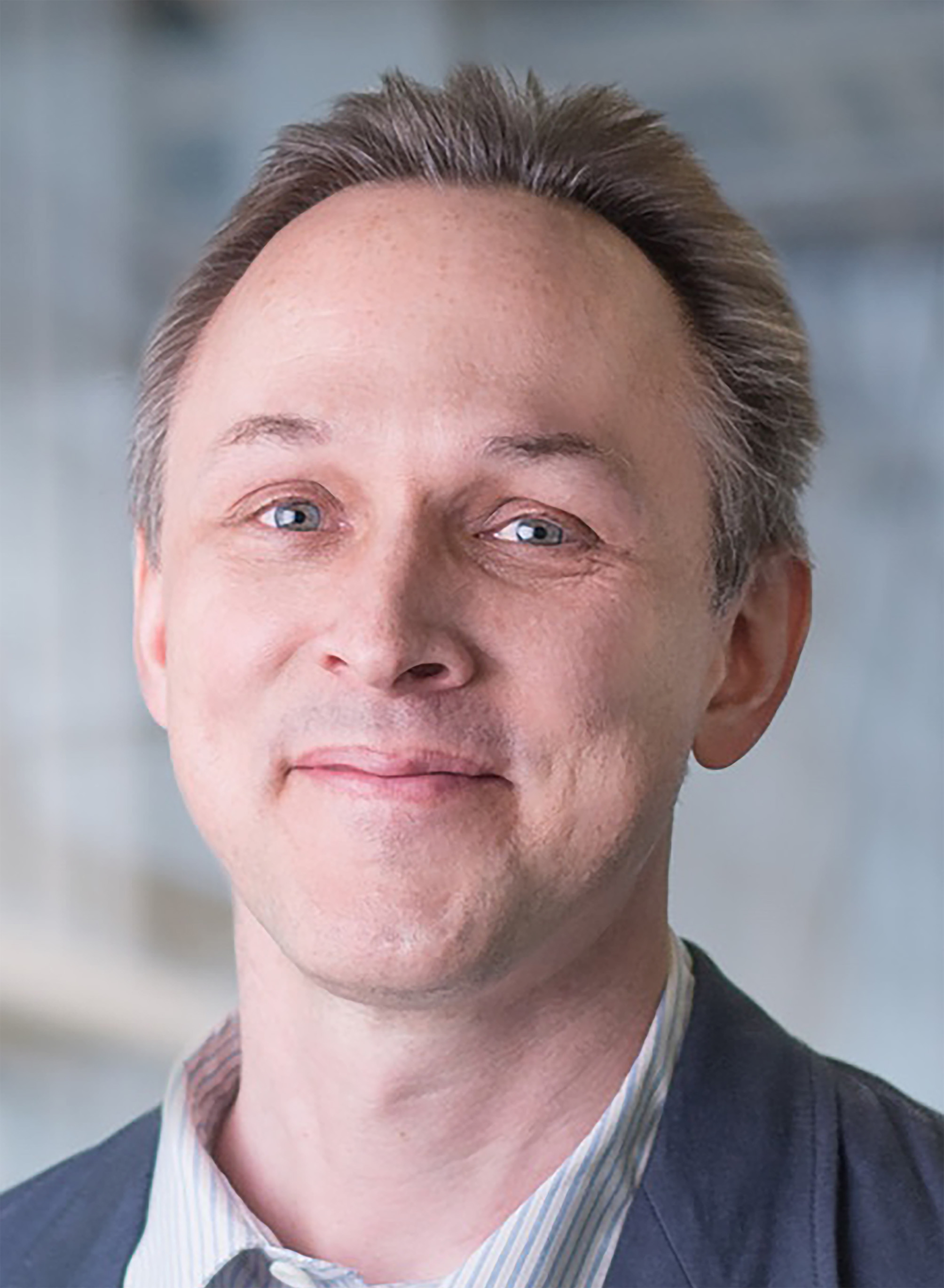
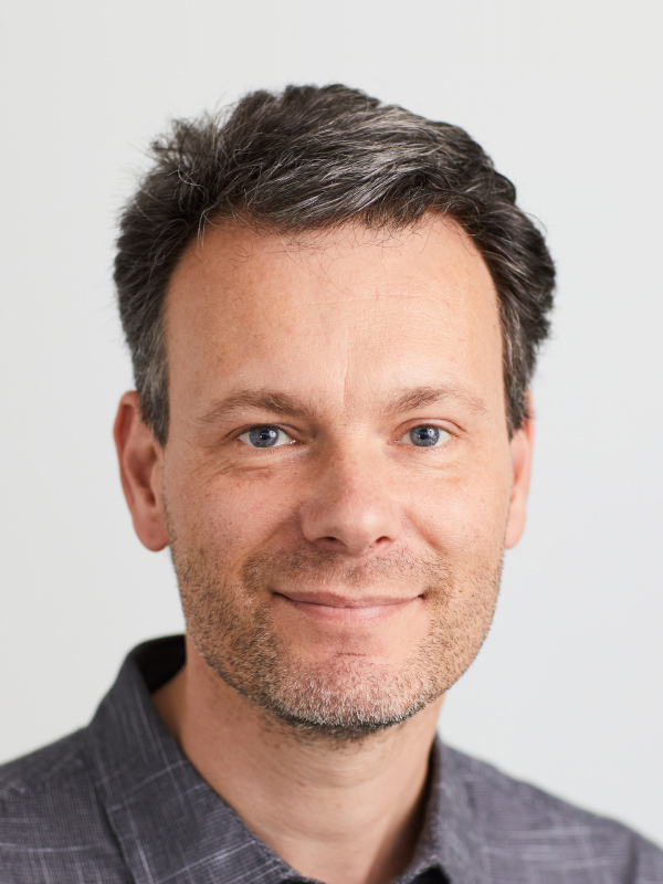
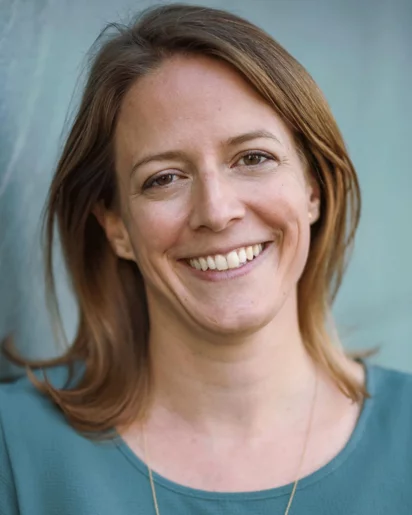
Daniel Sage is a Lecturer and Scientific Advisor at École polytechnique fédérale de Lausanne (EPFL) , affiliated with the Biomedical Imaging Laboratory (LIB) under the College of Engineering (STI) and School of Life Sciences (SV) . He specializes in bioimage informatics , structured-illumination microscopy , and deep learning applications for biomedical imaging. His work spans algorithm development for single-molecule localization microscopy (SMLM) , fluorescence imaging , and 3D reconstruction . His research group has developed open-source tools like FlexSIM for light inhomogeneity correction, DeepImageJ for integrating deep learning in ImageJ, and Steer'n'Detect for orientation-accurate template detection. His publications focus on correcting multiple-blinking artifacts in PALM, optimal transport metrics for SMLM evaluation, and contextual feature analysis for xenograft cell classification. He mentors PhD students and contributes to interdisciplinary education through courses such as Bioimage Informatics and Fundamentals of Image Analysis , emphasizing practical software solutions and Java programming for bioimage processing. His collaborations include institutions like Howard Hughes Medical Institute and Centre National de la Recherche Scientifique (CNRS) .
Professor Jeffrey W. Bode serves as Full Professor at the Department of Chemistry and Applied Biosciences at ETH Zurich, Switzerland, and maintains a secondary affiliation with the Institute of Transformative Biomolecules at Nagoya University, Japan. His internationally recognized research laboratory develops novel chemical reactions that operate under physiological conditions, bridging synthetic organic chemistry with biological applications. The Bode Research Group specializes in creating chemical methodologies that function in water and biological environments, including proteins, cells, and tissues. Their major research thrusts include acylboronate chemistry (particularly potassium acyltrifluoroborates or KATs), protein synthesis through ketoacid-hydroxylamine (KAHA) ligation, synthetic fermentation for drug discovery, and SnAP chemistry for N-heterocycle synthesis. These innovations enable applications in wound healing, drug delivery, cellular encapsulation, and artificial tissue development. The group's work on chemoselective ligation reactions has fundamentally advanced amide bond formation without traditional coupling reagents. Recent publications demonstrate a strong trajectory toward automated synthesis platforms, protein engineering, advanced bioconjugation techniques, and applications in chemical biology. The group has successfully commercialized SnAP chemistry through Sigma Aldrich and developed KAHA ligation into a robust method for synthesizing large proteins. Their research consistently focuses on creating molecules inaccessible through existing technologies, with particular emphasis on physiological compatibility and biological relevance. Professor Bode leads an international research team of approximately thirty PhD students and postdoctoral researchers from twenty different countries. The Bode Research Group maintains extensive collaborations across disciplines, contributing significantly to chemical biology, medicinal chemistry, and materials science. Their laboratory is equipped with advanced automation platforms for organic synthesis and maintains strong connections with pharmaceutical and biotechnology industries for translational applications of their chemical methodologies.
Michaël Unser is a Full Professor at École Polytechnique Fédérale de Lausanne (EPFL) in the School of Engineering , leading the Biomedical Imaging Laboratory . He serves as Academic Director for Imaging at EPFL and contributes to cross-departmental teaching in Microengineering , Mathematics , and Life Sciences Engineering . His research spans Image Processing , Medical Imaging , Wavelets , and Spline-based Modeling , with a focus on multiresolution analysis and single-molecule localization microscopy . He has mentored over 30 PhD students and supervised numerous research projects. Recent publications highlight advancements in super-resolution microscopy , deep learning integration , and inverse problem solving for biomedical imaging. His work emphasizes mathematical rigor and open-source software development for accessible bioimaging tools. IEEE Technical Achievement Award (2008) IEEE EMBS Career Achievement Award (2020) Three ERC Advanced Grants (FUNSP, GlobalBioIm, FunLearn) As Academic Director for Imaging , he leads EPFL's cross-disciplinary imaging initiatives. His teaching includes Fundamentals of Image Analysis and Signals and Systems courses.
Diego Ulisse Pizzagalli is an independent researcher in Computational Medicine affiliated with the Faculty of Biomedical Sciences at the Università della Svizzera italiana (USI), the Euler Institute, and the Institute for Research in Biomedicine (IRB). He holds roles as a lecturer at USI and teaching assistant, focusing on machine learning in medicine and signal processing. His work bridges immunology and AI, with projects involving wearable device monitoring in chronic diseases and the development of digital biomarkers. His research focuses on applying artificial intelligence to understand immune responses, tissue remodelling, and chronic diseases. Key areas include trajectory analysis of immune cells using intravital microscopy, predictive models for disease complications, and bioimage analysis. He leads the IMMUNEMAP initiative, an open immunology data platform, and collaborates on hardware prototyping for medical applications. Pizzagalli’s recent work emphasizes AI-driven medical imaging tools (e.g., CompositIA for CT scans, deep learning in forefoot morphology analysis) and trajectory-based studies of immune cell behavior. His projects integrate clinical data with computational models, such as predicting hospital admissions in chronic liver disease patients using wearable sensors. He co-leads projects funded by systemsx.ch, Apple Inc., and others. Supervises BSc/MSc students. The Digital Pathophysiology Lab at Euler Institute serves as his primary research base, promoting interdisciplinary collaboration in biomedical imaging, trajectory mining, and wearable sensor development.
Arne Seitz is a Lecturer at EPFL's School of Engineering (SV), holding academic roles in the departments of Life Sciences Engineering and Bioimaging Technology. As Unit Manager of the Bioimaging and Optical Imaging Technology Platform (PTBIOP), he oversees advanced microscopy infrastructure and supports research in biological imaging. His teaching spans courses like Bioimage informatics , Advanced Microscopy for Life Science , and Image Processing for Life Science , emphasizing practical computational tools for biological data analysis. Research focuses on developing open-source software tools for microscopy and bioimage analysis, including ABBA+, MarrowQuant, and QUAREP-LiMi initiatives. These projects address challenges in image registration, diagnostic workflows, and reproducibility standards in microscopy. His work bridges basic science and clinical applications, such as automated bone marrow analysis and molecular diagnostics for heart transplants. Active in multi-department collaborations, Seitz contributes to educational programs across SV-SSV, EDPO, EDMS, and EDMX engineering schools. His labs and platforms provide cutting-edge imaging solutions for EPFL researchers, emphasizing open-access methodologies and community-driven scientific standards.
Romain Guiet is a Scientist at the BioImaging and Optics Core Facility (BIOP) and a Lecturer in the EDMS program at École Polytechnique Fédérale de Lausanne (EPFL). He is affiliated with the School of Life Sciences (SV) and contributes to EPFL’s imaging community through technical support, teaching, and research. His roles span scientific support, education, and institutional service, including membership in the School Council SV. His research interests center on BioImage Analysis , with expertise in image acquisition (widefield, confocal, super-resolution), image processing , and quantitative data extraction . He develops open-source workflows using tools like ImageJ/Fiji, QuPath, Python, and R, promoting FAIR data principles. His background includes a PhD in Biology with a focus on immunology, cancer, 3D cell migration, and organoid systems. He supports experimental design and staining strategies for in vitro models including cell cultures and organoids. His recent publications highlight interdisciplinary work in organoid modeling, cardiogenesis, neural circuit imaging, and bioimage informatics. He co-developed DEVILS, a data visualization tool, and contributes to NEUBIAS training initiatives. His work emphasizes open science and community engagement through platforms like forum.image.sc. Metabolic reprogramming in osteoclasts Osteoclasts: Other functions RANKL-responsive epigenetic mechanism reprograms macrophages into bone-resorbing osteoclasts Nuclear receptors in osteoclasts WNT-modulating gene silencers as a gene therapy for osteoporosis, bone fracture, and critical-sized bone defects Romain Guiet advises on microscopy-based projects and mentors students through EPFL’s teaching programs. He is actively involved in grant-supported research and collaborative projects across EPFL. He is a key contributor to the BioImaging and Optics Core Facility, supporting a wide range of life science research. He also participates in the School Council SV, contributing to academic governance.
Erik Meijering is a Professor of Biomedical Image Computing at the School of Computer Science and Engineering of the University of New South Wales in Sydney, Australia. With a PhD and status as an IEEE Fellow (FIEEE), he leads research in bioimage analysis, neuron reconstruction, and cell tracking. His work bridges computer science, biomedical engineering, and neuroscience, with applications across multiple medical disciplines. Meijering's research focuses on developing computational methods for analyzing biological images, particularly in the areas of neuron tracing, cell tracking, and medical image segmentation. His work employs advanced techniques including deep learning, algorithm benchmarking, and large-scale image analysis. He has pioneered community efforts like the Cell Tracking Challenge and BigNeuron initiative, which have established standardized benchmarks for evaluating bioimage analysis algorithms. His research has significant implications for neuroscience, cancer diagnostics, and personalized medicine. His publication record shows a clear evolution from foundational work in image processing to cutting-edge applications of deep learning in bioimage analysis. Recent work emphasizes cross-domain generalizability of medical AI models, high-plexity microscopy techniques, and large-scale neuron reconstruction. Meijering's research consistently addresses the challenge of translating computational methods into practical biomedical applications. Distinguished Lecturer, IEEE Signal Processing Society (2022-2023) Guest Editor, Special Issue on Deep Learning in Biological Image and Signal Processing (2022) Chair, IEEE Technical Committee on Bio Imaging and Signal Processing (2018-2019) Associate Editor, IEEE Transactions on Medical Imaging (2004-2024) Meijering has secured substantial research funding from multiple sources including NHMRC, EU, NWO, and UNSW. His current projects include Revolutionizing Immunotherapy Response Prediction in Non-Small Cell Lung Cancer (2022-2025) and the Academic Start-Up Fund for Biomedical Image Computing (2019-2024). He has successfully led international collaborations through initiatives like BigNeuron and the Cell Tracking Challenge, which have brought together researchers from Australia, Europe, Asia, and the USA. His research group maintains active collaborations with numerous institutions worldwide, spanning neuroscience, medical imaging, and computational biology. Through his leadership of the BigNeuron project and Cell Tracking Challenge, Meijering has established international communities focused on advancing bioimage analysis. These initiatives have created standardized benchmarks and resources that accelerate progress in neuron reconstruction and cell tracking. His work has helped establish best practices for algorithm evaluation in bioimage analysis, significantly impacting how researchers develop and validate computational methods for biological imaging.
Marianne Liebi is a Tenure-Track Assistant Professor at the École Polytechnique Fédérale de Lausanne (EPFL), affiliated with the School of Engineering’s Institute of Materials. She leads the Structure and Mechanics of Advanced Materials group at the Paul Scherrer Institute (PSI) within the Photon Science Division. Her research focuses on developing advanced X-ray imaging techniques, particularly small-angle X-ray scattering (SAXS) tensor tomography, to study hierarchical materials such as biomimetic composites, cellulose-based materials, and bone tissues. She has held positions at Chalmers University of Technology (Sweden) and Empa, and her work bridges materials science, biomedical engineering, and nanotechnology. Education: PhD in Food Science from ETH Zurich (2013), followed by postdoctoral research at the Swiss Light Source. Professional roles include Assistant Professor at Chalmers University (2017–2020) and Scientific Group Leader at Empa (2020–2021). Research interests emphasize imaging complex structures across scales, with applications in 3D printing, industrial plastics, and biological tissues. Key methods include SAXS, ptychographic tomography, and X-ray fluorescence. Her group collaborates widely, addressing challenges in material design, medical diagnostics, and sustainable polymers. Advising and grants: While specific grants are not detailed, her leadership roles and publications indicate active research funding. Labs/teams: PSI’s Laboratory for Condensed Matter and Materials Science, with ongoing collaborations at synchrotron facilities like ESRF and MAX IV.