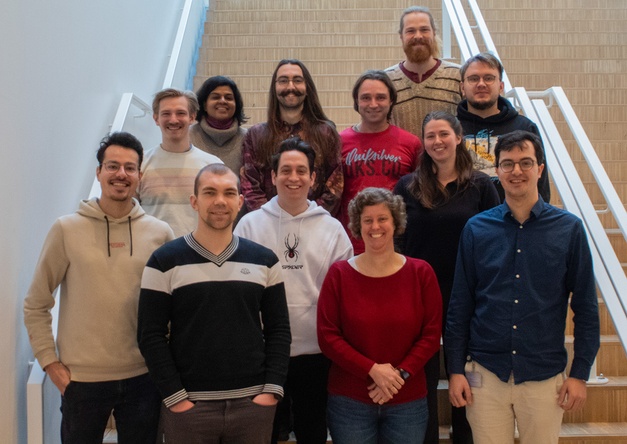
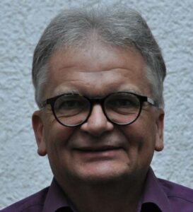
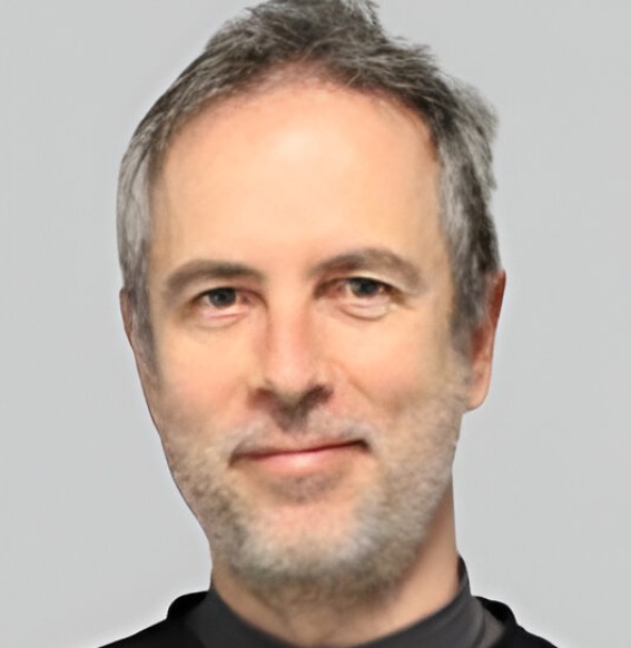
Tobias Baskin is a Professor in the Department of Biology at the University of Massachusetts Amherst, where he leads a research group focused on plant morphogenesis and growth regulation. His work integrates molecular genetics, cell biology, and computational approaches to understand how plant cells control shape and growth anisotropy. Education: B.Sc., Yale University, 1980 Ph.D., Stanford University, 1986 Postdoctoral Training, University of California, Berkeley (1987–1990) Postdoctoral Training, Australian National University (1990–1992) His research centers on plant morphogenesis , particularly how cell division , cytoskeleton dynamics , and cell wall mechanics coordinate to determine organ shape. He investigates the regulation of anisotropic cell expansion, using Arabidopsis thaliana as a model system. His lab employs quantitative imaging, genetic mutants, and in vitro assays to dissect molecular pathways governing growth. A key innovation includes developing algorithmic image processing tools for measuring growth profiles. The recent publications highlight a strong trend in understanding root development , environmental responses (e.g., thermomorphogenesis), and cell wall ultrastructure . His work spans molecular genetics, biomechanics, and computational biology, often involving interdisciplinary collaborations. While no formal awards are listed in the provided text, his research has been published in high-impact journals such as Development , Current Biology , and New Phytologist . Baskin advises students and collaborates widely, with recent work involving computer scientists and plant geneticists. His lab has secured research funding enabling long-term studies on growth regulation. He has contributed to methodological advances, such as the RTip automated root tracker, which supports precise phenotyping under perturbation. His laboratory is active in studying cortical microtubules and cellulose alignment, maintaining a focus on the mechanical basis of plant form. The team uses advanced microscopy techniques including atomic force microscopy and field-emission scanning electron microscopy to probe cell wall architecture.