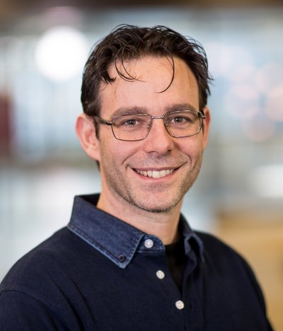
Massimo Mischi is a Full Professor at the Faculty of Electrical Engineering of the Eindhoven University of Technology (TU/e) and chairs the Signal Processing Systems (SPS) Division , the largest division at TU/e with over 250 researchers. He founded the Biomedical Diagnostics (BM/d) Lab in 2012, which now includes 180 researchers and clinical/industrial advisors, focusing on biomedical signal processing for diagnostics and monitoring.
Dr. James Ashton-Miller is a prominent faculty member in the Department of Mechanical Engineering at the University of Michigan, where he directs the Biomechanics Research Laboratory. He serves as a Center Member of the University of Michigan Injury Prevention Center and maintains affiliations with the Institute of Gerontology. His interdisciplinary work bridges engineering principles with medical applications, focusing on injury prevention across sports medicine, obstetrics, and geriatrics. Dr. Ashton-Miller's educational background includes: PhD from the University of Oslo, Oslo, Norway (1978-1983) MSME from M.I.T., Cambridge, MA, U.S.A (1972-1974) B.SC. (Hons) from the University of Newcastle-upon-Tyne, Newcastle-upon-Tyne, England (1967-1972) His research focuses on the biomechanics of injury prevention across multiple critical domains. In sports medicine, he has demonstrated that some ACL injuries are overuse injuries resulting from too many sub-maximal loading cycles that prevent healing of collagen damage. In women's health, his work on childbirth injuries addresses conditions that affect more women than breast cancer. His research on fall-related injuries in older adults reveals the dual threat of physical and cognitive factors. He also investigates sciatica, disc degeneration, and develops new medical devices for screening, diagnosis and treatment. Dr. Ashton-Miller's recent publications show a strong trend toward developing practical clinical applications from fundamental biomechanical research, with emphasis on advanced imaging methods, wearable sensors, and computational modeling for pelvic floor function assessment. His work consistently aims to translate engineering insights into clinical solutions for injury prevention. His research insights have earned him numerous national and international research awards, though specific awards aren't detailed in the available information. His work involves close collaboration with clinicians and surgeons who meet weekly to discuss progress and next steps. Dr. Ashton-Miller is deeply committed to mentoring, working with NIH K-series fellows along with 1-2 post-doctoral fellows, 3-5 PhD students, 2-4 M.S. students, 4-5 undergraduate students, and 2-4 young clinicians. His research is generously supported by the National Institutes of Health, National Science Foundation, National Basketball Association, Fortune 500 companies, and startup companies including Procter & Gamble and Hologic, Inc. He directs the Biomechanics Research Laboratory and co-leads the Pelvic Floor Research Group, where his teams develop new medical devices to improve screening, diagnosis, and treatment of various biomechanical conditions. These laboratories maintain strong clinical connections, ensuring research remains grounded in real-world medical challenges.
Indrani Bhattacharya, PhD, is an Assistant Professor in the Department of Biomedical Data Science and the Center for Precision Health and Artificial Intelligence (CPHAI) at Dartmouth College's Geisel School of Medicine. Her research focuses on developing human-centered AI systems for healthcare, particularly in multimodal medical imaging and behavioral health analytics. She holds a BS in Electrical Engineering from Jadavpur University (India), and MS/PhD from Rensselaer Polytechnic Institute (USA). Postdoctoral training at Stanford University's Department of Radiology further specialized her in biomedical imaging informatics. Research interests include: Integrating imaging and non-imaging data for precision medicine AI-driven prostate cancer detection/classification Multimodal behavior estimation for doctor-patient interactions Privacy-preserving sensor systems for group interaction analysis Her work bridges computer vision, medicine, and social science, with recent breakthroughs in MRI-ultrasound fusion AI outperforming radiologist interpretations in multi-center studies. Active in AI ethics and translational research, she leads teams developing clinical decision support tools for oncology and behavioral health. Key career milestones include: Postdoctoral scholar at Stanford University School of Medicine (2016-2021) Academic research staff at Stanford Radiology (2021-2022) Founding member of Dartmouth CPHAI precision health initiatives Labs/Teams: Leads the Biomedical AI for Healthcare group at Dartmouth, collaborating with Stanford and industry partners on AI-driven diagnostic systems.
Andrea Falini is a Full Professor of Neuroradiology at the Vita-Salute San Raffaele University (School of Medicine). He holds leadership roles including Head of the Neuroradiology Department at the Scientific Institute S. Raffaele Hospital and Head of the Advanced Diagnostic in Neuro-oncology Unit. His career spans clinical, academic, and administrative roles since 1991, with extensive experience in neuroradiology, neuro-oncology, and neuroimaging research. Education: MD (1986, University of Milan), PhD in Neurological Sciences (1989), and specialized training in Neurology and Radiology at leading institutions, including a visiting scholar position at UCSF (1996). Research interests focus on advanced MRI techniques, neuro-oncology, neurodegenerative diseases, and cognitive neuroscience. His work emphasizes functional MRI, diffusion tensor imaging, and clinical applications in glioma management and neurodegenerative disorders. Awarded the 1989 'De Visart' Prize and grants supporting his international collaborations. He has authored 218+ peer-reviewed articles, with an H-index of 40+, and actively contributes to academic committees and international conferences. Leadership roles include Vice-Director of the Neuroscience Division (2014–present) and coordination of the BraiMap Research Program. His academic teaching spans neuroradiology, biomedical engineering, and cognitive neuroscience courses.
Sandeep Kumar Mishra, PhD, is an Associate Research Scientist in the Department of Radiology & Biomedical Imaging at Yale School of Medicine, where he holds a primary appointment in the Magnetic Resonance Research Center within the Division of Bioimaging Sciences. He completed his doctoral training at Pondicherry University (2017) and finished post-doctoral research at Yale in 2024 before transitioning to his current research-intensive faculty role. Education: PhD, Pondicherry University – 2017 Post-doctoral Associate, Yale University – 2024 Research Focus: Mishra’s work integrates multinuclear magnetic resonance spectroscopy, responsive paramagnetic probes, and nano-constructs to quantify the tumor microenvironment. Major themes include in-vivo mapping of interstitial pH and sodium gradients in gliomas, development of Fe(II)/Co(II)/Ni(II)-DOTA tetraglycinate complexes for simultaneous pH–temperature sensing, and engineering dual-modal nano-agents that couple MR angiography with therapeutic delivery (chemo-photothermal, cryo-ablation, MMP inhibition). Publication Trends: Across 28 peer-reviewed articles (2016-2025) he demonstrates a sustained trajectory in cancer imaging, moving from theranostic nanoparticles toward sophisticated spectroscopic imaging of tumor acid–base and ionic homeostasis, with increasing translational orientation involving rodent glioma and hepatocellular carcinoma models. Collaborations & Affiliations: He is embedded in Yale’s inter-disciplinary MR research ecosystem, collaborating recurrently with faculty in Radiology, Biomedical Engineering, and the Magnetic Resonance Research Center (D. Coman, F. Hyder, P. Herman, J. Verhagen, J. Santana, A. Shewarega). Contact: sandeepkmishra11@gmail.com | Magnetic Resonance Research Center, 300 Cedar Street, New Haven, CT 06519, USA
Simon Brewster serves as Senior Research Fellow and Consultant Urological Surgeon at the University of Oxford's Hertford College, where he has been Clinical Tutor since 2002 after serving as Lecturer (2002-2011). His clinical work focuses on prostate cancer at Oxford University Hospitals NHS Trust, delivering exam-focused bedside teaching for Years 4-6 medical students. Education: BSc in Anatomy (1st class), Charing Cross Hospital Medical School, 1983 MBBS (Hons. Pathology), Charing Cross Hospital Medical School, 1986 FRCS exams and MD research, Bristol (1988-1998) Brewster's research program demonstrates sequential evolution from basic science (1998-2007) investigating IGF1R targeting and Wnt signaling in prostate cancer to clinical translation (2011-2022) evaluating multiparametric MRI integration in diagnostic pathways and active surveillance protocols. His work bridges laboratory discoveries with patient-centered outcomes, particularly examining how imaging advancements impact treatment decisions and quality of life. The multidisciplinary nature of his clinical research involves close collaboration between urologists, oncologists, radiologists, and pathologists to optimize prostate cancer management. Analysis of his 15 most recent publications reveals three dominant themes: (1) MRI-driven diagnostic refinement including pre-biopsy MRI implementation and active surveillance reclassification, (2) comparative treatment efficacy studies evaluating focal therapy versus radical prostatectomy, and (3) national guideline development through NICE recommendations. His work consistently addresses real-world clinical challenges in prostate cancer management, with strong emphasis on patient satisfaction metrics and practical implementation within the UK healthcare system. Scientific Awards: BJUI Article of the Week (November 18, 2018) for MRI impact on active surveillance As an educator, Brewster has co-supervised four higher research degrees and served as Clinical Supervisor for junior surgical trainees since 1998, including Intercollegiate ISCP Educational Supervisor role since 2009. His grant portfolio includes the UK PART trial (focal therapy vs radical prostatectomy, PI Prof Richard Bryant) and Oxfordshire MRI implementation study (2015-2019). He chairs the Hertford College/Vaughan Williams Prize for Excellence in Clinical Medicine. He leads a multidisciplinary research consortium including Dr. Val Macaulay (basic science) and Prof. W. Bodmer (Wnt pathway research), while coordinating clinical activities through Oxford University Hospitals NHS Trust. His work directly informs national prostate cancer pathways through British Association of Urological Surgeons committees and NICE guideline development.
Knut Håkon Hole is an Associate Professor at the University of Oslo's Department of Radiology and Nuclear Medicine. His research focuses on diagnostic imaging applications in oncology, particularly in prostate and rectal cancers. He specializes in MRI, PET, and radiogenomics techniques to assess tumor biology, treatment response, and recurrence. Expertise: Prostate cancer imaging, neoadjuvant therapy response, tumor hypoxia, and imaging biomarkers Key affiliations: Oslo University Hospital (Rikshospitalet), Radium Hospital Research interests include: Developing MRI and PET protocols for cancer staging and recurrence detection Integrating imaging with genomic data (radiogenomics) Optimizing therapeutic approaches using imaging biomarkers Recent work highlights: Prostate cancer radiogenomics and hypoxia biomarkers (2024) MRI/PET comparisons for tumor localization (2021-2023) Neoadjuvant therapy response assessment in rectal and breast cancers (2020-2023) Publications span over 50 peer-reviewed articles with a focus on translational imaging research. Collaborates extensively with oncology and urology teams.
Brent A. French is a Professor in the Department of Biomedical Engineering at the University of Virginia. His research focuses on integrating targeted drug/gene delivery with advanced imaging techniques (MRI, PET, ultrasound) to address cardiovascular disease mechanisms and therapy evaluation. He leads the Molecular Bioengineering Lab, emphasizing interdisciplinary collaboration and translational research. Education: B.S. in Biochemistry, Louisiana State University (1982) Ph.D. in Biochemistry, Louisiana State University (1987) Postdoctoral Fellowship at Baylor College of Medicine/NIH (1987–1991) Research Interests: Targeted drug and gene delivery systems Novel diagnostic imaging methods (e.g., ultrasound, MRI) Tissue engineering for cardiovascular applications Cardiovascular disease mechanisms and therapeutic strategies Grants & Projects: NIH R01 HL147193: $1.6M for developing molecularly targeted AAV for cardiac regeneration CHRB #207-10-19: $266K for in situ cardiac regeneration post-infarction NIH R01 HL147104: $2.99M for multiparametric MRI in coronary microvascular disease Labs/Teams: Molecular Bioengineering Lab (MR5 Building, Rooms 1205/1219), collaborating on interdisciplinary projects with cardiovascular and imaging experts.
Cornelius Faber is a University Professor in the Department of Radiology at the University of Münster, Germany, where he leads the Experimental Nuclear Magnetic Resonance research group. His work focuses on developing and implementing novel MRI techniques that extend the boundaries of magnetic resonance imaging in terms of spatial and temporal resolution, sensitivity, and specificity for physiological, structural, and molecular changes. He actively participates in the "Cells in Motion" interdisciplinary research initiative at the university. Professor Faber's research spans multiple critical areas in medical imaging and biomedical science. His primary expertise lies in MRI cell tracking , enabling visualization of cellular dynamics in vivo. He has made significant contributions to infection imaging , developing methods to detect and characterize microbial infections using MRI. His work on MR methodology development has advanced quantitative imaging techniques, while his research on multimodal integration in MR and MRI contrast mechanisms has provided deeper insights into molecular and cellular processes. His research bridges physics, engineering, and biomedical applications, with particular relevance to inflammation, cancer, neurological disorders, and cardiovascular disease. Analysis of Professor Faber's extensive publication record reveals a clear evolution from fundamental MRI technique development toward increasingly sophisticated applications in disease models. His recent work demonstrates a strong trend toward multimodal imaging approaches that combine MRI with complementary techniques such as mass spectrometry, optical imaging, and PET. This integration creates comprehensive diagnostic platforms that provide both anatomical and molecular information. A notable pattern is the focus on cellular dynamics, particularly immune cell behavior in inflammatory conditions and tumor microenvironments, with applications spanning neuroscience, oncology, and cardiology. Professor Faber leads a multidisciplinary research team of approximately 15 members, including scientists, doctoral students, technicians, and medical students. His laboratory is deeply integrated with the University of Münster's research infrastructure, particularly the Multiscale Imaging Centre. The group's work contributes significantly to advancing preclinical MRI methodologies while maintaining strong clinical relevance, with numerous publications in high-impact journals across medical imaging, neuroscience, and biomedical engineering disciplines.
Sameer Nath, MD, is an Associate Professor in the Department of Radiation Oncology at the University of Colorado Anschutz Medical Campus School of Medicine. He holds leadership roles as Vice Chair of Clinical Affairs, Clinical Practice Director at Anschutz Medical Campus, and Medical Director of Radiation Oncology at Highlands Ranch Hospital. Dr. Nath is board-certified in Radiation Oncology and practices at UCHealth Radiation Oncology and Rocky Mountain Gamma Knife Center. MD: University of California, San Diego School of Medicine (2010) Internship: University of California, San Diego (2011) Residency: Yale-New Haven Medical Center, Chief Resident in Radiation Oncology (2015) His research focuses on radiation therapy applications for prostate and lung cancers, brain metastases management, and immunotherapy-radiation combinations. He has pioneered work in stereotactic body radiation therapy (SBRT), tumor hypoxia, radiation pneumonitis prevention, and medical imaging innovations for treatment planning. Recent publications examine oligoprogressive prostate cancer management, radiation oncology nurse education, and deep learning applications in patient positioning. Dr. Nath's 15 most recent publications (2024-2017) demonstrate expertise in prostate cancer (4 articles), lung cancer (4 articles), brain metastases (4 articles), and medical imaging/education (3 articles). Key research themes include SBRT optimization, immunotherapy response monitoring, and radiation toxicity management. American Society of Radiation Oncology (ASTRO) Member Practice locations: UCHealth Radiation Oncology - Anschutz Medical Campus (Aurora, CO) and Rocky Mountain Gamma Knife Center - Anschutz Medical Campus (Aurora, CO) Hospital affiliations: UCHealth Highlands Ranch Hospital and University of Colorado Hospital
D.S. Fahmeed Hyder is a Professor of Biomedical Engineering at Yale University, with additional appointments in Radiology & Biomedical Imaging. He holds a Ph.D. from Yale University and leads the Hyder Lab, which focuses on advancing quantitative and translational imaging technologies using magnetic resonance methods to study brain function and dysfunction at the laminar level. His research integrates multidisciplinary approaches, including molecular imaging, neurophysiology, and material science. Research Interests: Neurovascular and neurometabolic coupling mechanisms Molecular imaging probes for disease diagnostics Functional MRI (fMRI) and metabolic imaging in health and disease Imaging applications for Alzheimer’s, stroke, and cancer Publications: Dr. Hyder’s recent work emphasizes cutting-edge imaging techniques, such as pH-sensitive biosensors, high-resolution fMRI, and multimodal optical imaging. His research highlights include studies on neurovascular dysfunction in Alzheimer’s models, therapeutic interventions for brain injury, and molecular imaging of tumor microenvironments. Awards: Niels Lassen Award (2003), for cerebral blood flow research Early Career Faculty Award (1998), NSF & NIH Pilot Awards from JDRF & Yale-UCL Collaborative (2008, 2013) Labs/Teams: The Hyder Lab collaborates with institutions like UCL and the James S. McDonnell Foundation, advancing translational imaging solutions for clinical applications.
Dr. Markus Zimmermann is a researcher at the Institute of Neuroscience and Medicine (INM-4: Physics of Medical Imaging) at the Research Center Jülich. His work focuses on advancing quantitative MRI techniques, particularly in water content mapping, multiparametric imaging, and ultrahigh-field MRI applications. He contributes to developing methods for eddy current characterization, multi-exponential relaxometry, and rapid whole-brain protocols. His research addresses neurological and medical imaging challenges, including cerebral pathologies and neurobiological implications. Key areas of expertise include MRI parameter estimation, medical imaging algorithms, and the integration of advanced imaging techniques for clinical and neuroscience applications. His projects often involve collaborations to validate methodologies using in vivo/ex vivo experiments and super-resolution reconstruction. Dr. Zimmermann’s work aims to enhance diagnostic precision and understanding of brain physiology through innovative MRI technologies.
Daniel M. Spielman is a Professor of Radiology and Electrical Engineering (courtesy appointment) at Stanford University. His office is located at the Lucas Center P-274. Dr. Spielman's research focuses on medical imaging, particularly magnetic resonance imaging and in vivo spectroscopy, with applications spanning cancer diagnosis, treatment monitoring, and neurodegenerative diseases. Dr. Spielman's research interests center on advancing magnetic resonance imaging (MRI) and spectroscopy techniques to provide clinically valuable metabolic imaging. His work addresses challenges of low metabolite concentrations, overlapping resonances, and field inhomogeneities through improved spectroscopic imaging, shimming methods, and optimal data quantification. Current applications include cancer diagnosis, treatment monitoring, prediction of therapy response, brain development in pediatric patients, and neurodegeneration associated with Alzheimer's disease, alcoholism, epilepsy, and aging. His publications demonstrate a strong focus on hyperpolarized carbon-13 MRI, metabolic imaging, and multimodal imaging approaches. The research trend shows increasing emphasis on clinical translation of advanced imaging techniques, particularly for oncology applications, with significant contributions to understanding metabolic processes in various disease states. Dr. Spielman collaborates extensively with faculty and staff across Stanford's Medical School and School of Engineering. He advises graduate students in Biophysics, Bioengineering, Electrical Engineering, and Medical Informatics programs. His research is conducted in collaboration with various departments at Stanford, focusing on developing novel imaging techniques that bridge engineering principles with clinical applications.
Bradley D Allen is an Assistant Professor in the Department of Radiology at Northwestern University Feinberg School of Medicine. He serves as Chief of Cardiovascular and Thoracic Imaging and is board-certified in Diagnostic Radiology by the American Board of Radiology. MD (2011) and MS (2015) from Northwestern University Feinberg School of Medicine Completed postgraduate training in Internal Medicine, Diagnostic Radiology, and Cardiovascular Imaging His research focuses on advancing cardiovascular and thoracic magnetic resonance imaging (MRI) techniques to improve disease diagnosis and treatment. Key areas include quantitative MRI, 4D flow imaging, hemodynamics, and applications in aortic aneurysms, lung cancer, and heart transplants. Recent publications highlight his work in applying 4D flow MRI to type B aortic dissection, Covid-19 vascular effects, automated MRI pipelines, and cardiac allograft monitoring. His awards include the Society of Cardiovascular Magnetic Resonance Fellowship (2023), multiple reviewer accolades from the Journal of Cardiovascular Magnetic Resonance, and recognition as Research Mentor of the Year (2022). Co-chair, American Heart Association CVRI Early Career Committee (2024–Present) Editorial Board Member, Canadian Association of Radiologists Journal (2025–Present) Member, American College of Radiology Cardiothoracic Committee (2022–Present)
Associate Professor Tan Cher Heng is an Associate Professor at the Lee Kong Chian School of Medicine (Nanyang Technological University Singapore) and Assistant Dean (Clinical Research). He serves as a Senior Consultant at Tan Tock Seng Hospital's Department of Diagnostic Radiology and holds leadership roles in the National Healthcare Group (NHG) as Group Chief Research and Innovation Officer and Director of the Academic Partnerships Office. His expertise spans radiology, artificial intelligence (AI) in healthcare, and clinical research innovation. He completed an MBBS (Singapore), FRCR (UK), and an Executive MBA from Insead. Education: MBBS, Singapore FRCR (UK) Abdominal Imaging Fellowship at MD Anderson Cancer Center Executive MBA from Insead Research Interests: His work focuses on advancing AI-driven diagnostics in radiology, optimizing imaging techniques for liver and prostate cancers, and translating clinical research into healthcare practice. He has pioneered initiatives like the national imaging AI platform (AimSG) and contributed to guidelines for imaging appropriateness and technology adoption. His research bridges clinical practice, innovation, and population health. Articles Summary: Recent publications emphasize AI applications in pneumonia diagnosis, radiology education, and liver/prostate cancer imaging. Themes include AI ethics, imaging cost-effectiveness, and guideline adaptation for diseases like tuberculosis and COVID-19. His work highlights leveraging technology to improve diagnostic accuracy and healthcare accessibility. Labs/Teams: He leads the Academic Partnerships Office and collaborates with global networks like the Asian Oceanian Society of Radiology. His roles in NHG and LKCMedicine drive cross-disciplinary projects in AI, clinical research, and medical innovation.