
Dr. Cassandra Sampaio Baptista is a Lecturer at the University of Glasgow's School of Psychology & Neuroscience. Her research focuses on brain plasticity in adulthood, particularly exploring how experiences like skill learning or rehabilitation influence structural and functional changes in the brain. She employs neuroimaging techniques such as fMRI neurofeedback and MRI to investigate mechanisms of myelin and white matter plasticity. Her work emphasizes translational applications, including stroke rehabilitation and promoting healthy aging. Key contributions include demonstrating myelin's role in motor learning and developing MRI protocols for white matter analysis. She collaborates on projects funded by the BIAL Foundation (2025–2026) and has supervised multiple postgraduate students. Recent publications highlight studies on oligodendrocyte dynamics, neurofeedback interventions for stroke survivors, and cross-species neuroscience approaches. While no specific awards are listed, her extensive publication record reflects her leadership in neuroplasticity research.
جستجوی استاد راهنمای دانشگاهی
برای مسیر پژوهشیتان، استاد راهنما، استاد دانشگاه و پژوهشگر مناسب را پیدا کنید و با آنها ارتباط بگیرید.
نمایش ۱-۱۵ از ۶۷۴ استاد راهنما
اطلاعات پروفایلها از صفحات عمومی دانشگاهها گرفته شده و ممکن است بهروز نباشد.
Thomas Perlmann is a Professor in Molecular Developmental Biology at the Karolinska Institutet , leading research at the Department of Cell and Molecular Biology and serving as Director of the Stockholm Branch of the Ludwig Institute for Cancer Research. He also holds the position of Secretary General of the Nobel Assembly and Nobel Committee for Physiology or Medicine since 2016. Ph.D. , Karolinska Institutet, 1991 M.Sc. , Stockholm University, 1987 Research Interests : The Perlmann lab investigates the specification and maintenance of dopamine neurons in the central nervous system, with a focus on transcriptional regulation , signaling pathways , and regenerative medicine applications for Parkinson’s disease and other neurodegenerative disorders. His work bridges developmental biology and neuroscience , emphasizing the role of transcription factors in neuronal identity and function. Recent Research Trends : Perlmann’s recent publications highlight the use of single-cell RNA sequencing to dissect dopamine neuron heterogeneity , epigenetic regulation during development, and transcriptomic changes in Parkinson’s disease models. His studies increasingly leverage multiomics and bioinformatics to map neuronal lineage trajectories and gene expression dynamics. Scientific Awards : Royal Medal by HM the King (2025) Nicholson Lecturer, Rockefeller University (2011) Göran Gustafsson Prize in Molecular Biology (1999) Eric K. Fernström Young Investigator Prize (1997) Advising & Collaborations : While no student names are explicitly listed, Perlmann collaborates extensively with researchers such as Malin Parmar , Agnete Kirkeby , and Per Svenningsson on projects related to neuronal development and cell therapy . His lab receives funding from institutions like the Ludwig Institute for Cancer Research . Labs & Teams : The Perlmann Lab at Karolinska Institutet includes researchers like Linda Gillberg , Laura Lahti , and Behzad Yaghmaeian Salmani , who work on mouse models , single-cell transcriptomics , and bioinformatics to study dopamine neuron biology.
Uwe Himmelreich is a Full Professor at the Faculty of Medicine, KU Leuven , leading the Biomedical MRI unit. He is actively involved in the Medical Imaging Division , KU Leuven Brain Institute , KU Leuven Institute for Integration of Micro- and Nano-scale Technologies , and KU Leuven Cancer Institute . Role: Full Professor and Head of Biomedical MRI Affiliations: Faculty of Medicine, Medical Imaging Division, LBI, LIMNI, LKI His research spans neuroscience , cardiovascular imaging , and nano/micro-scale technologies . Key projects include: MindMAP: Radiotherapy-induced neurotoxicity in juvenile brains Preclinical cancer models for oral tumors Quantitative T2 mapping of lung disease in murine models Neuroinflammation and cognitive decline in cryptococcosis Resistance training effects on cortical thickness in aging cohorts His work integrates MRI , multi-photon microscopy , and novel contrast agents for longitudinal in vivo studies. Methodologies include vascular density mapping , proton therapy verification , and preclinical radiotherapy evaluation . Notable contributions include: 2025: JAK/STAT inhibition in malaria-induced inflammation 2025: Manganese-enhanced MRI for cardiac injury 2025: Quantitative lung imaging at 9.4T 2024: IVIM as vascular density marker in rat brain 2024: Phase-change ultrasound agents for proton therapy
- Medical Imaging
- Radiology
- Neuroscience
- +۴ مورد دیگر

Dr. Teresa Puthussery is an Associate Professor in the School of Optometry & Vision Science at the University of California, Berkeley. Her research focuses on retinal neurobiology and neurophysiology, investigating how visual signals are encoded in healthy retinas and disrupted during degeneration. She uses advanced techniques like patch-clamp electrophysiology, immunohistochemistry, and microscopy to study retinal circuits, neurotransmitter receptors, and ion channels. Dr. Puthussery teaches courses on vision science anatomy, physiology, and problem-based learning, including VISION SCIENCE 206B/C and 260C. Her research explores questions such as how retinal neurons extract motion/spatial details, how photoreceptor mutations cause degeneration, and how inner retinal circuits adapt post-photoreceptor loss. Recent work includes studies on ON-type direction-selective ganglion cells, optogenetic therapy for vision restoration, and calcium dynamics in foveal ganglion cells post-degeneration. She collaborates on projects involving primate and rodent models, contributing to understanding retinal disease mechanisms and therapeutic targets. Dr. Puthussery’s lab (retinalab.berkeley.edu) emphasizes translational research, bridging basic science and clinical applications. Her work has been published in journals like Nature and Cell Reports , with a focus on retinal degeneration, synaptic plasticity, and optogenetic interventions. She actively participates in training future vision scientists through Berkeley’s Optometry program and oversees GSI affairs as a faculty advisor.
Juan Carlos Cerpa Gilvonioمشاهده پروفایل
پژوهشگر ارشد
Juan Carlos Cerpa Gilvonio is a Research Fellow at the Oxford Department of Experimental Psychology, University of Oxford. His research focuses on neural circuits in the basal ganglia, their neuromodulation, and decision-making processes. He investigates noradrenergic systems in the prefrontal cortex and corticosteroid effects on memory consolidation in rodents. He contributes to the Neurochemistry, Action and Control research group, exploring interdisciplinary neuroscience topics. His recent publications address noradrenergic innervation patterns and corticosteroid impacts on memory, reflecting his commitment to advancing neurochemical mechanisms understanding.
- Neuroscience
- Neurochemistry
- Decision-Making
- +۳ مورد دیگر
Andrew J. Todd is a Professor and Honorary Fellow in the School of Psychology & Neuroscience at the University of Glasgow. His research focuses on neurochemistry and synaptic connections in the mammalian spinal cord, particularly the organization of neuronal circuits underlying pain and itch perception. He employs techniques like immunocytochemistry, confocal microscopy, and electron microscopy. Collaborations include researchers from institutions such as UCL, Saga University, and the University of Pittsburgh. His work is funded by the Wellcome Trust and BBSRC. Roles: Professor, Honorary Fellow Affiliations: School of Psychology & Neuroscience, University of Glasgow Research Interests Dr. Todd investigates spinal dorsal horn circuits, including projection neurons, interneurons, and synaptic plasticity. Key topics include: Neurochemical characterization of spinal neurons Role of neuropeptides like substance P and gastrin-releasing peptide Mechanisms of neuropathic pain and spinal circuit adaptations Functional roles of specific neuron populations in laminae I-III Articles Overview Recent work includes studies on spinal projection neuron markers (e.g., Tacr1, Gpr83), synaptic circuits involving GRP-expressing neurons, and interneuron subtypes' roles in pain/itch. Notable findings include the absence of neuronal loss in neuropathic pain models and the identification of novel spinal circuits. Grants & Funding Funded by the Wellcome Trust and BBSRC . Collaborations span international institutions, emphasizing spinal neurobiology and sensory processing.
- Neurochemistry
- Neuroscience
- Pain Research
- +۳ مورد دیگر

Kimberly Keil Stietz is an Assistant Professor in the Department of Comparative Biosciences at the University of Wisconsin-Madison School of Veterinary Medicine. Her research focuses on environmental toxicology, neurotoxicology, and urology, particularly examining how developmental exposure to polychlorinated biphenyls (PCBs) affects urinary function. Education: B.S. in Biology, St. Norbert College (2010) Ph.D. in Comparative Biosciences, University of Wisconsin-Madison (2014) Postdoctoral research in neurotoxicology, University of California-Davis (2015-2019) Stietz's lab investigates the effects of environmental contaminants on the lower urinary tract, emphasizing PCB exposure during development. Using in vitro and in vivo mouse models, the lab studies disruptions in bladder epithelium organization, nerve fiber patterning, inflammation, and peripheral-central nervous system crosstalk. Key projects include analyzing PCB impacts on bladder barrier function, innervation patterns, inflammatory responses, and dorsal root ganglia signaling. Recent publications highlight her work on PCB effects across multiple domains: adult female bladder contractility (2023), prostatic collagen changes (2023), machine learning-aided metabolite analysis (2023, 2022), and developmental PCB exposure links to voiding physiology (2022). Her 2021 studies explored behavioral phenotypes in PCB-exposed mice, dendritic effects, and bladder inflammation, while 2019 work included host-microbe interactions and open-source uroflowmetry tools.
- Environmental Toxicology
- Neurotoxicology
- Urology
- +۴ مورد دیگر

Carlos Brody is the Wilbur H. Gantz III '59 Professor of Neuroscience at Princeton University, where he leads a research group at the Princeton Neuroscience Institute. His laboratory employs a unique combination of computational, behavioral, and electrophysiological techniques to investigate the neural mechanisms underlying cognitive abilities. Dr. Brody's research focuses on understanding how the brain processes information during cognitive tasks, particularly examining short-term memory, decision-making, and time perception. His lab trains rats to perform complex cognitive tasks while recording neural activity, and develops computational models to explain the experimental findings. They have pioneered the study of 'internal signals' in neural activity that constitute 'the internal conversation of the mind,' with their key discovery being 'nTc' (Neurally-inferred Time of Commitment), a biomarker that indicates decision commitment before overt behavioral responses. Dr. Brody's laboratory has been continuously supported by HHMI (Howard Hughes Medical Institute) with renewal until 2032. They are currently conducting groundbreaking research using multiple Neuropixels probes for large-scale recordings across the brain while rats perform cognitive behaviors, representing what Dr. Brody considers the future of cognitive systems neuroscience that combines advanced recording technology, AI-based analysis, and well-controlled behavioral paradigms. Scientific Awards HHMI Investigator (renewed until 2032) Advising and Research Support Dr. Brody has mentored numerous successful researchers who have secured faculty positions and leadership roles: Marino Pagan (Nature publication, SFARI Bridge to Independence Award) Edward Nieh (faculty position at University of Virginia) Manuel Schottdorf (Nature publication) Sue Ann Koay (publications in Neuron and eLife, Group Leader at Janelia) Brian DePasquale (faculty position at Boston University) Emily Dennis (Group Leader at HHMI's Janelia) Ahmed El Hady (Group Leader at Max Planck Institute) Abby Russo (joined CTRL-Labs startup) Diksha Gupta (Best Paper Award at RLDM conference) His lab is currently supported by HHMI funding and is planning to implement next-generation Neuropixels probes in Spring 2025 to record from 6,000-12,000 neurons simultaneously across multiple brain regions. Research Team and Facilities The Brody Lab features a diverse team ranging from purely computational to purely experimental researchers. The lab emphasizes minimizing barriers between computational and experimental approaches, encouraging researchers to move freely along this spectrum based on their interests. They maintain state-of-the-art facilities for behavioral training, electrophysiological recordings, and computational analysis, with plans to implement next-generation Neuropixels recording technology in Spring 2025.

Claire Acevedoمشاهده پروفایل
استادیار
Claire Acevedo is an Assistant Professor in the Department of Mechanical and Aerospace Engineering at the University of California San Diego (UCSD), affiliated with the Jacobs School of Engineering. Her lab, the Fracture and Fatigue of Skeletal Tissues Laboratory (F² Lab), focuses on understanding mechanisms of deformation, fracture, and biological responses in skeletal tissues and biomaterials across molecular to macro scales. She holds a Ph.D. from the Swiss Federal Institute of Technology Lausanne (EPFL) and completed postdoctoral research at UC San Francisco and UC Berkeley/Lawrence Berkeley National Laboratory. Dr. Acevedo’s research is funded by the National Science Foundation (NSF), National Institutes of Health (NIH), and the Advanced Light Source. Her work bridges biomechanics, materials science, and high-energy X-ray physics to address bone fragility in aging and diabetes. Key projects include investigating collagen cross-linking effects on bone mechanics and developing novel imaging techniques like deep learning-enhanced synchrotron micro-CT. Education: Ph.D., Swiss Federal Institute of Technology Lausanne (EPFL) Postdoctoral Research: UC San Francisco & UC Berkeley/Lawrence Berkeley National Lab Previous Faculty Position: University of Utah (Mechanical Engineering) Recent contributions include the NSF CAREER Award for studying fracture mechanisms in fragile bones and an NIH R21 grant to explore collagen-level diabetes impacts. Her lab collaborates with the University of Utah Tanner Dance Program to develop K-12 educational initiatives linking dance with biomechanics. Publications span topics like synchrotron imaging innovations, diabetes-induced bone fragility, and collagen nanomechanics. Students in her lab have contributed to advancements in fatigue testing, cross-link analysis, and imaging algorithms. Awards: NSF CAREER Award (2024) NIH R21 Grant (2023) Alice L. Jee Award (2022) Nikon Small World Image of Distinction (2024) The F² Lab hosts a dynamic team with ongoing projects on glycemic effects, synchrotron techniques, and biomaterial design. Future work emphasizes translating findings into clinical fracture prevention strategies and educational outreach.
- Biomechanics
- Fracture Mechanics
- Skeletal Tissues
- +۵ مورد دیگر
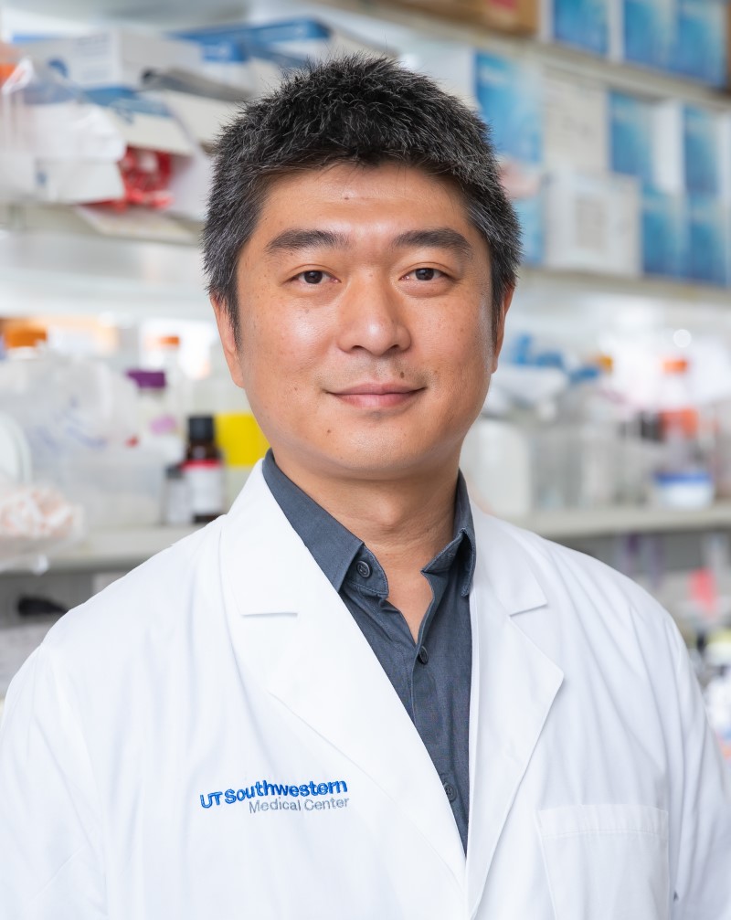
Jun Wuمشاهده پروفایل
استادیار
Dr. Jun Wu is an Assistant Professor in the Department of Molecular Biology at UT Southwestern Medical Center. He holds a PhD in Life Science from the University of Tennessee and completed postdoctoral training at the University of Southern California and the Salk Institute. His research focuses on stem cell biology, genome editing, and interspecies chimeras to advance regenerative medicine and developmental biology. Education: Bachelor of Medicine, Shandong University School of Medicine (China) PhD in Life Science, University of Tennessee (2013) Postdoctoral Fellowships: USC (2013-2015) and Salk Institute (2015-2017) Research Interests: Generation of pluripotent stem cells with distinct molecular/phenotypic features Development of interspecies blastocyst complementation systems Modeling peri-implantation human development using stem cell embryo models Study of species-specific developmental barriers and evolutionary biology Application of chimeras to study cancer resistance and organ size determination Lab Activities: The Wu Lab develops stem cell models to study mammalian development and create regenerative therapies. Key projects include: Creation of interspecies chimeras (human-monkey, rat-mouse) Development of blastoids and peri-gastruloids Analysis of species-specific cell competition mechanisms Engineering cross-species organogenesis systems Grant Activities: Focuses on NIH-funded projects related to stem cell biology, interspecies chimerism, and regenerative medicine applications. Collaborates with institutions like Salk Institute and University of California campuses. Lab Team: Includes postdoctoral researchers like Dr. Yi Ding and a multidisciplinary team of molecular biologists, bioengineers, and computational biologists.

Heidi Johansen-Berg is Pro-Vice Chancellor (Strategic Initiatives) at the University of Oxford and Associate Head (Research and Innovation) in the Medical Sciences Division. She holds a Professorship in Cognitive Neuroscience and a Wellcome Principal Research Fellowship at the Nuffield Department of Clinical Neurosciences, where she leads the Plasticity Group at the Oxford Centre for Functional MRI of the Brain (FMRIB). Her research centers on neuroplasticity mechanisms in the sensorimotor system, with emphasis on white matter plasticity, activity-dependent myelination, and implications for stroke rehabilitation and age-related brain decline. She integrates multimodal neuroimaging with behavioral studies to investigate how the brain adapts to learning, experience, and damage, translating findings into therapeutic interventions for neurological conditions. Recent publications reveal strong thematic trends in sleep-motor interactions post-stroke, exercise-induced neuroprotection in aging and adolescence, and experience-dependent white matter remodeling. Her work demonstrates how physical activity modulates brain structure-function relationships across the lifespan, with direct applications for neurorehabilitation protocols. Scientific recognition includes: Fellow of the Royal Society (FRS) Fellow of the Academy of Medical Sciences (FMedSci) Wellcome Principal Research Fellowship Professor Johansen-Berg directs the WIN Plasticity Group and co-leads the WIN Neuroplastics Network and Oxford University Centre for Integrative Neuroimaging (OxCIN). Her research program drives translational initiatives in stroke recovery and brain health maintenance, with ongoing projects examining digital sleep therapies, myelin dynamics, and exercise neuroscience through large-scale clinical trials and advanced imaging methodologies.

Katrina Choeمشاهده پروفایل
استادیار
Katrina Choe serves as Assistant Professor in the Department of Psychology, Neuroscience & Behaviour at McMaster University and holds a Tier 2 Canada Research Chair in Neurobiology of Social Behaviour. Her research program investigates the multi-level neurobiological mechanisms underlying psychiatric disorders, with primary focus on autism spectrum disorders (ASD) and oxytocin signaling pathways. Her academic training includes: PhD in Neuroscience from McGill University (2013) Honours BSc in Zoology from University of Toronto (2002-2006) Postdoctoral Fellowship at UCLA (2013-2020) Dr. Choe's research employs an integrative approach spanning molecular, cellular, circuit, and network levels to examine how ASD-associated gene mutations disrupt social behavior. Current work centers on oxytocin signaling mechanisms in ASD, convergent neurobiological pathways across psychiatric disorders, and the role of glial cells in neural circuit function. Her lab utilizes advanced techniques including optogenetic fMRI, single-cell RNA sequencing, and multi-level behavioral assays in genetic mouse models. Analysis of her 15 most recent publications reveals a clear research trajectory: early work (2015-2020) established foundational knowledge in vasopressin neuron regulation and salt homeostasis, while recent publications (2022-2025) demonstrate a focused shift toward ASD mechanisms, oxytocin signaling, and social circuit dysfunction using the Cntnap2 knockout model. This evolution reflects her transition from postdoctoral training to independent research leadership. Her scientific recognition includes: Tier 2 Canada Research Chair in Neurobiology of Social Behaviour (2022) NIMH K99/R00 Award CIHR Postdoctoral Fellowship Dr. Choe actively mentors six graduate students across PhD and MSc programs while leading a dynamic research team comprising postdoctoral fellows, laboratory technicians, and undergraduate researchers. Her program receives substantial support from major grants including a 5-year CIHR Project Grant and NSERC Discovery Grant focused on 'The role of CASPR2 in central oxytocin system development.' The Choe Lab maintains active collaborations with leading neuroscience groups including the Bourque, Prager-Khoutorsky, and Cunningham labs, as evidenced by participation in the 4th 1000 Islands/Gananoque Meeting on Hypothalamic Mechanisms. Her laboratory, established in 2020, operates as a multidisciplinary hub utilizing molecular biology (qPCR, RNA-seq), advanced imaging (lightsheet, confocal), electrophysiology (in vitro and in vivo), and behavioral neuroscience approaches to investigate social behavior mechanisms. Current projects examine microglia-astrocyte-neuron interactions in social circuit function and the therapeutic potential of oxytocin for ASD-related social deficits.
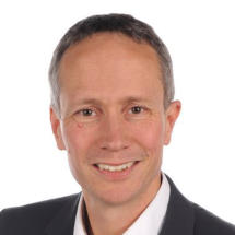
Cornelius Faber is a University Professor in the Department of Radiology at the University of Münster, Germany, where he leads the Experimental Nuclear Magnetic Resonance research group. His work focuses on developing and implementing novel MRI techniques that extend the boundaries of magnetic resonance imaging in terms of spatial and temporal resolution, sensitivity, and specificity for physiological, structural, and molecular changes. He actively participates in the "Cells in Motion" interdisciplinary research initiative at the university. Professor Faber's research spans multiple critical areas in medical imaging and biomedical science. His primary expertise lies in MRI cell tracking , enabling visualization of cellular dynamics in vivo. He has made significant contributions to infection imaging , developing methods to detect and characterize microbial infections using MRI. His work on MR methodology development has advanced quantitative imaging techniques, while his research on multimodal integration in MR and MRI contrast mechanisms has provided deeper insights into molecular and cellular processes. His research bridges physics, engineering, and biomedical applications, with particular relevance to inflammation, cancer, neurological disorders, and cardiovascular disease. Analysis of Professor Faber's extensive publication record reveals a clear evolution from fundamental MRI technique development toward increasingly sophisticated applications in disease models. His recent work demonstrates a strong trend toward multimodal imaging approaches that combine MRI with complementary techniques such as mass spectrometry, optical imaging, and PET. This integration creates comprehensive diagnostic platforms that provide both anatomical and molecular information. A notable pattern is the focus on cellular dynamics, particularly immune cell behavior in inflammatory conditions and tumor microenvironments, with applications spanning neuroscience, oncology, and cardiology. Professor Faber leads a multidisciplinary research team of approximately 15 members, including scientists, doctoral students, technicians, and medical students. His laboratory is deeply integrated with the University of Münster's research infrastructure, particularly the Multiscale Imaging Centre. The group's work contributes significantly to advancing preclinical MRI methodologies while maintaining strong clinical relevance, with numerous publications in high-impact journals across medical imaging, neuroscience, and biomedical engineering disciplines.
- MRI cell tracking
- infection imaging
- MR methodology
- +۶ مورد دیگر
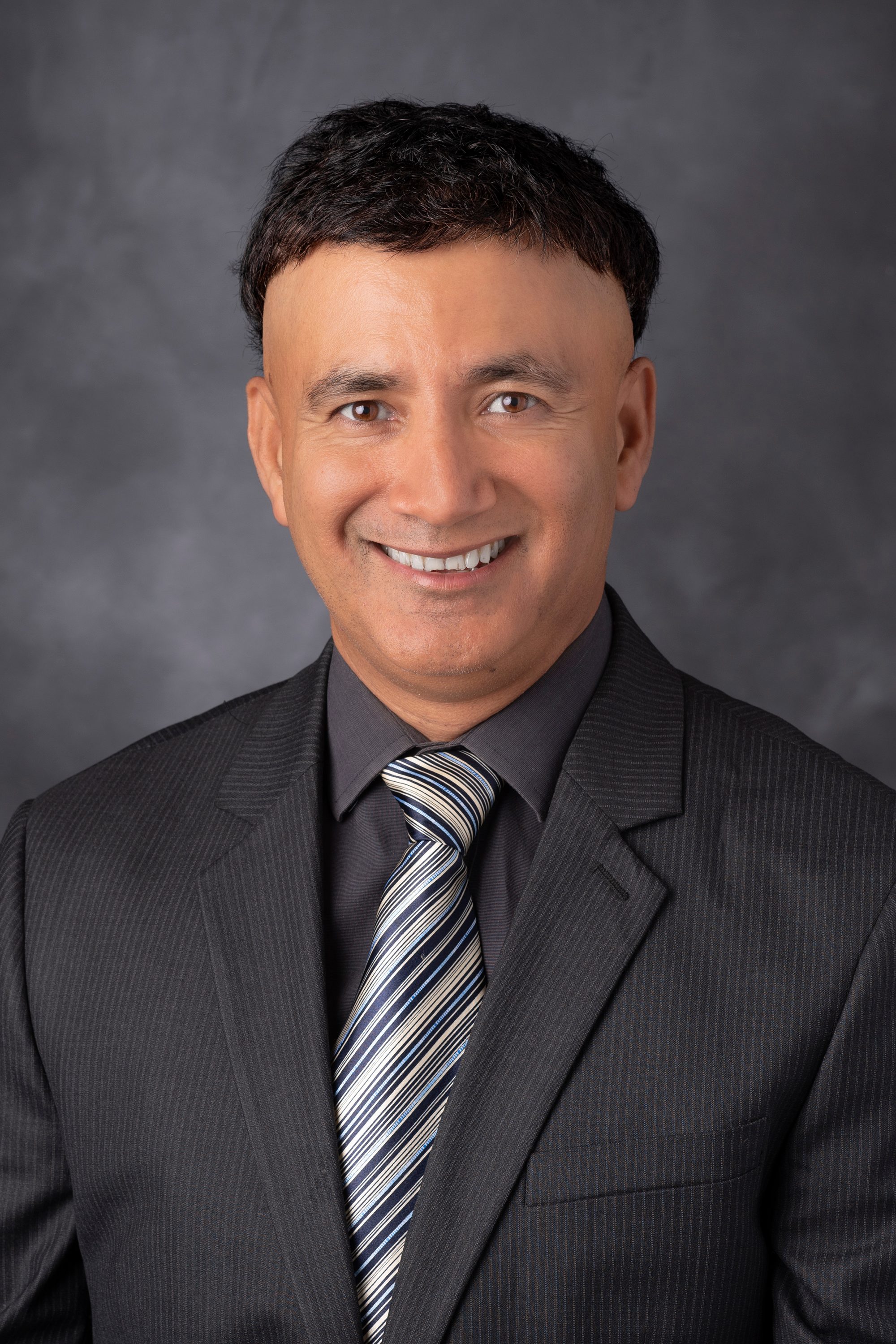
Pratip K. Bhattacharya, Ph.D. , is an Associate Professor in the Department of Cancer Systems Imaging and the Department of Imaging Physics at The University of Texas MD Anderson Cancer Center, with a joint appointment in the Graduate School of Biomedical Sciences at The University of Texas Health Science Center. He is a principal investigator leading the Bhattacharya Laboratory, dedicated to advancing magnetic resonance imaging (MRI) through hyperpolarization techniques for applications in cancer and cardiovascular diseases. His research focuses on developing real-time metabolic and molecular imaging methods using hyperpolarized 13 C and 15 N-labeled compounds and silicon nanoparticles. These innovative probes significantly enhance MRI sensitivity, enabling non-invasive assessment of tissue metabolism and targeted imaging. His lab's work spans three primary areas: real-time metabolic MR imaging, targeted molecular MR imaging with functionalized silicon nanoparticles, and high-resolution MR metabolomics. These efforts are aimed at improving disease diagnosis and therapy monitoring. Analysis of his recent publications reveals a strong and consistent focus on hyperpolarized MRI, particularly using silicon particles and metabolic tracers like succinate, to visualize cancer metabolism and cardiovascular conditions in vivo. His research integrates physics, chemistry, and biomedical engineering to create novel imaging tools with direct clinical translational potential. PHIP Hyperpolarization Dynamic Nuclear Polarization (DNP) Hyperpolarized Silicon Nanoparticles Real-Time Metabolic Imaging Cancer and Cardiovascular Imaging Theranostic Applications Dr. Bhattacharya actively mentors graduate students and postdoctoral fellows, including Saleh Ramezani, Jose Enriquez, Dontrey Bourgeois, and Kang-Lin Hsieh. He collaborates closely with physician-scientists, radiologists, and oncologists to ensure his imaging science innovations address critical clinical needs. His laboratory is supported by grant funding, facilitating the development of cutting-edge imaging technologies. The Bhattacharya Laboratory is a key component of the Division of Diagnostic Imaging at MD Anderson, fostering a collaborative environment for interdisciplinary research. The lab focuses on translating fundamental discoveries in hyperpolarization physics into practical tools for improving cancer care.
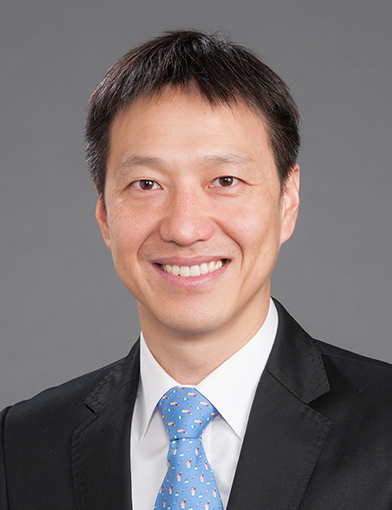
Bumsoo Ahnمشاهده پروفایل
استادیار
Bumsoo Ahn, PhD, is an Assistant Professor in the Department of Gerontology and Geriatric Medicine at Wake Forest University School of Medicine. He is also affiliated with the Wake Forest Institute for Regenerative Medicine, Center for Redox Biology and Medicine, Sticht Center for Healthy Aging and Alzheimer’s Prevention, Cardiovascular Sciences Center, Comprehensive Cancer Center, Center for Precision Medicine, and the Center for Vaccines at the Extremes of Aging. His educational background includes a BS from Yonsei University (2004), an MS from the University of North Carolina at Chapel Hill (2011), and a PhD from the University of Florida (2015). Dr. Ahn's research focuses on the mechanisms of muscle aging and wasting, particularly sarcopenia and cancer cachexia. His work emphasizes mitochondrial redox biology, oxidative stress, and the role of reactive oxygen species in muscle dysfunction. He employs transgenic mouse models to investigate causal relationships and test potential interventions, including unacylated ghrelin. His research spans molecular, cellular, and physiological levels, aiming to extend active and functional lifespans. His recent publications highlight investigations into mitochondrial hydrogen peroxide scavenging, SERCA activity restoration, neuronal SOD1 expression, and pharmacological protection against diaphragm dysfunction. These studies reflect a strong interdisciplinary focus on redox biology, muscle physiology, aging, and translational therapeutics. Scientific Awards: No awards listed in the provided text. Dr. Ahn contributes to graduate education through the Molecular Medicine and Translational Science PhD program and the Integrative Physiology and Pharmacology PhD program. His laboratory operates at the intersection of aging, regenerative medicine, and redox biology, engaging in collaborative research with multiple centers. While specific grants are not mentioned, his affiliations suggest involvement in federally and institutionally funded research initiatives focused on healthy aging and disease mitigation. The Ahn Lab investigates mitochondrial redox biology and muscle contractile properties in the context of aging and chronic diseases, utilizing animal models to identify therapeutic strategies for preserving muscle function.
- Mitochondria
- Aging
- Muscle
- +۱۲ مورد دیگر