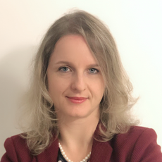
Neda Haj Hosseini is a Senior Lecturer and Associate Professor in Biomedical Engineering at Linköping University's Department of Biomedical Engineering (IMT) . She contributes to teaching courses like TBMT56 - Medical Technology and TBME08 - Biomedical Modeling and Simulation , while leading research initiatives in AI-driven cancer diagnostics and biomedical optics. Research Focus: Development of AI methods for cancer diagnostics, optical coherence tomography (OCT) applications, and fluorescence spectroscopy in surgical guidance Affiliations: Center for Medical Image Science and Visualization (CMIV) , Analytic Imaging Diagnostic Arena (AIDA) , Swedish Medical Technology Association Recent Research Trends demonstrate expertise in applying deep learning to: Pediatric brain tumor classification using multimodal imaging Optical biopsy techniques for intraoperative decision support Automated biomarker quantification in histopathology Medical imaging data integrity and algorithm validation Scientific Awards include grants from: Joanna Cocozza Foundation (2022) Swedish Childhood Cancer Foundation (2024) Academic Leadership involves mentoring students in projects such as: "Multiple Instance Attention-based Learning for Brain Tumor Classification" "Vision Transformers for Multiclass Brain Tumor Tissue Classification" "Reaction-diffusion Models for Image-driven Tumor Simulation"
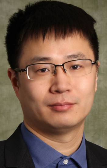
Wenfeng Zhao is an Assistant Professor in the Department of Electrical and Computer Engineering at Binghamton University. He holds a PhD from the National University of Singapore (2014) and BS/MS degrees from Huazhong University of Science and Technology (2007-2009). Prior to this role, he conducted postdoctoral research at the University of Minnesota's Biomedical Engineering Department. His research focuses on neural engineering, compressed sensing, ultra-low-power VLSI systems, and in-memory computing. Key areas include hardware security, biomedical signal processing, and energy-efficient computing architectures. His work spans applications in neural interfaces, cryptographic hardware, and IoT edge devices. Recent publications highlight advancements in block-cipher-in-memory architectures, emotion recognition via EEG analysis, and energy-efficient FPGA accelerators for neural networks. His research also addresses challenges in cryogenic memory systems and MRI-compatible neural recording devices. Zhao's contributions emphasize interdisciplinary approaches at the intersection of hardware design, signal processing, and cybersecurity. His lab develops novel solutions for low-power embedded systems and trustworthy IoT infrastructure.
Andrew Godley, Ph.D., is the Associate Vice Chair of Clinical Physics Operations and Quality and an Associate Professor of Radiation Oncology at UT Southwestern Medical Center. He is part of the Department of Radiation Oncology’s Division of Medical Physics and Engineering. Dr. Godley holds a Texas Medical Physics License and is board-certified in therapeutic radiologic physics by the American Board of Radiology. Education: Received his Ph.D. in high-energy physics from the University of Sydney as part of the NOMAD experiment at CERN. Completed a postdoctoral fellowship at the University of South Carolina with the MINOS experiment at FermiLab. Transitioned to medical physics at the Medical College of Wisconsin. Research Interests: Focus on advanced radiation therapy techniques including brachytherapy, SBRT, Gamma Knife, MR-linac integration, and adaptive radiotherapy. His work emphasizes improving treatment accuracy, patient-specific quality assurance, and innovative approaches to personalized oncology care. Publications: Over 118 peer-reviewed articles, with recent work emphasizing adaptive radiotherapy strategies, MR-guided therapies, and computational tools for dose verification. Professional Contributions: Active in developing clinical protocols for radiation physics operations, including machine QA/commissioning and program development for new technologies like MR-linac systems.
Prof. George Magoulas is a Professor of Computer Science at the University of London's School of Computing and Mathematical Sciences and Director of the Birkbeck Knowledge Lab. He specializes in machine intelligence, machine learning algorithms, and AI system architectures, with applications in healthcare (e.g., neurodegenerative disease diagnosis) and educational technologies. His research has received awards from IEEE, ACM, and others. He holds a PhD in Nonlinear Optimization for Neural Networks and a PGCE in Higher Education. Education: BEng/MEng (Integrated Master's in Systems & Control Engineering), University of Patras, Greece PhD in Nonlinear Optimization for Neural Networks Learning, University of Patras, Greece PGCE in Teaching and Learning (Higher Education) Research & Leadership: He leads the Birkbeck Knowledge Lab, focusing on AI's impact on learning and communication. His work includes designing learning algorithms for psychophysiological data modeling and developing the cloudUPDRS app for Parkinson's disease assessment. He has supervised over 12 PhD students and contributed to 200+ publications. Awards & Recognition: Stanford’s “World’s top 2% of Scientists” (2024) Best Paper Awards at IEEE, ACM, and EUNITE Keynote speaker at major AI and e-learning conferences Honorary membership in the Hellenic Artificial Intelligence Society Administrative Roles: Director of Teaching & Learning Quality (2016–2023) Chair of Postgraduate Programmes Exam Board (2010–2022) Editor-in-Chief, International Journal on Artificial Intelligence Tools Teaching: He teaches courses on Artificial Intelligence, Neural Networks, and Project Management at both undergraduate and postgraduate levels. Labs & Collaborations: He directs the Birkbeck Knowledge Lab and is a member of the Data Science and AI Research Group. His projects include analyzing violent cycles using AI and collaborating on EU-funded initiatives.
Prof. Markus Axer is a Professor and Deputy Head of the Structural and Functional Organisation of the Brain (INM-1) at the Institute of Neuroscience and Medicine (INM) within Forschungszentrum Jülich. His research focuses on connectomics, neuroimaging technologies (e.g., 3D-Polarized Light Imaging), and high-performance computing applications in brain architecture analysis. He leads the 'Fiber Architecture' working group, advancing microscopy techniques like scattered light imaging and MRI-histology correlation for studying brain microstructure. His work bridges experimental neuroscience with computational methods, aiming to decode brain organization at meso- and macroscales. Key achievements include developing the HippoMaps atlas of the human hippocampus and improving fiber orientation mapping in brain tissue. Awards include Fellowship in the Royal Netherlands Academy of Arts and Sciences (2024). Research emphasizes cross-modal data integration, with applications in Alzheimer’s disease biomarker validation and primate brain evolution studies. He collaborates with academic institutions like the University of Wuppertal and contributes to international initiatives like the BigBrain Analytics Learning Laboratory.
Nico Buls is a Researcher in the Department of Radiology at Universitair Ziekenhuis Brussel (UZ Brussel). His work focuses on translational projects in medical imaging physics, radiation dosimetry, and engineering, with an emphasis on advanced imaging technologies for diagnostic and interventional radiology. Key research areas include imaging physics, spectral CT techniques, iterative reconstruction in CT, radiation dosimetry, neuro MRI applications, and applied statistics in medical imaging. He leads projects such as the PAD flow study (quantitative blood flow assessment via 4D CT) and the evaluation of lung ventilation using Xenon gas-enhanced CT. Buls collaborates internationally, with active research in Belgium and beyond. Affiliations: UZ Brussel, Research Centre for Digital Medicine. Grants/Projects: 12 active projects including OZR4357 (PhD stipend), PAD flow, and Xenon gas imaging studies. Scientific Awards: Editor's Recognition Award (2014, 2016) Radiological Society of North America (RSNA) Fellowship (2011) Young Physicist Grant (2001) Advising/Grants: Supervises 20+ research projects and students, with notable contributions to 4D CT applications and radiation safety protocols. His lab, the Research Centre for Digital Medicine, drives innovation in clinical imaging technologies.
Li Min is an Associate Professor at the University of California, Los Angeles (UCLA), affiliated with the Department of Anthropology. His research focuses on Chinese prehistoric and Bronze Age archaeology, emphasizing state formation, social memory, and climatic responses. He also studies maritime archaeology of the Asiatic Trade in the Early Modern Era, using ceramic analysis to trace global trade impacts. Li teaches graduate seminars in archaeology theories and undergraduate courses on Chinese civilizations, collaborating across Anthropology, Asian Languages and Cultures, and the Interdepartmental Program of Archaeology. He co-directs the Wen-Si River Basin archaeological project with Chinese institutions. His 2018 book, Social Memory and State Formation in Early China , is a key contribution to the field. His education includes a Ph.D. from the University of Michigan (2008). Research interests further encompass landscape archaeology, integrating ceramics analysis with remote sensing and historical records. Subfield expertise includes social archaeology, material culture studies, and historical anthropology. Recent publications (2023–2025) concentrate on neuro-oncology imaging innovations, including MRI techniques for glioma characterization, adaptive clinical trials (e.g., GBM AGILE), and biomarker development. These studies highlight advanced applications of AI in medical imaging and molecular targeting therapies for brain tumors. Despite no listed awards, his work on imaging biomarkers and tumor response assessment has advanced clinical neuro-oncology standards. He advises on interdisciplinary collaborations, such as the Wen-Si project, and participates in global clinical trials for glioblastoma therapies.
Jingjing Zou is an Assistant Professor in Residence at the Herbert Wertheim School of Public Health & Human Longevity Science, University of California San Diego. Her research focuses on integrating statistical methodologies with medical imaging and public health challenges, particularly in cancer prevention, physical activity analysis, and radiomics. Education: PhD in Statistics, Columbia University MA in Statistics, Columbia University BS in Statistics, Peking University Research Interests: Dr. Zou's work emphasizes functional data analysis, accelerometer-based physical activity monitoring, and MRI techniques for cancer treatment evaluation. She develops statistical models to analyze longitudinal health data, with applications in cardiovascular disease prevention and oncology. Her studies often bridge clinical outcomes with advanced imaging biomarkers. Grants: NIH/NHLBI (Principal Investigator): Examining Longitudinal Changes in Accelerometer-Measured Physical Activity in Preventing Cardiovascular Disease (2024-2028) NIH R37CA249659 (Co-Investigator): Advanced diffusion MRI for cervical cancer treatment evaluation (2021-2026) NIH R01CA255780 (Co-Investigator): Whole-body radiomics for gynecologic cancers (2020-2024) Labs/Teams: Collaborates with interdisciplinary teams in radiology, oncology, and biostatistics, including co-authors like Loki Natarajan (UCSD) and Loren Mell (UCSD).
Dr. Shahram Shirani is a Professor and holds the L.R. Wilson/Bell Canada Chair in Data Communications in the Department of Electrical & Computer Engineering at McMaster University. He also serves as Acting Chair of the department. His research focuses on multimedia communications, image/video processing, medical imaging, and hardware architectures. He teaches courses like Image Processing (COMPENG 4TN4) and 3D Image Processing and Computer Vision (ECE 736). Shirani earned his B.Sc. from Isfahan University of Technology (1989), M.Sc. from Amirkabir University of Technology (1994), and Ph.D. from the University of British Columbia (2000). His achievements include the Faculty of Engineering Leadership Fellowship (2014–15) and leadership roles in editorial boards for IEEE Transactions on Multimedia and Circuits and Systems for Video Technology. Research interests include video quality assessment, biomedical signal processing, and edge computing for traffic monitoring. His lab develops algorithms for multimedia representation, compression, and hardware implementation. Recent work includes AI-driven medical sound datasets, real-time noise removal in MRI, and efficient CNN pruning techniques. He advises over 15 graduate students and collaborates on projects like the HLS-CMDS dataset and cardiac segmentation reviews. His lab’s contributions span biomedical engineering, autonomous systems, and smart sensor technologies.
Prof. Anna Franklin is a Professor of Visual Perception and Cognition at the University of Sussex's School of Psychology. She leads the Sussex Colour Group and Sussex Baby Lab , focusing on human color perception, development, and neural representation. Her research combines cognitive psychology, developmental science, and neuroscience to explore how color perception develops, influences aesthetics, and relates to conditions like autism. Key projects include ERC-funded initiatives studying environmental impact on color perception and developing the ColourSpot diagnostic app for childhood color vision deficiency. She also serves as Deputy Director of Research and Knowledge Exchange for the School of Psychology. Education includes a BA from the University of Nottingham and a PhD from the University of Surrey, followed by a postdoctoral fellowship. Her work spans 107+ publications, emphasizing interdisciplinary methods like hyperspectral imaging and fMRI. Collaborations include industry partnerships with ETTA LOVES and COSATTO to apply infant visual preference research in product design. Research grants include European Research Council awards (Starting Grant 2012-2017, Consolidator Grant 2018-2025) and a Proof of Concept grant for ColourSpot . Her labs investigate cross-cultural color categorization, infant aesthetics, and neural correlates of color processing. Future work focuses on calibrating visual systems to environmental statistics and improving early childhood color vision screening.
Srinivas Sridhar is a University Distinguished Professor of Physics, Biomedical Engineering, and Chemical Engineering at Northeastern University, with a secondary appointment as Lecturer on Radiation Oncology at Harvard Medical School. He previously served as Vice Provost for Research at Northeastern University (2004–2008), overseeing its research portfolio. As an elected Fellow of the American Physical Society and the American Institute of Medical and Biological Engineering, his research spans nanomedicine, neurotechnology, drug delivery, and quantitative MRI, with over 450 publications and patents. He founded the Nanomedicine Innovation Center and directs major NIH/NSF programs like CaNCURE and IGERT, focusing on undergraduate and graduate training in nanomedicine, particularly for underrepresented communities. His research interests include Nanomedicine Neurotechnology Quantitative MRI Drug Delivery Systems Metamaterials and Nanophotonics Quantum Chaos Superconductivity . Recent work involves machine learning-enhanced diagnostics for glaucoma, engineered nanoparticles for BRCA-deficient cancers, and portable neuro-ophthalmic devices. His publications from 2025–2017 reflect interdisciplinary applications in oncology, neurology, and materials science, with a focus on therapeutic and diagnostic innovation. Scientific accolades include the 2016 Biomedical Engineering Society Diversity Award University Distinguished Professorship . As an educator and entrepreneur, he has trained over 120 researchers, developed first-of-their-kind nanomedicine courses, and founded companies commercializing technologies like QUTE-CE MRI. His lab leads projects on cancer nanomedicine, quantitative imaging, and nanoscale magnetism, supported by grants from NIH, NSF, DoD, and private foundations.
Christine Tardif is an Assistant Professor in the Department of Biomedical Engineering and the Department of Neurology and Neurosurgery at McGill University. As head of the McConnell Brain Imaging Centre lab at the Montreal Neurological Institute, she develops advanced MRI techniques for in-vivo brain imaging, focusing on quantitative mapping of myelin and cortical microstructure. Her work spans methodological innovation (e.g., multi-modal biophysical modeling) and translational applications across preclinical (7 Tesla) and clinical (3 and 7 Tesla) systems. Undergraduate: B.Eng. in Computer Engineering, McGill University (2004) Master's: M.Sc. in Bioengineering, Imperial College London (2006) PhD: Biomedical Engineering, McGill University (2011) Her research explores myelin dynamics in health and disease, emphasizing its role in neural conduction, brain plasticity, and cognitive functions. The lab investigates dysmyelination in psychiatric disorders (e.g., bipolar disorder) and neurodegenerative conditions (e.g., multiple sclerosis) using relaxometry , magnetization transfer , and diffusion-weighted imaging . Recent methodological work includes 3D MERMAID sequences for motion-insensitive diffusion imaging and optimization of magnetization transfer saturation maps. Current projects integrate ultra-high field MRI with histological validation in preclinical models (e.g., marmoset brain sections), aiming to bridge microstructural metrics with macro-scale brain function. Applications span Alzheimer's disease risk assessment via white matter alterations, synaptic density mapping in psychosis, and cortical laminar differentiation studies.
Prof. Dr.-Ing. Maria Francesca Spadea serves as Director of the Institute of Biomedical Engineering (IBT) at Karlsruhe Institute of Technology (KIT), part of the Helmholtz Association. Her leadership role includes overseeing research initiatives, teaching activities, and administrative responsibilities within the institute. Located in space 512, she maintains regular consultation hours on Wednesdays from 10:30-11:30 am by appointment. Professor Spadea's research spans several cutting-edge areas in biomedical engineering, with particular focus on medical image processing, artificial intelligence applications in healthcare, and radiomics. Her work bridges computational techniques with clinical applications, emphasizing practical solutions for medical imaging challenges. She has pioneered approaches in federated learning for medical image translation, particularly in CT/MRI synthesis for radiation therapy applications. Her research also extends to cancer cell analysis, vascular biomechanics, and medical robotics, demonstrating a broad yet cohesive research portfolio that addresses critical challenges in modern healthcare. Analysis of Professor Spadea's recent publications reveals a strong emphasis on AI-driven medical imaging solutions, particularly in the translation between different imaging modalities (like MRI-to-CT) using federated learning approaches that preserve patient privacy. Her work demonstrates growing specialization in radiation therapy applications, with multiple publications addressing synthetic CT generation for treatment planning. There's also a clear trajectory toward multi-institutional collaboration, as evidenced by her involvement in projects spanning multiple research centers across Europe. Professor Spadea actively mentors numerous students, including M. Krohmer Zabaleta, N. Skupien, and M. Destito, who have completed bachelor's and master's theses under her supervision. Her research group appears well-integrated within the broader Institute of Biomedical Engineering, collaborating extensively with colleagues like P. Zaffino and C.B. Raggio on multiple projects. The group maintains strong connections with clinical partners, as evidenced by publications addressing real-world medical challenges in radiation therapy, cardiology, and neurosurgery. The research activities of Professor Spadea's team are centered within the Institute of Biomedical Engineering at KIT, with particular focus on medical imaging processing and AI applications. Her laboratory appears to specialize in developing computational tools for medical image analysis, with recent work emphasizing privacy-preserving federated learning frameworks that enable multi-institutional collaboration without sharing sensitive patient data. The team maintains active collaborations with clinical departments, particularly in radiation oncology, as evidenced by numerous publications addressing CT synthesis for radiation therapy planning.
Xiaobo Li is a Professor in the Department of Bio-Medical Engineering at New Jersey Institute of Technology. Holding a Ph.D. in Computer Aided Geometric Design from the University of Birmingham and a B.S. in Automation from Nanjing University of Aeronautics, their research bridges computational methods with neuroimaging and psychiatric disorder analysis. Ph.D., University of Birmingham (Computer Aided Geometric Design, 2004) B.S., Nanjing University of Aeronautics (Automation, 1999) Dr. Li’s work focuses on applying machine learning and graph theory to understand brain network abnormalities in conditions like ADHD , schizophrenia , and traumatic brain injury . Their studies analyze structural-functional connectivity , reward processing , and gut-brain axis interactions using fMRI , fNIRS , and diffusion tensor imaging . Recent publications highlight their development of tools like the GAT-FD MATLAB toolbox for brain network analysis and their exploration of multimodal MRI in schizophrenia diagnosis. They also investigate the neurobiological effects of photobiomodulation and vision therapy interventions.
Edward Delp is the Charles William Harrison Distinguished Professor of Electrical and Computer Engineering at Purdue University's College of Engineering. He holds affiliations with both the Department of Electrical and Computer Engineering and the Department of Biomedical Engineering. His research spans computer vision, medical imaging, and data forensics with a focus on synthetic media detection, deep learning applications, and healthcare technologies. Education: Not explicitly listed in the provided text. His work includes developing algorithms for speech forensics, microscopy image analysis, and food/nutrition assessment systems. He leads projects on synthetic speech detection, medical image segmentation, and automated crop disease measurement using RGB imaging. Delp collaborates across disciplines, integrating machine learning with healthcare and agricultural challenges. Recent work emphasizes ethical AI through fairness in synthetic media detection and explainable artifacts in biomedical imaging. He contributes to large-scale datasets like MetaFood3D and 3D nuclear segmentation frameworks for microscopy analysis. His grants and advising focus on interdisciplinary applications, though specific grant details are not provided. Delp is affiliated with the Purdue School of Biomedical Engineering and maintains active collaborations in medical imaging, computer vision, and aerospace anomaly detection.


.png)