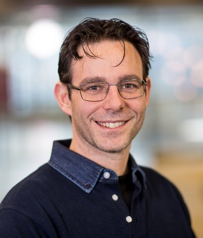
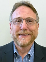
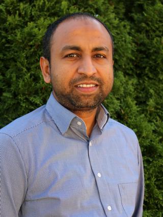
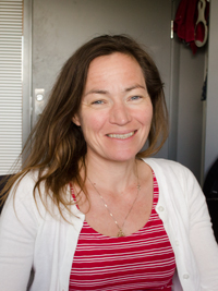
Massimo Mischi is a Full Professor at the Faculty of Electrical Engineering of the Eindhoven University of Technology (TU/e) and chairs the Signal Processing Systems (SPS) Division , the largest division at TU/e with over 250 researchers. He founded the Biomedical Diagnostics (BM/d) Lab in 2012, which now includes 180 researchers and clinical/industrial advisors, focusing on biomedical signal processing for diagnostics and monitoring.
Craig H. Meyer is a Professor in Biomedical Engineering and Radiology & Medical Imaging at the University of Virginia. He holds a Ph.D. from Stanford University and leads the Rapid MRI Research Group, focusing on developing advanced MRI techniques for cardiovascular disease, neural disorders, and pediatrics. His work integrates physics, signal processing, and machine learning to improve MRI acquisition and processing speed. Education: Ph.D. in Biomedical Engineering, Stanford University. Research Interests: Medical and Molecular Imaging, Signal and Image Processing, Biomedical Data Sciences, Biomechanics, and Cardiovascular Engineering. His innovations include fast spiral imaging, conjugate phase reconstruction, and machine learning-enhanced MRI denoising. Awards: Notably includes the Dean’s Award for Excellence in Team Science (2014), Fellowships from NAI (2021), AIMBE (2015), and ISMRM (2013). He also authored two landmark MRI papers recognized as pivotal in the field. Teaching: Courses include BME 6310 (Computation and Modeling in Biomedical Engineering) and BME 8782 (Magnetic Resonance Imaging). He emphasizes translational research, with applications in clinical MRI advancements and collaborative interdisciplinary projects. Labs/Groups: Rapid MRI Research Group focuses on cutting-edge MRI technologies, including real-time cardiac imaging and artifact reduction through deep learning.
Brian W. Pogue, Ph.D., is the Robert A. Pritzker Chair in Biomedical Engineering at Dartmouth College's Thayer School of Engineering, with a joint appointment as an Honorary Fellow in Medical Physics at the University of Wisconsin-Madison. His academic background includes a Ph.D. in Medical/Nuclear Physics from McMaster University and a Research Fellowship at Harvard Medical School's Wellman Center for Photomedicine. He has led significant administrative roles, including Dean of Graduate Studies at Dartmouth (2008–2012) and Chair of Medical Physics at Wisconsin (2022–2025). Research Focus : Dr. Pogue pioneers Optics in Medicine , specializing in cancer imaging, photodynamic therapy, and surgical guidance. His work integrates fluorescence imaging, radiation therapy monitoring, and molecular diagnostics to improve cancer treatment precision. Key innovations include Cherenkov imaging for radiotherapy dosimetry and hypoxia-sensitive probes for tumor resection. Publication Trends : Recent articles (2023–2025) emphasize real-time surgical guidance, hypoxia quantification, and multimodal imaging systems. Dominant themes include fluorescence tomography, radiation dosimetry, and low-cost diagnostic devices, reflecting a translational focus from preclinical validation to clinical applications. Awards & Honors : Fellow, Optica (formerly OSA) Fellow, American Institute for Medical and Biological Engineering (AIMBE) Fellow, American Association of Physicists in Medicine (AAPM) Fellow, SPIE (International Society for Optics and Photonics) Funding & Innovation : Continuously funded by the NIH since 2001 ($52M+ total), Dr. Pogue founded three startups: DoseOptics LLC (radiotherapy dose imaging) and Hypoxia Surgical LLC (tissue hypoxia cameras), bridging academic research to clinical tools.
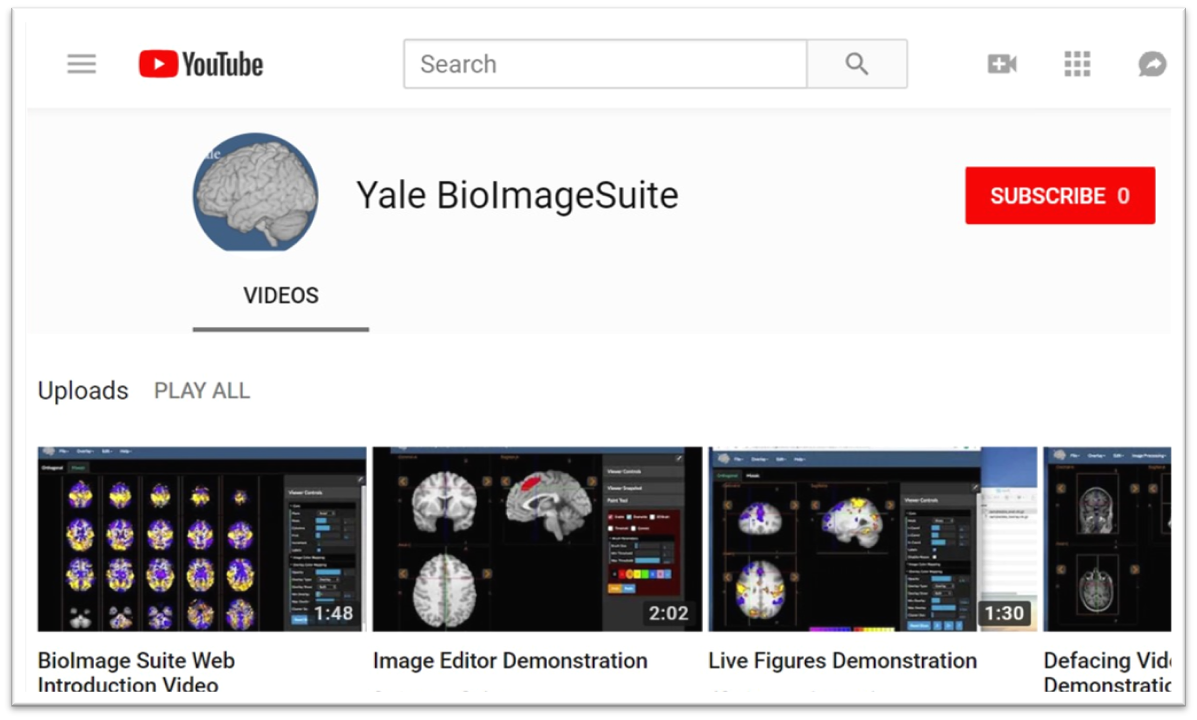
Xenophon Papademetris is a Professor of Biomedical Informatics & Data Science and Radiology & Biomedical Imaging at Yale School of Medicine. He serves as Associate Director of Biomedical Imaging Data Sciences at Yale Biomedical Imaging Institute and directs the Medical Software and Medical Artificial Intelligence Certificate Program. PhD in Electrical and Information Sciences from Yale University (2000) BA from Cambridge University (1994) Postdoctoral Fellowship at Yale University (2002) His research focuses on medical image analysis, machine learning, and biomedical software development. He has developed tools like BioImage Suite Web and contributed to standards committees at the Association for the Advancement of Medical Instrumentation (AAMI). His work spans modalities including MRI, CT, PET, and optical imaging. Recent publications emphasize neuroimaging analysis, explainable AI in healthcare, and multimodal data integration across species. He leads NIH-funded research under the BRAIN Initiative (R24 MH114805) and has authored a textbook on Medical Software published by Cambridge University Press. IEEE Senior Member Yale Brown-Coxe Postdoctoral Fellowship Harding Bliss Prize for Excellence in Engineering He directs the BioImage Suite Project, creating web-based image analysis tools using JavaScript and WebAssembly. His teaching includes both academic courses and a Coursera program on Medical Software with over 14,000 enrollments.
Professor Fernando Calamante is a Professor of Biomedical Engineering at The University of Sydney and Director of Sydney Imaging Core Research Facility. He leads the National Imaging Facility node and focuses on advanced MRI methodologies, particularly Diffusion and Perfusion MRI, to study brain connectivity and neurological disorders. His work includes developing the MRtrix software, widely used in diffusion MRI analysis. He holds extensive funding (~$50M) and has been recognized with awards like ISMRM Fellowship and NHMRC grants. His research spans super-resolution imaging, brain connectomics, and clinical applications in stroke and tumors. Education: BSc (Physics, Argentina), PhD (Magnetic Resonance Imaging, University College London). Career highlights include leadership roles at The Florey Institute and ISMRM presidency (2021-2022). Research interests include: Novel MRI methods for brain connectivity and super-resolution imaging Applications of Diffusion and Perfusion MRI in neurology Integration of structural and functional connectomics Key achievements: Over 200 publications, software innovations, and leadership in global MRI societies.
Volker J Schmid is a Professor of Bayesian Imaging and Spatial Statistics at the Department of Statistics, Ludwig Maximilian University of Munich. He leads the Bayesian Imaging and Spatial Statistics group and contributes to interdisciplinary initiatives like the Munich Center of Machine Learning. His work bridges statistical theory with applications in medical imaging and biology. PhD in Statistics (2004), LMU Munich Diploma in Statistics (2000), LMU Munich Abitur, Joseph-von-Fraunhofer-Gymnasium Cham (1993) His research focuses on Bayesian computational methods for high-dimensional data, particularly in medical imaging (MRI, DCE-MRI) and biological microscopy (e.g., 3D nuclear architecture analysis via super-resolution microscopy). Key applications include disease mapping , image segmentation , and spatio-temporal modeling . His software tools (e.g., nucim , bioimagetools , BAMP ) enable quantitative analysis in nuclear imaging and age-period-cohort modeling. His 15 most recent publications span Bayesian modeling for medical imaging , spatio-temporal epidemiology , and computational biology . Topics include co-localization metrics in fluorescence microscopy, nuclear architecture analysis, and dynamic Bayesian frameworks for MRI data. Collaborations extend to neuroimaging, oncology, and nuclear biology.
Professor Denis Doorly is a Professor of Fluid Mechanics in the Department of Aeronautics at Imperial College London's Faculty of Engineering. His research focuses on biomedical fluid mechanics, particularly respiratory and cardiovascular systems, with expertise in computational fluid dynamics (CFD) and aerosol transport. He has published extensively on nasal airflow modeling, cardiovascular MRI simulations, and aerosol dynamics in medical contexts. Key contributions include CFD cohort studies on nasal decongestion effects, benchmarking models for SARS-CoV-2 transmission, and ventilator strategies during the pandemic. Research interests span biological fluid mechanics, biomedical flows, and medical device design. His work integrates computational modeling with clinical applications, addressing issues like tracheal compression, myocardial perfusion, and aerosol extraction during surgeries. Collaborations include studies on isolated heart models and particle deposition in respiratory systems. Affiliations include the Biological Fluid Mechanics and Biomedical Flows groups at Imperial. His publications (139+ articles) highlight interdisciplinary applications of fluid mechanics to healthcare challenges.
Brad Sutton is a Professor of Bioengineering at the University of Illinois Urbana-Champaign and Technical Director of the Biomedical Imaging Center at Beckman Institute. He holds affiliate roles in the Neuroscience Program, Department of Electrical and Computer Engineering, and is a Health Innovation Professor at the Carle Illinois College of Medicine. His roles also include fellowship positions with the National Center for Supercomputing Applications and the CZ Biohub Chicago. Education: Ph.D. in Biomedical Engineering from the University of Michigan (2003). Research Interests: Focus on advanced MRI techniques for structural and functional brain imaging, including diffusion-weighted imaging, dynamic imaging, and neuromuscular coupling studies. His work emphasizes multi-scale bioimaging to understand brain function across interventions, aging, and disease. Publications: Over 180 peer-reviewed articles in 2025-2024 highlight innovations in MRI technology and applications in neuroscience, including breakthroughs in laminar fMRI specificity, myelin development modeling, and Alzheimer’s biomarker studies. Recent work extends to clinical applications like aortic imaging automation and mixed reality training tools. Awards: AIMBE and ISMRM Fellowships (2017/2024), Abel Bliss Scholar (2014-), and over 9 patents in imaging techniques. Labs & Teams: Leads the Magnetic Resonance Functional Imaging Lab. Collaborates with interdisciplinary teams across engineering, medicine, and computational science to advance imaging technologies and their clinical translation.
Associate Professor Andre Kyme is an academic staff member in the School of Biomedical Engineering at The University of Sydney. His research focuses on developing enabling technologies for biomedical imaging, including motion compensation in MRI/PET, robotic platforms for image-guided therapy, and cross-disciplinary applications like plant salt uptake analysis using PET. He collaborates with institutions globally and advises students on projects like lameness detection in horses and AI-based motion correction. Research Interests: Kyme's work spans motion correction in medical imaging modalities, medical robotics integration with imaging systems, and innovative applications of imaging technologies in non-traditional fields. His team emphasizes leveraging advancements in computer vision, machine learning, and instrumentation to improve imaging performance and accessibility. Recent Projects: Current research includes MRI-compatible robotic platforms for therapy applications, AI-driven lameness detection in horses, and pediatric neuroimaging improvements. He leads the BREEZE initiative to enhance MRI accessibility for children with cerebral palsy through eye-gaze communication technology. Publications: His work spans 20+ years with over 50 peer-reviewed publications in journals like Physics in Medicine and Biology and IEEE Transactions. Key areas include PET/SPECT/CT motion correction algorithms, robotic systems for medical imaging, and novel imaging applications in plant science. Teaching: Kyme instructs core biomedical engineering courses including thesis supervision and capstone projects at both undergraduate and postgraduate levels. Labs/Teams: Active in the Brain and Mind Centre and Biomedical Imaging, Visualisation and Information Technologies groups at Sydney. Collaborates with industry partners like TeleMedVet and academic institutions including University of California Davis and Chinese University of Hong Kong.
Daniel B. Vigneron, PhD is a Professor at the University of California, San Francisco (UCSF) Department of Radiology and Biomedical Imaging. He serves as Director of the Hyperpolarized MRI Technology Resource Center (HMTRC), Director of Human Imaging Core Services, Director of Advanced Imaging Technologies SRG, and Operations Director of the Surbeck Laboratory for Advanced Imaging. As a core member of the UCB/UCSF Graduate Group in Bioengineering, Vigneron has established himself as a leader in molecular imaging research with over three decades of experience at UCSF. Vigneron's research focuses on developing advanced functional and metabolic MRI techniques, particularly hyperpolarized carbon-13 technology, for studying prostate cancer, brain tumors, and other diseases. His work enables non-invasive imaging of metabolic processes, allowing clinicians to monitor therapy effectiveness and guide treatments. The HMTRC, which he founded in 2011 with NIH funding and recently secured a 5-year renewal for, has supported 20 external projects domestically and 15 internationally, produced 239 publications, and trained 149 researchers. Vigneron's lab develops novel coil and software techniques for high-field MRI, MR spectroscopy, and diffusion imaging at 3T and 7T for studying brain, prostate cancer, and other organs. His recent publications demonstrate a clear trajectory toward clinical translation of hyperpolarized carbon-13 MRI across multiple organ systems. The research spans abdominal imaging with advanced denoising techniques, cardiac metabolism studies, whole-brain coverage applications, and cerebral perfusion analysis. This work represents a significant shift from basic science toward practical clinical applications in oncology, cardiology, and neurology, with particular emphasis on standardization for multi-center studies. Scientific Awards: 2022 Outstanding Faculty Mentoring Award from UCSF Department of Radiology and Biomedical Imaging Vigneron has mentored 149 trainees throughout his career, with several former students now serving as faculty members including Duan Xu, Peder Larson, and Susan Noworolski. As Principal Investigator overseeing eight grants, he has secured significant NIH funding for the HMTRC and other research initiatives. His administrative leadership extends to co-chairing the department's Safety and Compliance Committee, where he has helped establish robust safety protocols for PET-MR programs. Vigneron's mentoring philosophy emphasizes adapting to individual needs at different career stages, moving from instructor to coach to manager to cheerleader as trainees progress. The Vigneron Lab, located in Byers Hall on the UCSF Mission Bay campus, operates within the Surbeck Laboratory for Advanced Imaging. The lab group develops novel acquisition techniques and hardware for multinuclear MR spectroscopy, with particular focus on hyperpolarized carbon-13 metabolic imaging. The HMTRC serves as a hub for team science, bringing together researchers from diverse disciplines to advance metabolic imaging technology and its clinical applications.
Dr. Tanzil M. Arefin is an Assistant Professor of Neuroscience at the University of Rochester School of Medicine and Dentistry and Associate Director of the Preclinical Imaging Core at the Center for Advanced Brain Imaging and Neurophysiology (CABIN). His research focuses on developing neuroimaging techniques to study brain functions and microstructures in animal models of human disorders, including neurodegenerative and psychiatric illnesses. He holds affiliations with the Del Monte Institute for Neuroscience and the Neuroscience Ph.D. Program. **Education**: Ph.D., Neuroscience, University of Freiburg and University of Strasbourg (2017) M.Sc., Biomedical Engineering, Czech Technical University and University of Groningen (2012) B.Sc., Electrical and Electronic Engineering, Islamic University of Technology (2007) **Research Interests**: Dr. Arefin's lab employs multimodal MRI methodologies (resting-state fMRI, diffusion MRI, ASL perfusion MRI, MR spectroscopy) alongside optogenetics and chemogenetics to elucidate molecular mechanisms impairing brain plasticity. Current projects include studying cerebellar connectivity's role in non-motor behaviors and developing interventions for alcohol-dependent brains. **Awards**: Magna cum Laude, Summa Cum Laude, Erasmus Mundus Fellowships (both Doctoral and Masters). **Grants & Advising**: Not explicitly listed in texts, but lab activities suggest involvement in NIH-funded projects. Advising details are pending explicit student listings. **Lab & Affiliations**: Arefin Lab focuses on translational imaging tools. Affiliated with UR CABIN and URMC's Neuroscience programs. Location: 430 Elmwood Ave, Rochester, NY.
Brent A. French is a Professor in the Department of Biomedical Engineering at the University of Virginia. His research focuses on integrating targeted drug/gene delivery with advanced imaging techniques (MRI, PET, ultrasound) to address cardiovascular disease mechanisms and therapy evaluation. He leads the Molecular Bioengineering Lab, emphasizing interdisciplinary collaboration and translational research. Education: B.S. in Biochemistry, Louisiana State University (1982) Ph.D. in Biochemistry, Louisiana State University (1987) Postdoctoral Fellowship at Baylor College of Medicine/NIH (1987–1991) Research Interests: Targeted drug and gene delivery systems Novel diagnostic imaging methods (e.g., ultrasound, MRI) Tissue engineering for cardiovascular applications Cardiovascular disease mechanisms and therapeutic strategies Grants & Projects: NIH R01 HL147193: $1.6M for developing molecularly targeted AAV for cardiac regeneration CHRB #207-10-19: $266K for in situ cardiac regeneration post-infarction NIH R01 HL147104: $2.99M for multiparametric MRI in coronary microvascular disease Labs/Teams: Molecular Bioengineering Lab (MR5 Building, Rooms 1205/1219), collaborating on interdisciplinary projects with cardiovascular and imaging experts.
Prof. Dr. Kirsten Jung is a faculty member at the Department of Microbiology , Faculty of Biology , Ludwig Maximilian University of Munich . Her research focuses on bacterial signal transduction, stress response mechanisms, and systems biology approaches to understand microbial regulatory networks. Key research areas include stress-dependent gene expression in bacterial populations Structural and functional analysis of membrane-integrated receptors Metabolism-based chemical communication in bacteria Integration of experimental and computational systems biology Recent publications highlight her lab's work on Escherichia coli epitranscriptomic modifications under heat stress, m 5 C rRNA dynamics, and the role of RNA methylation in host-pathogen interactions. Collaborative studies address bacterial acid stress responses and their implications for antibiotic tolerance. Her interdisciplinary work bridges microbiology with ecological studies, as evidenced by research on biodiversity conservation in forest and urban ecosystems. Publications also demonstrate expertise in advanced imaging techniques (e.g., arterial spin labeling for glioma analysis) and bioinformatics approaches. Current advisees include Gloria Gessinger and Tania P. Gonzalez-Terrazas . She can be contacted at jung@lmu.de .
Kristine Beate Walhovd is a Professor at the Department of Psychology , University of Oslo, and co-leader of the Center for Lifespan Changes in Brain and Cognition (LCBC) . She is affiliated with the UiO:Life Science initiative and has led major projects like Lifebrain (€2.5M) and Neurocognitive Plasticity (ERC Starting Grant, €1.5M). Current roles: Professor (since 2016), Co-leader of LCBC Key affiliations: University of Oslo, Lifebrain Consortium, UiO:Life Science Her research focuses on lifespan brain and cognitive changes , examining both positive/negative developmental trajectories from age 4 to 90. She studies interactions between biomedical risks (e.g., Alzheimer's, fetal drug exposure) and cognitive outcomes, employing MRI , DTI , and ERP methodologies. Recent publications highlight work on Alzheimer's biomarkers , sleep-brain interactions , memory consolidation , and fetal brain influences . Her 2025 papers in Scientific Reports and Neurobiology of Aging examine hippocampal stability and brain network segregation. Awards include: 2015 University of Oslo Research Prize (shared with Anders Fjell) 2011 Member, Norwegian Academy of Sciences and Letters 2006 His Majesty the King's Gold Medal for best doctoral thesis She supervises doctoral candidates and teaches cognitive neuroscience , experimental methods , and thesis writing .
Professor Sungheon Gene Kim holds a faculty position at the Weill Cornell Medicine Graduate School of Medical Sciences within the Department of Radiology . His research focuses on quantitative MRI methodology for oncological applications , particularly in breast cancer and head and neck cancer . Kim's lab develops advanced dynamic contrast-enhanced MRI (DCE-MRI) and diffusion MRI (dMRI) techniques to assess tumor microenvironment and treatment response . Key research areas include: Tumor vascular properties via 3D UTE-GRASP MRI Cellular microstructural analysis through POMACE framework Adipose-tissue cancer interaction via MR spectroscopic imaging His lab has received continuous funding from the National Cancer Institute (R01CA219964, UG3/UH3CA228699, R01CA160620). Recent publications demonstrate technical advancements in ultrafast MRI reconstruction , deep learning-enhanced perfusion analysis , and multi-parametric tumor characterization . Collaborations with the National Institutes of Health Quantitative Imaging Network have produced novel cellular water exchange rate measurements that correlate with patient survival outcomes .