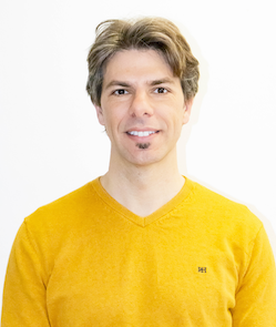
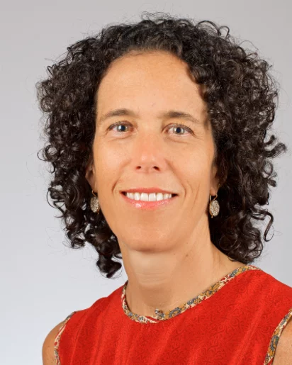
Jing Wang, Ph.D., is a Professor of Radiation Oncology at UT Southwestern Medical Center, affiliated with the Department of Radiation Oncology’s Division of Medical Physics and Engineering. His research bridges medical imaging, machine learning, and radiation therapy optimization. B.Sc. in Material Physics, University of Science and Technology of China M.A. and Ph.D. in Physics, Stony Brook University Postdoctoral training in Radiology (Stony Brook) and Radiation Physics (Stanford) Dr. Wang’s work focuses on enhancing medical imaging quality for quantitative applications in image-guided radiation therapy (IGRT) and adaptive radiation therapy (ART) . Key areas include CT/MRI/PET reconstruction , deep learning for tumor localization , and radiomics-based survival prediction . His AIRT Lab develops AI algorithms for treatment outcome modeling and real-time imaging. Recent publications highlight advancements in transformer networks for anatomy prediction, uncertainty-aware segmentation , and delta radiomics for surgical margin analysis. Journals span Medical Physics , Physics in Medicine and Biology , and International Journal of Radiation Oncology .
Professor Steven Lee is a leading figure in biophysical chemistry at the University of Cambridge , where he leads the TheLeeLab in the Yusuf Hamied Department of Chemistry . His research focuses on developing advanced single-molecule fluorescence and multidimensional super-resolution imaging techniques to probe fundamental biological processes at unprecedented spatial precision. Developed novel super-resolution microscopy approaches for 2D/3D visualization of T-cell membrane proteins and histone assembly in fission yeast nuclei Pioneer of 15-20nm resolution imaging strategies through fluorophore kinetics and image reconstruction algorithms Recipient of the 2017 Marlow Prize in Physical Chemistry , Lee's lab produces cutting-edge tools with applications in immunology , neurodegeneration , and cellular biophysics . His team maintains active collaborations with Prof Klenerman (FRS MedSci) and Prof Moerner (Nobel Chemistry 2014). Research Highlights : Molecular origins of immunity through T-cell membrane protein interactions 3D histone dynamics during DNA replication/repair Amyloid aggregate quantification for neurodegenerative disease diagnosis Volumetric imaging innovations via vLUME virtual reality platform
Dr. Thomas Smits is a faculty member at the Faculty of Humanities of the University of Amsterdam , specializing in Digital Humanities , Visual Culture , and Computational History . His research bridges artificial intelligence with historical image analysis, focusing on media history, colonial visual representations, and algorithmic approaches to archival studies. Recent publications highlight his work on multimodal datasets, generative AI in protest memory reconstruction, and the ethical implications of machine learning for historical research. Smits employs computational methods like CLIP and computer vision to analyze large-scale visual collections, including magic lantern slides, historical advertisements, and news images. Key research areas: Digital Humanities, Visual Memory, Algorithmic Analysis, Colonial Media, Gender Studies, Historical Epistemology. Methodological focus: Stochastic modeling, multimodal machine learning, distant reading of visual datasets, and AI-driven archival enhancement.
Gregg Trahey is the Robert Plonsey Distinguished Professor of Biomedical Engineering at Duke University, with additional appointments in Radiology. He leads pioneering research in medical ultrasound imaging and serves as a Bass Fellow, reflecting his significant contributions to both research and education. B.S. from University of Michigan, Ann Arbor (1975) M.S. from University of Michigan, Ann Arbor (1979) Ph.D. from Duke University (1985) Dr. Trahey's research focuses on medical ultrasound, image guided surgery, adaptive imaging, imaging of tissue's mechanical properties, and radiation force imaging . His laboratory develops and evaluates novel ultrasonic imaging methods with current projects involving high resolution imaging of the breast and mechanical characterization of both breast and cardiovascular systems. They conduct comprehensive testing through phantom models, animal trials, ex vivo experiments, and human clinical trials, with current clinical applications focusing on vascular plaque imaging and breast lesion characterization. Analysis of Dr. Trahey's recent publications (2022-2025) reveals a strong emphasis on spatial coherence techniques, adaptive ultrasound imaging systems, and quantitative tissue characterization. His work bridges engineering innovation with clinical applications, particularly in cardiac and breast imaging, with key themes including clutter reduction, real-time adaptive systems, and mechanical property assessment of tissues. Fellow, Institute of Electrical and Electronics Engineers (IEEE), 2022 MERIT Award, National Institutes of Health, 2009 Fellows, American Institute for Medical and Biological Engineering, 1999 Dr. Trahey has taught courses including MEDPHY 738: Radiology in Practice, ECE 392: Projects in Electrical and Computer Engineering, and BME 848L: Radiology in Practice. His research is supported by significant funding, particularly from the National Institutes of Health as evidenced by his prestigious MERIT Award, which provides extended grant support to researchers with exceptional performance. Dr. Trahey leads an active research laboratory that conducts comprehensive studies from phantom development through clinical trials. His team collaborates extensively with clinicians for translational research applications, particularly in cardiology and radiology. Current projects focus on high-resolution imaging techniques, mechanical tissue characterization, and development of novel ultrasound methods for improved diagnostic capabilities while maintaining patient safety.
Adam Wax is a Professor of Biomedical Engineering and Professor of Physics at Duke University , where he leads cutting-edge research in optical spectroscopy for cancer detection , novel microscopy techniques , and low-coherence interferometry . As a member of both the Duke Cancer Institute Faculty Network and Duke Institute for Brain Sciences , he bridges engineering and biomedical applications. Education: B.S. from Rensselaer Polytechnic Institute (1993), M.A. (1996) and Ph.D. (1999) from Duke University Honors: Fellow, American Institute for Medical and Biological Engineering (2014); Fellow, SPIE (2010); Fellow, OSA (2010); NSF CAREER Award (2004) His research focuses on biomedical optics , particularly optical coherence tomography (OCT) and quantitative phase microscopy , with applications in early cancer detection , subcellular imaging , and clinical diagnostics . Recent work emphasizes low-cost, portable OCT systems for point-of-care applications and multimodal imaging combining spectroscopy with phase analysis. Scientific awards include: Fellow, American Institute for Medical and Biological Engineering (2014) Fellow, International Society for Optics and Photonics (2010) Fellow, Optical Society of America (2010) National Science Foundation CAREER Award (2004)
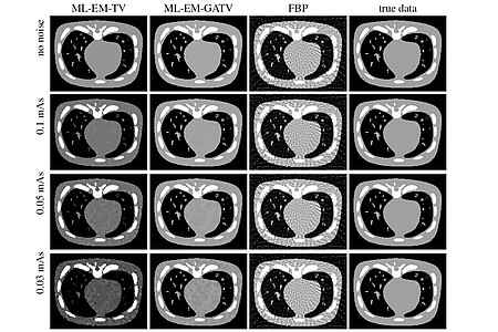
Jürgen Hesser is a Professor at the Mannheim Medical Faculty , Heidelberg University, specializing in Experimental Radiotherapy and Medical Imaging . His research focuses on solving inverse problems in imaging, particularly for CT reconstruction , brachytherapy planning , and low-dose imaging . Current affiliations: Clinic for Radiotherapy and Radiooncology, Mannheim University Hospital Collaborative ties: Interdisciplinary Center for Scientific Computing (IWR) and Center for Bioinformatics (ZITI) at Heidelberg University Research interests center on anisotropic total variation techniques for medical and industrial applications, including MR-guided interventions and real-time radiation therapy . His work has led to a 1000x speed improvement in brachytherapy planning algorithms. Recent publications highlight expertise in image reconstruction (CT/X-ray), noise optimization , machine learning for cancer classification, and big data management solutions. His methods are applied to both clinical and industrial imaging challenges. Additional contributions include scientific data infrastructure development and variance stabilization techniques for medical sensors. The research group maintains strong interdisciplinary links with Physics, Mathematics, and Computer Science faculties.
Xavier Décoret is a researcher at INRIA since October 2003, specializing in computer graphics with core expertise in real-time rendering, visibility algorithms, and level-of-detail techniques. He teaches courses at Grenoble (Master IVR program) and École Polytechnique, including Java programming, virtual image creation, and advanced image synthesis. His educational background includes: PhD in Computer Graphics under François Sillion Post-doctoral position at MIT under Frédo Durand Décoret's research spans non-photorealistic rendering, shadow computation, and GPU-accelerated algorithms. He develops practical graphics tools including XdkWRL (VRML parsing), Argstream (command-line arguments), and XdkBibTeX (BibTeX handling), emphasizing real-world implementation for interactive systems. His 2008 publications reveal trends toward efficient interactive rendering techniques, with contributions in dynamic stylization, label placement, soft shadows, and GPU voxelization—highlighting innovations in plausible visual effects for real-time applications. He is affiliated with the Artis research team at INRIA, focusing on virtual reality and computer graphics advancements.
Rafael Sebastian is a Full Professor at Universitat de Valencia and General Director for Science and Research of the Generalitat Valenciana. He leads the Computational Multiscale Simulation Lab (CoMMLab) and collaborates with institutions like Oxford University and Yale University. Department of Computer Science, Universitat de Valencia CoMMLab Founder Spanish Network of Excellence in Cardiac Modeling His research focuses on multi-scale computational models and artificial intelligence for patient-specific cardiac simulations , aiming to improve arrhythmia risk stratification and therapy planning . Key topics include cardiac conduction system modeling , scar-related ventricular tachycardia , and machine learning pipelines for clinical applications. Recent publications emphasize automata-based simulations for atrial arrhythmias, machine learning in arrhythmia localization, and 3D geometric characterization of aortic diseases. Trends show integration of computational modeling with clinical data and medical imaging . Scientific Awards: Best Poster Award, Functional Imaging and Modeling of the Heart (2021) Cum Laude Award, SPIE Medical Imaging (2009) Student Presentation Award (2011) He has supervised 7 PhD/Master students and led grants exceeding €1 million, including projects like iSARC-GENETICS and iCardioTwins , focusing on digital twin technology and cardiac disease stratification .
Prof. Dr. Ben Jeurissen is an Associate Research Professor at the Department of Physics, University of Antwerp , Belgium, specializing in Medical Image Computing , Quantitative MRI , and Diffusion MRI . He received an ERC Consolidator Grant (2023) and FWO Senior Postdoctoral Fellowship (2018–2021) , with a focus on data-driven approaches to study brain microstructure. Education : MSc in Computer Science (2004), Biomedical Imaging (2006), PhD in Science (2012) from University of Antwerp. Research : His work bridges Neuroscience , Medical Imaging , and Computational Methods , particularly in Diffusion MRI and Quantitative MRI for applications in Alzheimer’s disease , spaceflight effects on the brain, and knee imaging . Awards : ERC Consolidator Grant (2023) Australian Museum Eureka Prize Finalist (2024) Multiple Magna/Summa Cum Laude Merit Awards at ISMRM conferences Prize Robert Oppenheimer (2015) Publications span NeuroImage , Human Brain Mapping , Journal of Alzheimer's Disease , and Investigative Radiology , with key contributions to super-resolution MRI , fiber tracking , and brain microstructure analysis . He serves as a contributor to MRtrix and advisor for PhD theses in Computational Anatomy and Medical Imaging .
Francisco Santibanez is a Research Assistant Professor in the Department of Biomedical Engineering at the University of North Carolina. He holds a Ph.D. in Physics from Universidad de Santiago de Chile (2010). His research focuses on experimental physics, biomedical imaging, and non-linear wave propagation in complex media. Since joining the Pinton Lab in 2018, he has investigated ultrasound super-resolution, functional imaging, and shear wave dynamics in soft tissues. His work spans advanced ultrasound techniques, including transcranial imaging, volumetric imaging, and real-time monitoring of neurovascular responses. Key projects include developing sparse ultrasound arrays and addressing image-degrading effects in clinical settings. His contributions extend to interdisciplinary areas like granular dynamics and acoustic sensing for pest control. Notable research trends include advancing super-resolution imaging capabilities and exploring shock wave behavior in biological tissues. His publications reflect expertise in medical imaging modalities, wave propagation modeling, and hardware innovations for diagnostic applications. Labs/Teams: Active member of the Pinton Lab, collaborating on biomedical engineering and medical imaging projects. His work emphasizes translational research with potential for clinical impact in neuroimaging and oncology monitoring.
Marinko Sarunic is an Adjunct Professor at the School of Engineering Science , Simon Fraser University . He holds a PhD in Biomedical Engineering from Duke University and has been recognized as a Michael Smith Foundation for Health Research Scholar . His research focuses on biomedical imaging , particularly optical coherence tomography (OCT) , microscopy , and low-coherence interferometry , with applications in diabetic retinopathy , Alzheimer’s disease , and age-related macular degeneration . Dr. Sarunic's work spans adaptive optics , deep learning , and sensorless OCT systems , emphasizing clinical translation and open-source software development (e.g., OCTAVA ). His Google Scholar publications highlight multimodal imaging , vascular heterogeneity analysis , and AI-driven diagnostics for retinal diseases. His contributions include the Michael Smith Foundation for Health Research Scholar award. Though not currently teaching courses, his collaborations and leadership in retinal imaging and medical device innovation are pivotal for advancing non-invasive diagnostics in neurodegenerative and diabetic conditions .
Federica Marone Welford is a Beamline Scientist at the TOMCAT beamline of the Swiss Light Source (SLS) at the Paul Scherrer Institute (PSI) . She holds an Earth Sciences degree with a focus on seismology and a PhD in seismology from ETH Zurich , following a postdoctoral fellowship at the Berkeley Seismological Laboratory . Her work centers on advancing tomographic reconstruction algorithms, mitigating artifacts, and optimizing computational infrastructure for high-speed X-ray imaging at the TOMCAT beamline. Research Focus: X-ray tomography methodology, data compression, real-time reconstruction systems, and applications in paleontology, earth sciences, additive manufacturing, and energy research. Collaborations: Engages with global researchers and industry partners, particularly in battery/fuel cell analysis and laser powder bed fusion. Teaching: Lectures at ETH Zurich on X-ray imaging techniques. Publications highlight her contributions to X-ray scattering tensor tomography, dynamic process visualization, and computational advancements in imaging systems.
Roger Fulton is a Professor of Medical and Preclinical Imaging at the University of Sydney and a Principal Nuclear Medicine Physics Specialist at Westmead Hospital's Department of Medical Physics. He is part of the Brain and Mind Centre and leads the Physics and Biomodelling team. His roles include Conjoint Professor in Medical Radiation Sciences, RHD student supervision, and coordination of the MRTY5134 (Computed Tomography Theory) and MRTY2107 (Imaging Technology 2) units of study. He holds a PhD in Applied Physics. His career has spanned collaborations with institutions like the Research Center Julich (Germany), Massachusetts General Hospital (USA), and the International Atomic Energy Agency (Austria). Professional recognitions include Fellow of ACPSEM, Senior IEEE Membership, and Chair of the Nuclear Medical Imaging Sciences Council (IEEE). Research interests focus on motion tracking/correction for SPECT, PET, and CT imaging, including patent innovations and leadership in radiation dose reduction techniques. He has secured $9.2M in competitive grants since 2004 and contributed to global standards through IAEA initiatives. Grants and advising: Over $9.2M in grants since 2004, advising PhD student Bader ALOUFI on liver imaging kinetics. His work bridges hardware engineering (e.g., Intel RealSense depth cameras) with software solutions (neural networks, Bayesian methods) to enhance imaging accuracy and reduce patient motion artifacts. Scientific Awards: US Patent (2020) ARC College of Experts (2016–2018) ACPSEM Fellowship IEEE Senior Membership IEEE Nuclear and Plasma Sciences Society leadership Labs/Teams: Physics and Biomodelling team at the Brain and Mind Centre, collaborating with international groups on preclinical imaging systems and awake-animal PET methodologies.
B. F. Spencer Jr. is the Nathan M. and Anne M. Newmark Endowed Chair in Civil Engineering at the University of Illinois at Urbana-Champaign, where he directs the Multi-Axial Full-Scale Sub-Structured Testing & Simulation Facility and the Smart Structures Technology Laboratory. He joined the university in 2002 after serving as Leo E. and Patti Ruth Linbeck Professor of Engineering at the University of Notre Dame (1985-2002). Education includes: Ph.D. in Theoretical and Applied Mechanics, University of Illinois at Urbana-Champaign (1985) M.S. in Theoretical and Applied Mechanics, University of Illinois at Urbana-Champaign (1983) B.S. in Mechanical Engineering, University of Missouri-Rolla (1981) His research focuses on pioneering innovations in structural health monitoring, stochastic mechanics, and smart sensor technologies. Key areas include development of wireless sensor networks for real-time infrastructure assessment, seismic hazard mitigation strategies, and AI-driven damage detection systems. His work bridges theoretical computational mechanics with practical civil engineering applications to enhance resilience against natural disasters. Recent publications emphasize digital twins, UAV-based structural inspection, machine learning for damage identification, and advanced sensor networks. Trends show strong integration of AI, 3D visualization, and edge computing for rapid post-disaster evaluation and predictive maintenance of critical infrastructure. Major scientific honors: ASCE Housner Medal (2015) J.M. Ko Medal (2014) Foreign Member of Polish Academy of Sciences (2005) Structural Health Monitoring Person of the Year (2011) JSPS Fellowships (1999, 2000) He leads significant infrastructure projects including NSF-funded facilities and industry collaborations. Laboratory initiatives involve full-scale testing of bridges, gates, and seismic mitigation systems. Educational outreach includes K-12 STEM programs like 'Shakes and Quakes' to inspire future engineers.
W. Clem Karl is Professor and Chair of the Electrical and Computer Engineering Department at Boston University's College of Engineering, with additional appointments in Biomedical Engineering and Systems Engineering. Education: PhD in Electrical Engineering and Computer Science, Massachusetts Institute of Technology, 1991 Research Interests: Professor Karl's work focuses on computational imaging, statistical signal processing, inverse problems, and biomedical applications. His research develops advanced algorithms for image reconstruction in medical imaging, synthetic aperture radar, and remote sensing, emphasizing statistical methods for signal estimation and detection. Scientific Awards: Fellow of IEEE Fellow of AIMBE Fellow of BMES IEEE Vice President of Publication Services and Products (2025) Inaugural Editor-in-Chief of IEEE Transactions on Computational Imaging (2014-2017) Editor-in-Chief of IEEE Transactions on Image Processing (2012-2014) ECE Award for Excellence in Teaching (2000) Teaching: EC 401 Signals and Systems EC 416 Introduction to Digital Signal Processing EC 505 Stochastic Processes EC 717 Image Reconstruction and Restoration Advising: Due to high email volume, Professor Karl does not respond to individual student inquiries; prospective students must apply directly through College of Engineering degree programs. Labs and Affiliations: Director of the Information & Data Sciences Laboratory, with affiliations in the Center for Information and Systems Engineering, Center for Space Physics, Neurophotonics Center, Center for Nanoscience and Nanobiotechnology, and Division of Systems Engineering.