Dr. Ahmed El-Sayed is an Associate Professor in the Department of Electrical and Computer Engineering at the School of Engineering, University of Bridgeport. He holds a Ph.D. in Computer Science and Engineering (2016) and M.Sc. degrees in Computer Engineering (2011) and Engineering Mathematics (2006). Ph.D. in Computer Science and Engineering, University of Bridgeport (2016) M.Sc. in Computer Engineering, University of Bridgeport (2011) M.Sc. in Engineering Mathematics, Alexandria University (2006) B.Sc. in Electrical Engineering, Alexandria University (2003) His academic work focuses on robotics , artificial intelligence , and computer vision , with specific emphasis on fuzzy systems , machine learning , and soft computing . His research spans theoretical developments and practical implementations in autonomous systems , medical imaging , and industrial automation . The 15 most recent publications highlight his contributions to generative models , medical diagnostics , disassembly sequencing , and high-dimensional feature analysis . These works intersect disciplines such as robotics , machine learning , and sustainable manufacturing . Professional Memberships: IEEE ASEE Phi Kappa Phi Upsilon Pi Epsilon Egyptian Engineering Syndicate (lifetime member) He teaches courses including Embedded Systems Design , Computer Vision , and Introduction to Autonomous Vehicles .
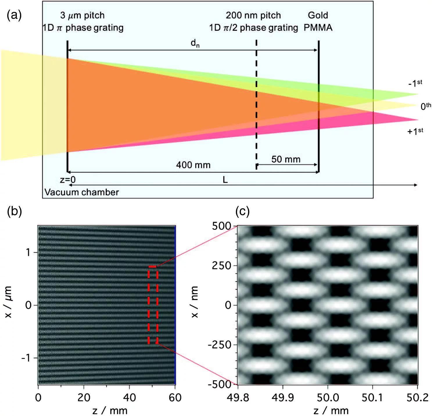
Benedikt Rösner is a researcher at the Paul Scherrer Institute (PSI) in the Laboratory for Non-linear Optics under the Center for Photon Science. He specializes in X-ray optics, orbital angular momentum (OAM) of light, and nanofabrication techniques for advanced microscopy. Education : PhD in Physical Chemistry from Friedrich-Alexander University Erlangen-Nürnberg (FAU Erlangen-Nürnberg). Rösner's research focuses on developing diffractive X-ray optics for high-resolution imaging, achieving a world-record 7 nm spatial resolution in soft X-ray microscopy. His work explores light-matter interactions with OAM beams, demonstrating novel phenomena like helical dichroism and magnetic helicoidal dichroism . He also investigates ultrafast processes via X-ray transient grating spectroscopy and time-streaking experiments at free-electron lasers. Key trends in his publications include advancements in X-ray microscopy resolution, OAM-based spectroscopies, and magnetic dynamics studies using femtosecond pulses. Collaborations span institutions like CNRS, University Pierre and Marie Curie, and FERMI free electron laser. His technical contributions involve designing optical elements for beamline upgrades in the SLS 2.0 project.
Iacopo Mochi is a Group Leader for Advanced Lithography and Metrology and beamline scientist at the Paul Scherrer Institute (PSI), specifically working at the Laboratory for X-ray Nanoscience and Technologies within the PSI Center for Photon Science. He has extensive experience in EUV lithography, X-ray optics, and semiconductor metrology, with a career spanning multiple prestigious research institutions including Lawrence Berkeley National Laboratory and imec. Dr. Mochi received his physics degree from the University of Florence and earned a PhD in Methods and Technologies for Environmental Monitoring. His career path has included significant contributions to LIDAR systems, astrophysical instrumentation, and ultimately EUV technologies for semiconductor manufacturing. At PSI since 2016, he has established himself as a leading researcher in advanced lithography techniques. His primary research focuses on the development of EUV instrumentation for semiconductor metrology, particularly the XIL-II metrology end station - a lensless microscope dedicated to EUV photomask inspection. His work spans nano-imaging, interferometry techniques in the X-Ray spectrum, and the characterization of advanced materials for semiconductor manufacturing. Dr. Mochi has made significant contributions to the field through the development of RESCAN, an actinic pattern inspection platform based on coherent diffraction imaging. Analysis of his publication record reveals a strong trajectory of innovation in EUV lithography, with recent work pushing resolution limits to 5 nm, developing novel metrology techniques, and applying machine learning approaches to improve image reconstruction and defect detection. His research directly addresses critical challenges in semiconductor manufacturing as the industry moves toward smaller technology nodes. Dr. Mochi's scientific contributions include groundbreaking work on EUV pellicles for mask protection, characterization of absorber and phase defects on EUV reticles, and advancements in interference lithography techniques. His collaborative research demonstrates strong partnerships with industry and academic institutions worldwide. As the beamline scientist for the XIL-II beamline, Dr. Mochi coordinates and supervises experiments that support cutting-edge research in semiconductor manufacturing and nanotechnology. His leadership extends to mentoring junior researchers and contributing to the development of next-generation scientists in the field of advanced lithography.
Yang Cao is a Professor at the University of Science and Technology of China , Department of Automation, Hefei, China. He holds a PhD from Northeastern University (2004, Shenyang, China) and has active affiliations with institutions like Virginia Tech and Huazhong University of Science and Technology. Research Focus: Spatiotemporal modeling, event-based vision, 3D human-object interaction, and industrial defect detection. Publications: 15 recent articles highlight his work in diffusion models, transformers, and state-space networks for tasks like traffic emission imputation, eye tracking, and PCB defect detection. Collaborative Work: Co-authored with Zheng-Jun Zha, Wei Zhai, Yu Kang, and others in journals like IEEE Transactions on Neural Networks and CVPR Workshops. Scientific Contributions: His research bridges computer vision, machine learning, and industrial applications, emphasizing real-world challenges such as low-light enhancement and sensor fusion.
Jichun Li is a Professor in the Department of Mathematical Sciences at the University of Nevada Las Vegas, with a prolific research career spanning computational mathematics, image processing, and computer vision. His work bridges theoretical mathematics with practical applications in medical imaging, environmental science, and biometrics. Li's research interests center on computational mathematics with particular focus on partial differential equations and finite element methods, alongside significant contributions to image processing and computer vision. His work demonstrates a unique integration of mathematical theory with practical applications, particularly in medical imaging where his techniques enable improved early cancer diagnosis through advanced lesion segmentation. In environmental science, his research on land surface albedo dynamics provides critical insights into climate change impacts in sensitive regions like the Tibetan Plateau. His recent work on face recognition with synthetic data addresses contemporary challenges in biometric security systems. Analysis of Li's publication trends reveals a strategic evolution from foundational mathematical research toward interdisciplinary applications. While maintaining strong theoretical contributions in computational mathematics, particularly in PDEs and finite element methods, he has increasingly focused on medical imaging applications since 2020, developing novel techniques for cancer diagnosis and ultrasmall object detection in CT scans. His 2022-2025 publications show growing emphasis on synthetic data applications, particularly in face recognition challenges, demonstrating adaptability to emerging AI trends. Professor Li maintains an extensive collaborative network, with frequent co-authorship patterns suggesting mentorship relationships with researchers like Bo Yan, Weimin Tan, Guannan Chen, and Encai Zhang across multiple publications. His work appears in leading journals including IEEE Transactions on Neural Networks and Learning Systems, IEEE Transactions on Multimedia, and Computational Mathematics and Applications, reflecting both the theoretical depth and practical relevance of his research.
Dr. Karol Tylmann is an Assistant Professor in the Department of Geomorphology and Quaternary Geology at the University of Gdańsk's Faculty of Oceanography and Geography. He leads research at the Laboratory of Geomorphological Reconstructions, focusing on the dynamics of Pleistocene ice sheets and landscape evolution in Northern Europe. His research integrates glacial geomorphology , Quaternary stratigraphy , and geochronological methods (e.g., cosmogenic nuclide dating, gamma-ray spectrometry) to reconstruct deglaciation patterns, ice-marginal processes, and sedimentary environments across Poland and the Baltic region. Key themes include: Ice sheet behavior during the Last Glacial Maximum and Younger Dryas Subglacial deformation mechanisms Coastal and fluvial responses to climatic shifts Geoheritage conservation in postglacial landscapes Recent publications (2018–2023) demonstrate a methodological emphasis on high-resolution geospatial analysis (LiDAR, object-based image processing), advanced dating techniques (10Be exposure, Bayesian modeling), and sedimentological diagnostics . Predominant research clusters include glacial landform mapping, deglaciation chronology, and geomorphic responses to paleoenvironmental change. Dr. Tylmann contributes to geotourism initiatives by evaluating the educational value of Poland's glacial landscapes. He maintains no listed research grants, awards, or supervised students in the available data.
Nathan Sherer is a Professor in the Department of Medical Microbiology and Immunology at the University of Wisconsin–Madison, with affiliations in the Bacteriology Department and the UW Carbone Cancer Center. His research integrates live-cell imaging and biochemical approaches to study virus-host interactions, focusing on HIV-1, Hepatitis B virus (HBV), and Human Papillomavirus (HPV). Institution: University of Wisconsin–Madison School: School of Medicine and Public Health Department: Department of Medical Microbiology and Immunology Additional Affiliation: UW Carbone Cancer Center Nathan Sherer earned his BA in Biology from Grinnell College in 1997, followed by a PhD in Microbiology from Yale University in 2006. He completed postdoctoral training at King’s College London before establishing his independent research program at UW-Madison. Dr. Sherer's research centers on the molecular virology of retroviruses, particularly HIV-1. His lab investigates how viral particles assemble and spread through host cells, with a focus on the post-transcriptional journey of HIV-1 genomic RNAs (gRNAs) from the nucleus to the plasma membrane. They utilize cutting-edge live-cell fluorescence microscopy, genetic manipulations, and biochemical assays to dissect virus-host interactions. Key areas include viral RNA transport, ribosomal frameshifting, virological synapse formation, and the role of host proteins in viral replication. The lab has expanded its scope to include HBV and HPV, exploring mechanisms of genome packaging, capsid trafficking, and viral oncogenesis. The recent publications highlight a strong emphasis on imaging technologies, RNA biology, and host factor identification. Articles span super-resolution microscopy development, single-cell analysis of viral gene expression, and proteomic mapping of viral RNA interactomes. Collectively, the work reveals sophisticated strategies viruses use to hijack cellular machinery, offering insights for novel antiviral therapies. Dr. Sherer mentors a dynamic team of graduate students and postdoctoral researchers from programs such as Molecular and Cellular Pharmacology, Cancer Biology, and Cellular and Molecular Biology. His alumni have pursued successful careers in academia, biotechnology, and pharmaceutical research. While no specific scientific awards are listed, his consistent high-impact publications reflect significant contributions to virology. The lab also benefits from collaborative grants and institutional support, enabling advanced imaging and proteomic studies. Dr. Sherer leads an active research laboratory with ongoing projects in HIV-1, HBV, and HPV virology. The team employs a multidisciplinary approach, combining molecular biology, imaging, and biochemistry to understand viral replication mechanisms. Lab members include senior scientists, postdocs, and graduate students working on various aspects of viral assembly, RNA regulation, and host interactions.
Dr. Ahmet Coskun is an Assistant Professor of Biomedical Engineering at Georgia Institute of Technology and Emory University, where he holds the Bernie-Marcus Early-Career Professorship. He directs the Single Cell Biotechnology and Spatial Omics Laboratory, an interdisciplinary program focused on multiparameter imaging of single cells within their spatial context. His work bridges the fields of bioengineering, computational biology, and systems biology to address fundamental challenges in cancers, immunology, and pediatric diseases. Dr. Coskun received his PhD from the University of California, Los Angeles (UCLA) working with Aydogan Ozcan. He completed postdoctoral training at the California Institute of Technology with Long Cai and served as an Instructor at Stanford University with Garry Nolan. His educational background has provided him with a strong foundation in both engineering principles and biological systems. Dr. Coskun's research lies at the nexus of multiplex bioimaging, microfluidic biodynamics, and big data biocomputation. His laboratory pursues three main research thrusts: spatial genomics (using seqFISH and correlation FISH methods), spatial proteomics (using CODEX technology combined with super-resolution imaging), and spatial metabolomics (using computational and isotope barcoding approaches with MIBI). His team develops machine learning algorithms to analyze the resulting high-dimensional imaging datasets, creating image-based 'omic technologies to reveal the spatial nature of biological systems. Their work has significant implications for understanding therapeutic response variability and cellular organization in health and disease. NSF CAREER Award 2024 NIH R35 MIRA Award 2023 BMES-CMBE Rising Star Award 2023 American Lung Association Innovation Award 2022 Student Recognition of Excellence in Teaching: Class of 1934 CIOS Award NIH K25 Award Burroughs Wellcome Fund CASI Award Dr. Coskun leads an interdisciplinary research team comprising PhD students from Bioengineering, Electrical and Computer Engineering, Mechanical Engineering, and Biomedical Engineering programs. His lab has been supported by numerous federal and private grants, including funding from multiple NIH institutes (NIA, NIAID, NCI, NIDCR, OD, and ORIP), Wellcome LEAP, Burroughs Wellcome Fund, NSF CMaT, American Cancer Society IRG, Multi-cellular engineered living systems (M-CELS), and Regenerative Medicine Center. In addition to his research, Dr. Coskun leads outreach programs through BioCrowd Studio, which engages K12 and undergraduate students through interactive virtual media and distributed biokits. The Single Cell Biotechnology and Spatial Omics Laboratory is strategically positioned at the forefront of spatial biology research. The lab benefits from advanced technologies including super-resolution microscopy, imaging mass spectrometry, combinatorial molecular barcoding, and machine learning to enhance the information capacity of cellular data. The team's innovative approaches to spatial multi-omics profiling have positioned them as leaders in understanding cellular heterogeneity and organization within tissues.
Dr. Aimo Winkelmann is a Visiting Professor in the Department of Physics at the University of Strathclyde, United Kingdom. His research is centered on advanced electron microscopy techniques, particularly electron backscatter diffraction (EBSD), for the structural characterization of semiconductor thin films and microstructures. His research interests lie at the intersection of materials science, solid-state physics, and microstructural analysis. He specializes in: High-resolution crystallographic imaging Strain and defect mapping in semiconductors Development of EBSD and transmission Kikuchi diffraction methods Analysis of GaN and silicon-based thin films Simulation and interpretation of electron diffraction patterns His recent publications demonstrate a consistent focus on pushing the limits of diffraction imaging in scanning electron microscopes. The work spans from fundamental simulations of Kikuchi patterns to applied studies on strain in silicon membranes and luminescence in GaN microstructures, indicating a strong trend toward quantitative microstructural analysis in functional materials. Dr. Winkelmann has delivered invited and plenary talks at major international conferences, including the XVIIIth International Conference on Electron Microscopy (2024) and the Materials Research Society Fall Meeting (2023), highlighting his recognition in the field. He actively collaborates with a core research team at Strathclyde, including Prof. Carol Trager-Cowan, Dr. Jochen Bruckbauer, and Dr. Ben Hourahine, and contributes to open datasets supporting reproducibility in EBSD research. While no formal students are listed, his role in mentoring through collaborative research is evident.
Jan Huisken is a Humboldt Professor for Multiscale Biology at the Georg-August-Universität Göttingen, affiliated with the Johann Friedrich Blumenbach Institute of Zoology and Anthropology. His research focuses on advanced light sheet microscopy techniques for biomedical and developmental biology applications. Role: Humboldt Professor University: Georg-August-Universität Göttingen Department: Johann Friedrich Blumenbach Institute of Zoology and Anthropology Research interests include light sheet microscopy , biomedical imaging , and developmental biology with a strong emphasis on zebrafish models. He develops tools for tissue clearing , image processing , and 3D microscopy . The 15 most recent publications analyze innovations in light sheet microscopy, tissue clearing protocols, and computational methods for image restoration. These works span fields such as optical imaging , developmental cardiology , computational biology , and biomedical instrumentation . Huisken contributes to open-source microscopy systems like 'Flamingo' and 'BigFUSE,' aiming to democratize access to advanced imaging technologies. His work integrates engineering, computer science, and biology to solve complex imaging challenges.
Bahram Javidi is a Professor in the Department of Biomedical Engineering at the University of Connecticut. His research focuses on advanced optical imaging technologies, including real-time 3D sensing, visualization, and information processing. He integrates nanotechnology, biomedical photonics, and quantum optics into novel imaging systems for medical and underwater applications. Key Research Areas: 3D integral imaging, digital holography, compressive sensing, optical encryption, and biomedical imaging. Recent Publications: Highlight innovations in underwater signal detection, lensless imaging for disease screening, and adversarial attack defense using optical systems. Technological Impact: Develops portable, low-cost medical diagnostic tools and augmented reality visualization systems. Collaborations: Works with interdisciplinary teams in biomedical engineering, computer science, and optical physics.
Pan Pan is a Professor in the Department of Biomedical Engineering at Huazhong University of Science and Technology, with extensive research contributions spanning medical image analysis, computer vision, and underwater wireless communications. Their work demonstrates strong interdisciplinary collaboration between biomedical engineering and computer science, with significant industry partnerships including Alibaba. Research interests focus on medical image analysis (particularly automatic breast ultrasound systems), deep learning applications in healthcare diagnostics, and secure underwater communications . Their work bridges theoretical advances with practical clinical applications, developing innovative segmentation algorithms, tumor detection systems, and secure communication protocols for specialized environments. Analysis of recent publications reveals a strong trend toward integrating multi-modal data fusion techniques with uncertainty-aware deep learning models for medical diagnostics. The research spans both fundamental algorithm development (novel segmentation networks, feature matching optimization) and domain-specific applications (ABUS tumor detection, ICU mortality prediction, underwater sensor networks). Pan Pan maintains active collaborations with major Chinese technology companies and academic institutions, evidenced by the consistent publication record in top-tier conferences including CVPR, ICCV, and NeurIPS. While specific awards aren't documented in the provided materials, the research impact is demonstrated through numerous high-impact publications across computer vision and biomedical engineering venues. The research program shows particular strength in translating computer vision techniques to medical applications, with significant contributions to semi-supervised learning approaches for medical image segmentation where labeled data is scarce. Recent work also demonstrates growing interest in secure communications for specialized environments like underwater sensor networks.
Sabrina Pacor is an Associate Professor in Applied Biology (BIO/13) at the University of Trieste , where she teaches Pharmacology in the Pharmacy LM and STB BSc programs. With over 30 years of research experience in experimental oncology and host defense peptides, she has made significant contributions to studying antimicrobial peptides (AMPs) and their interactions with bacterial membranes. Her research focuses on: Direct antimicrobial activity of AMPs against Gram-positive and Gram-negative bacteria Indirect immunomodulatory effects of host defense peptides Development of drug delivery systems using nanomaterials (carbon nanotubes, gold nanoparticles) Mechanistic studies of ruthenium-based antimetastatic drugs She leads extensive cytofluorimetry research using flow cytometry platforms, particularly for evaluating: Cytotoxicity (necrosis/apoptosis, proliferation index) Modulation of host biological responses (chemotaxis, phagocytosis, ROS production) Peptide-bacterial membrane interactions through fluorescent labeling Her recent work demonstrates trends in: Proline-rich antimicrobial peptides against ESKAPE pathogens Hybrid antibiotic design (peptide-aminoglycoside conjugates) Structure-activity relationships in membranolytic peptides Evolutionary insights into defensin and cathelicidin families Prof. Pacor has co-authored over 100 peer-reviewed publications and actively mentors students, having supervised: 70 experimental/thesis reviews for Pharmacy/CTF Master's students 20 Bachelor's degree theses
Xiang Wang serves as an Associate Professor in the Department of Chemistry at Xiamen University's College of Chemistry and Chemical Engineering, maintaining his office in Room 434 of the Chemical Building since 2020. His research program bridges experimental nanoscience and electrochemistry with strong institutional ties to Xiamen University's PCOSS research group. His academic journey began with BS (2007) and PhD (2013) degrees from Xiamen University, followed by research assistantship (2014-2016) and postdoctoral training (2016-2020) at the same institution. This continuous affiliation demonstrates deep integration within Xiamen's chemical research ecosystem. Dr. Wang's research centers on advanced nanoscale spectroscopic techniques , particularly Tip-enhanced Raman spectroscopy (TERS) and Surface-enhanced Raman spectroscopy (SERS) . His work explores Nano-optics , Nanospectroelectrochemistry , and Nanoscale in-situ characterization to investigate 2D materials, electrocatalytic processes, and biomolecular structures at unprecedented resolution. This interdisciplinary approach merges physical chemistry with materials engineering to solve fundamental interface science problems. Analysis of his 2019-2024 publications reveals dominant themes in nanoscale characterization of 2D materials (particularly MoS 2 ), electrocatalyst evolution monitoring , and novel spectroelectrochemical methodologies . His work consistently appears in high-impact journals including Nature Catalysis , Chemical Society Reviews , and Advanced Materials , demonstrating significant contributions to nanospectroscopy and materials characterization fields.
Venkatesh Murthy serves as the Raymond Leo Erikson Life Sciences Professor of Molecular & Cellular Biology at Harvard University and co-directs the Harvard Brain Science Initiative. His laboratory is housed within the Faculty of Arts and Sciences at Harvard's Biological Laboratories in Cambridge, Massachusetts. His research investigates the neural and algorithmic basis of odor-guided behaviors in terrestrial animals, primarily using mouse models. The Murthy Lab develops naturalistic behavioral paradigms that enable simultaneous electrophysiological recordings, high-resolution optical imaging, and optogenetic manipulation. Key research areas include neural circuit dynamics in the olfactory system, modification of circuits through behavioral state and learning, and development of computational models to explain neural observations. The lab maintains strong interdisciplinary collaborations with theoretical neuroscientists. Recent publications (2023-2025) reveal consistent focus on olfactory navigation mechanisms, neural signal processing algorithms, circuit-level analysis of social behaviors, and biomimetic applications for electronic sensing systems. Work spans experimental techniques including multi-animal pose estimation (using DeepLabCut), neural deconvolution methods, and analysis of fluctuating odor environments. The Murthy Lab operates within Harvard's Department of Molecular & Cellular Biology as part of the broader Harvard Brain Science Initiative ecosystem, contributing to research in Cognitive and Behavioral Neuroscience, Theory and Computation, and Sensory and Motor Systems.