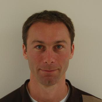
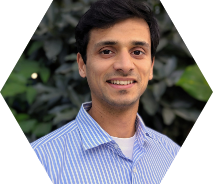
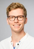
Prof. Dr. Dennis Säring is a faculty member at the University of Applied Sciences Wedel , specifically affiliated with the School of Engineering. His academic and research activities focus on Deep Learning , Medical Image Analysis , and applications of Artificial Intelligence in healthcare and biomedical imaging. He has led seminars on Deep Learning topics and supervised student projects in Autonomous Driving at Audi's AADC 2018 competition. Research Highlights : Cardiovascular imaging, forensic age estimation via MRI, neural network-based bone segmentation, and cerebrovascular aneurysm analysis. Technical Expertise : Cardiac MRI, 3D/4D image processing, parametric mapping, and spatiotemporal data fusion. His recent publications (2018-2023) emphasize 3D MR segmentation for age assessment, CMR strain analysis in athletes, and T1/T2 mapping for myocarditis. Key collaborations include institutions like the University Medical Center Hamburg-Eppendorf and Wedler Hochschulbund, with funding for autonomous vehicle research. While no explicit scientific awards are listed, his work spans clinical cardiology, forensic radiology, and AI-driven medical diagnostics.
Jeremy Dahl is a Professor of Radiology (Pediatric Radiology) at Stanford University School of Medicine. He directs the Ultrasound Imaging & Instrumentation Lab and serves as Director of Research Academic Affairs in the Department of Radiology since 2020. He holds multiple affiliations across Stanford including Bio-X, the Cardiovascular Institute, Wu Tsai Human Performance Alliance, Maternal & Child Health Research Institute, Stanford Cancer Institute, and Wu Tsai Neurosciences Institute. Dr. Dahl received his B.S. in Electrical Engineering from the University of Cincinnati (1999) and Ph.D. in Biomedical Engineering from Duke University (2004). His research focuses on developing ultrasonic beamforming and image reconstruction methods for diagnostic imaging applications, particularly techniques that generate high-quality images in difficult-to-image patients. His laboratory specializes in B-mode and Doppler imaging techniques that utilize additional information from ultrasonic wavefields to improve image quality and develop real-time imaging systems for clinical applications including cardiac, liver, and fetal imaging. Dr. Dahl's research has led to significant advancements in ultrasound molecular imaging platforms, sound speed estimation, aberration correction, and reverberation noise suppression. His work often bridges engineering innovation with clinical applications for cancer detection and other diseases. His recent publications demonstrate strong focus on machine learning applications in ultrasound, distributed aberration correction, and molecular imaging techniques. Fellow, American Institute of Ultrasound in Medicine (2021) Senior Member, Institute of Electrical and Electronics Engineers (2020) Distinguished Investigator Award, The Academy for Radiology & Biomedical Imaging Research (2018) Outstanding Paper Award, IEEE Ultrasonics, Ferroelectrics, and Frequency Control Society (2011) Dr. Dahl serves in editorial roles for major journals including IEEE Transactions on Medical Imaging (2017-2024) and IEEE Transactions on Ultrasonics, Ferroelectrics, and Frequency Control (2013-Present). His laboratory has successfully translated numerous innovations into clinical applications, with multiple patents including recent developments in pulsed focused ultrasound therapy and speed of sound quantification.
Professor Guy Williams is a leading academic at the University of Cambridge with a focus on imaging science and clinical neurosciences, affiliated with Downing College and the Wolfson Brain Imaging Centre . Holding a PhD in Physics from his initial Natural Sciences degree, he specializes in nuclear magnetic resonance (NMR) and MRI techniques for brain imaging. Education: BA, PhD in Physics His research centers on non-invasive imaging of brain structure and function, particularly in traumatic brain injury (TBI) and dementia. His work involves developing novel MRI pulse sequences and advanced data analysis algorithms, including AI-based diagnostic tools. He leads studies on white matter integrity post-trauma, longitudinal dementia assessment, and applications of MRI in disorders of consciousness and addiction. Recent publications highlight collaborations in traumatic brain injury outcomes, AI-guided dementia prediction, and neuroimaging of post-COVID cognitive deficits. His team's work on ultra-high field laminar fMRI and distortion correction methods has advanced clinical neuroscience applications. Key techniques include diffusion tensor imaging (DTI), 7 Tesla MRI, and positron emission tomography (PET/MR). His research spans from basic NMR physics to clinical translation, with a strong emphasis on multi-site studies and real-world diagnostic implementation.
Brian W. Pogue, Ph.D., is the Robert A. Pritzker Chair in Biomedical Engineering at Dartmouth College's Thayer School of Engineering, with a joint appointment as an Honorary Fellow in Medical Physics at the University of Wisconsin-Madison. His academic background includes a Ph.D. in Medical/Nuclear Physics from McMaster University and a Research Fellowship at Harvard Medical School's Wellman Center for Photomedicine. He has led significant administrative roles, including Dean of Graduate Studies at Dartmouth (2008–2012) and Chair of Medical Physics at Wisconsin (2022–2025). Research Focus : Dr. Pogue pioneers Optics in Medicine , specializing in cancer imaging, photodynamic therapy, and surgical guidance. His work integrates fluorescence imaging, radiation therapy monitoring, and molecular diagnostics to improve cancer treatment precision. Key innovations include Cherenkov imaging for radiotherapy dosimetry and hypoxia-sensitive probes for tumor resection. Publication Trends : Recent articles (2023–2025) emphasize real-time surgical guidance, hypoxia quantification, and multimodal imaging systems. Dominant themes include fluorescence tomography, radiation dosimetry, and low-cost diagnostic devices, reflecting a translational focus from preclinical validation to clinical applications. Awards & Honors : Fellow, Optica (formerly OSA) Fellow, American Institute for Medical and Biological Engineering (AIMBE) Fellow, American Association of Physicists in Medicine (AAPM) Fellow, SPIE (International Society for Optics and Photonics) Funding & Innovation : Continuously funded by the NIH since 2001 ($52M+ total), Dr. Pogue founded three startups: DoseOptics LLC (radiotherapy dose imaging) and Hypoxia Surgical LLC (tissue hypoxia cameras), bridging academic research to clinical tools.
Karthik Menon serves as an Assistant Professor with a joint appointment in the Woodruff School at Georgia Institute of Technology and the Coulter Department of Biomedical Engineering. His research integrates fluid mechanics, computational modeling, and data-driven methodologies to address critical challenges in healthcare, renewable energy, and bio-inspired engineering systems. His academic credentials include: Ph.D. in Mechanical Engineering, Johns Hopkins University (2021) M.S. in Mechanical Engineering, Johns Hopkins University (2019) B.E. in Mechanical Engineering, Birla Institute of Technology and Science, Pilani, India (2015) Menon's research program centers on three interconnected domains: cardiovascular flows for personalized treatment of heart disease, fluid-structure interactions in biological systems like heart valves and bio-mimetic robots, and vortex-dominated flows for renewable energy applications. His approach combines high-fidelity computational modeling with machine learning to uncover fundamental physics and develop clinical solutions, such as cardiovascular digital twins for non-invasive risk assessment. Current projects focus on patient-specific hemodynamics using CT imaging and uncertainty quantification to improve surgical planning. Analysis of his 15 most recent publications (2023-2025) reveals a dominant focus on advancing multi-fidelity computational frameworks for cardiovascular applications. Key trends include Bayesian uncertainty quantification, zero-dimensional solver development, and integration of clinical imaging data to create predictive digital twins. His work bridges fluid dynamics with clinical cardiology, targeting improved outcomes in coronary artery disease and Kawasaki-related complications through physics-informed machine learning. Menon's scholarly contributions have been recognized through competitive awards: WCCM-PANACM 2024 Travel Award, U.S. Association for Computational Mechanics (2024) Future Faculty Symposium Travel Award, Society of Engineering Science Conference (2023) Mark O. Robbins Prize in High-performance Computing, Johns Hopkins University (2021) Corrsin-Kovasznay Outstanding Paper Award, Johns Hopkins University (2020) Prosperetti Travel Award, Johns Hopkins University (2017) Mechanical Engineering Departmental Fellowship, Johns Hopkins University (2016) As principal investigator of the ComBiNE Fluid Dynamics Lab, Menon mentors graduate students in developing computational tools for fluid-structure interaction problems. His collaborative projects with cardiologists at Stanford and Emory hospitals translate engineering principles into clinical applications for cardiovascular disease management. Current grant activities focus on NSF and NIH-funded initiatives for uncertainty-aware cardiovascular modeling and bio-inspired flow energy harvesting. The ComBiNE Fluid Dynamics Lab operates as an interdisciplinary hub where engineers, clinicians, and data scientists collaborate on fluid mechanics challenges. Current lab initiatives include developing real-time hemodynamic simulators for surgical planning, creating reduced-order models for cardiac device optimization, and investigating vortex dynamics in fish schooling for underwater vehicle design. The lab maintains strong partnerships with Children's Healthcare of Atlanta and the Parker H. Petit Institute for Bioengineering and Bioscience.
Adam de la Zerda is an Associate Professor at Stanford University's Department of Structural Biology (School of Medicine) and Electrical Engineering (by courtesy). He develops advanced optical molecular imaging technologies combining nanoparticle contrast agents and adaptive OCT systems for cancer and ophthalmic disease research. Technion-Israel Institute of Technology (BSc, 2005) Stanford University (PhD, 2011) UC Berkeley (Postdoctoral Fellowship) Research Themes : Virtual biopsy using machine learning-enhanced OCT Gold nanorod-based molecular contrast agents Speckle noise reduction for cellular resolution Needle beam optical coherence tomography angiography His 15 most recent publications demonstrate technical innovations in: Metasurface optics for extended depth-of-field Spectral deconvolution of multiple contrast agents Speckle modulation for improved diagnostic clarity Noninvasive lymphatic system mapping Scientific Honors : Pew-Stewart Scholar for Cancer Research AFOSR Young Investigator NIH Early Independence Award Forbes 30 Under 30 (x2) Chan Zuckerberg BioHub Investigator His lab team has developed clinical prototypes including OcuBell Inc. 's ophthalmic imaging systems and Visby Medical 's diagnostic platforms. Current research spans from in vivo glycoprotein imaging to de novo biosensor development for real-time disease monitoring in awake animal models.
Professor Fernando Calamante is a Professor of Biomedical Engineering at The University of Sydney and Director of Sydney Imaging Core Research Facility. He leads the National Imaging Facility node and focuses on advanced MRI methodologies, particularly Diffusion and Perfusion MRI, to study brain connectivity and neurological disorders. His work includes developing the MRtrix software, widely used in diffusion MRI analysis. He holds extensive funding (~$50M) and has been recognized with awards like ISMRM Fellowship and NHMRC grants. His research spans super-resolution imaging, brain connectomics, and clinical applications in stroke and tumors. Education: BSc (Physics, Argentina), PhD (Magnetic Resonance Imaging, University College London). Career highlights include leadership roles at The Florey Institute and ISMRM presidency (2021-2022). Research interests include: Novel MRI methods for brain connectivity and super-resolution imaging Applications of Diffusion and Perfusion MRI in neurology Integration of structural and functional connectomics Key achievements: Over 200 publications, software innovations, and leadership in global MRI societies.
Prof. Dr. med. Franz Lennard Ricklefs is a Senior Physician and Head of the Working Group at the Department of Neurosurgery, University of Hamburg Faculty of Medicine. He is a Medical Specialist in Neurosurgery with cross-disciplinary expertise in neuro-oncology, molecular pathology, and extracellular vesicle research. Affiliations: University Medical Center Hamburg-Eppendorf (UKE), European Liquid Biopsy Society (ELBS), International Consortium on Meningiomas (ICOM) Research Interests: His work focuses on neurosurgical oncology, particularly glioblastoma and meningioma pathobiology. He investigates DNA methylation patterns, extracellular vesicle biomarkers, and liquid biopsy implementation in clinical neuro-oncology. Additional interests include surgical outcomes for epilepsy and aneurysm management. Article Trends: Over the last decade, Dr. Ricklefs has published extensively on: Extracellular vesicle applications as liquid biopsy markers DNA methylation subclasses for glioblastoma and meningioma Multicenter surgical outcome benchmarking Immune evasion mechanisms in neuro-oncology Technological innovations in neurosurgical visualization Molecular characterization of rare CNS tumors Professional Contributions: He co-authored the MISEV2023 guidelines for extracellular vesicle studies and participates in international consensus reviews for meningioma classification. His collaborations span institutions across Europe and North America.
Prof. Dr. Roland Zengerle serves as Full Professor for Application Development at the Institute of Microsystems Technology within the Faculty of Engineering at Albert Ludwigs University of Freiburg, concurrently holding the position of Director at Hahn-Schickard Institute for Microanalysis Systems in Freiburg. His academic leadership spans microsystems engineering with a focus on translational research bridging fundamental science and clinical applications. Zengerle's research expertise centers on Microfluidics, Lab-on-a-Chip systems, Bio-MEMS, Electrochemical Energy Systems, and Tomographic Reconstruction of Mesoporous Materials. He pioneers hybrid manufacturing techniques integrating molten metal printing with polymer processing to develop point-of-care diagnostic platforms and advanced energy systems. Current projects include UTI-Diag for urinary tract infection diagnosis and PhotonMed, a 32-million-euro medical technology initiative where his MEMS Applications Laboratory develops centrifugal microfluidic solutions. Analysis of his recent publications reveals a dominant trend toward multi-technology integration: centrifugal microfluidics combined with 3D bioprinting for organoid-based drug testing, molten metal printing for flexible electronics, and bead-based immunoassays for infectious disease detection. The work demonstrates strong clinical translation focus, particularly in cancer diagnostics (circulating tumor cell isolation), infectious disease testing (TB diagnostics), and regenerative medicine (spheroid/organoid handling). His laboratory has secured significant funding for high-impact projects including: UTI-Diag: Molecular diagnostics for urinary tract infections PhotonMed: Medical technology innovation consortium livMatS: Living, Adaptive and Energy-autonomous Materials Systems Zengerle actively mentors researchers through Freiburg's Master Lab program and Writer's Studio initiative while promoting young talent via Bootcamp training. His group maintains strategic alliances with Hahn-Schickard spin-offs and industry partners, leveraging university cleanroom facilities and specialized service centers for microfabrication. The MEMS Applications Laboratory operates as a hub for interdisciplinary innovation, combining microfabrication expertise with clinical insights to develop commercializable diagnostic solutions. Current infrastructure supports centrifugal microfluidic cartridge development, 3D-bioprinting of tissue models, and electrochemical sensor integration, with ongoing work focused on automating complex biological workflows for point-of-care applications.
Dr. Ulas Bagci is an Associate Professor at Northwestern University's Feinberg School of Medicine, Department of Radiology. He holds courtesy appointments in Biomedical Engineering (BME), Electrical and Computer Engineering (ECE) at Northwestern, and Computer Science at the University of Central Florida. As the director of the Machine and Hybrid Intelligence Lab, his research focuses on AI and machine learning applications in biomedical and clinical imaging. Education: BS: Bilkent University (2003) MS: Koç University (2005) Fellow: University of Pennsylvania (2009) PhD: University of Nottingham (2010) ISTP Fellow: NIH (2012) Research Interests: Dr. Bagci’s work spans artificial intelligence, machine learning, and their integration into medical imaging workflows. His lab develops algorithms for tumor segmentation, radiomics analysis, and ethical AI frameworks in healthcare. Notable projects include large-scale MRI segmentation of cirrhotic livers and predictive models for clinical outcomes in oncology and cardiology. Publications: His recent work emphasizes AI-driven solutions for challenges in radiology, including lung disease detection, pulmonary embolism mortality prediction, and ethical considerations in foundational AI models. His articles reflect a focus on bridging clinical needs with advanced computational methods. Lab & Affiliations: The Machine and Hybrid Intelligence Lab collaborates with the Robert H. Lurie Comprehensive Cancer Center. Research themes include federated learning, medical image synthesis, and AI ethics in clinical decision-making.
Dr. Robert A. Goldberg is a Professor at the Jules Stein Eye Institute, University of California, Los Angeles (UCLA), holding the Bert O. Levy Endowed Chair in Orbital and Ophthalmic Plastic Surgery. His work spans orbital decompression , thyroid eye disease , facial aging quantification , and complications of hyaluronic acid fillers . Research Focus: Orbital and ophthalmic plastic surgery, tumor histopathology, AI applications in facial aging, filler-related blindness mechanisms. Clinical Expertise: Management of proptosis, asymmetric ptosis, orbital vascular malformations, and thyroid-associated orbitopathy. Recent Trends: Publications emphasize multimodal therapies (e.g., Teprotumumab + decompression), nonoperative oncology (Bleomycin palliation), and dynamic imaging techniques (high-resolution ultrasound, CT volumetry). Collaborations: Extensive work with Rootman DB on orbital anatomy, Douglas RS on thyroid eye disease, and Ugradar S on filler complications. Technical Innovations: Described techniques for autologous tarsus banking , orbital volume expansion , and endoscopic orbital approaches .
Dr. Sung Sik Lee serves as a Lecturer in the Department of Materials at ETH Zurich, Switzerland. Affiliated with ScopeM (Scientific Center for Optical and Electron Microscopy), he develops microfluidic platforms for real-time cellular analysis at the HPM C 52.2 facility (Otto-Stern-Weg 3, Zürich). His research bridges engineering and biology to investigate cellular responses to mechanical and chemical stimuli. His primary research domains include: Microfluidics : Design of microfabricated devices for cell stretching, particle separation, and dynamic stimulation Cellular Aging : Mechanisms of chromosome loss and nuclear pore complex reorganization in yeast models Nanotoxicology : Impact of nanoplastics on macrophage inflammation and intestinal barrier integrity Advanced Imaging : Application of holotomography and Raman spectroscopy for label-free cellular analysis His work consistently targets translational applications in disease modeling and diagnostics. Analysis of his 50+ publications reveals strong interdisciplinary integration, particularly the convergence of machine learning with microscopy (e.g., automated vacuole quantification in yeast) and the development of open-access resources like MicrobioRaman. Recent trends emphasize nanoparticle-cell interactions and microfluidic solutions for inflammatory conditions including IBD and acute kidney injury. Dr. Lee actively contributes to ScopeM's mission of advancing microscopy techniques, maintaining collaborations across ETH Zurich's research ecosystem. His laboratory focuses on microfluidic device fabrication, cellular mechanotransduction studies, and biophysical characterization of particles and cells, with ongoing projects extending through 2025.
Paul A. Yushkevich is a Professor of Radiology at the Perelman School of Medicine, University of Pennsylvania , with affiliations in the Bioengineering Graduate Group . He leads the Penn Image Computing and Science Laboratory (PICSL) , focusing on advanced biomedical image analysis techniques. Developed first-of-its-kind computational atlas of the hippocampal formation Led NIH R01-funded research on MRI-derived biomarkers for Alzheimer's disease Created open-source software tools: ITK-SNAP and Convert3D Expert in statistical shape modeling and histology-MRI co-registration Research Focus: Specializes in hippocampal segmentation using high-resolution MRI, with applications in Alzheimer's disease research and cardiac imaging . His work combines differential equations and machine learning for accurate image analysis. Scientific Achievements: First-place MICCAI segmentation challenges (2012, 2013) Over 169 PubMed publications in neuroimaging and computational anatomy Developed DTI-TK toolkit for diffusion MRI analysis Collaborations: Works with Alzheimer's Disease Neuroimaging Initiative (ADNI) and multiple international institutions. Supervises graduate students in biomedical image analysis.
James Corbett serves as Professor of Radiology and Professor of Internal Medicine at the University of Michigan Medical School, with dual appointments reflecting his interdisciplinary expertise. He is an active Center Member of the Samuel and Jean Frankel Cardiovascular Center, contributing to one of the nation's leading cardiovascular research and clinical care institutions. Professor Corbett's research program centers on nuclear cardiology innovation, particularly PET-based myocardial blood flow quantification. His work bridges technical imaging optimization (e.g., Rb-82 dosing protocols, temporal sampling methods) with clinical applications in arrhythmia risk assessment, diastolic dysfunction, and pulmonary hypertension management. He has significantly influenced global standards through leadership in the IAEA Nuclear Cardiology Protocols Study (INCAPS) and SNMMI/ASNC clinical guidelines. His publication record from 2016-2019 reveals a strategic focus on advancing quantitative cardiac imaging while addressing real-world clinical challenges. The research demonstrates consistent methodology development paired with outcome studies, often through large multi-center collaborations that enhance generalizability of findings. As an active mentor (explicitly noted in his profile), Professor Corbett guides trainees through the University of Michigan's robust medical education system. His involvement in major international studies indicates substantial grant funding history, though current specific awards aren't detailed in the provided materials. The Samuel and Jean Frankel Cardiovascular Center affiliation provides Professor Corbett's team with state-of-the-art imaging facilities and access to diverse cardiovascular patient populations, enabling translational research from technical innovation to clinical implementation.
Bjoern Menze is a Professor and Rudolf Mößbauer Tenure Track Chair at the Technical University of Munich (TUM), leading the Image-based Biomedical Modeling Group within the Munich School of Bioengineering. His research focuses on medical image computing, tumor growth modeling, and computational physiology, with applications in clinical neuroimaging and personalized radiotherapy design. He holds a Ph.D. in Computer Science from Heidelberg University and has held positions at ETH Zurich, INRIA Sophia Antipolis, MIT, and Harvard Medical School. His academic journey includes a postdoc at MIT’s CSAIL and Harvard Medical School, followed by roles at ETH Zurich and INRIA. His work bridges biomedical imaging with machine learning, emphasizing model-driven analysis of physiological processes. He has been a visiting professor at Maastricht University and contributes to initiatives like the Center for Translational Cancer Research at TUM. Key research areas include tumor growth modeling, quantitative imaging biomarkers, and integrating mathematical models with clinical data. His awards include the MICCAI Young Scientist Award (2014), Leopoldina Fellowship (2009), and DFG Research Fellowship (2008). He advises on medical AI, leads interdisciplinary projects, and publishes extensively in top journals like Nature Neuroscience and IEEE Transactions on Medical Imaging. His lab’s work spans applications such as glioblastoma radiotherapy optimization, whole-body bone lesion detection, and neural connectivity imaging. Collaborations include institutions like Harvard, MIT, and ETH Zurich. He emphasizes translating computational methods into clinical practice for personalized healthcare solutions.