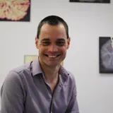
Massimo Mischi is a Full Professor at the Faculty of Electrical Engineering of the Eindhoven University of Technology (TU/e) and chairs the Signal Processing Systems (SPS) Division , the largest division at TU/e with over 250 researchers. He founded the Biomedical Diagnostics (BM/d) Lab in 2012, which now includes 180 researchers and clinical/industrial advisors, focusing on biomedical signal processing for diagnostics and monitoring.
Brendan A. Harley is the Robert W. Schaefer Professor in Chemical and Biomolecular Engineering at the University of Illinois at Urbana-Champaign (UIUC), with a joint appointment in the Carl R. Woese Institute for Genomic Biology. His research focuses on developing biomaterials to replicate complex tissue microenvironments, enabling insights into cell behavior during development, disease, and regeneration. He holds leadership roles in academic and professional organizations, including editorial positions for Science Advances and Tissue Engineering . Harley earned his SB from Harvard University (2000), SM/ScD from MIT (2002, 2006), and completed postdoctoral studies at Boston Children’s Hospital. **Education**: Sc.D., Massachusetts Institute of Technology, 2006 S.M., Massachusetts Institute of Technology, 2002 S.B., Harvard University, 2000 **Research Interests**: Engineering dynamic, spatially-patterned biomaterials to mimic extracellular matrices Regenerative repair of musculoskeletal tissues Biomaterial models of cancer microenvironments (e.g., glioblastoma) Artificial bone marrow systems for hematopoietic stem cell studies Harley’s work has produced over 100 peer-reviewed articles and co-authored a textbook Cellular Materials in Nature and Medicine . His lab develops materials for clinical applications, including osteochondral defect repair and craniofacial bone regeneration. Notable honors include the NSF CAREER Award (2013), AAAS Fellowship (2014), and AIMBE Fellowship (2019). **Awards/Recognition**: Fellow, AAAS (2014) Young Investigator Award, Society for Biomaterials (2014) Campus Distinguished Promotion Award, UIUC (2018) His research group emphasizes translational outcomes, with projects spanning biomaterial design, cancer modeling, and stem cell engineering. Collaborations include industry partners like Orthomimetics (acquired by TiGenix) and foundational studies on tumor microenvironments.
Dr. Sonit Singh is a Lecturer at the School of Computer Science and Engineering at the University of New South Wales (UNSW), Sydney, Australia. Prior to this role, he served as a Postdoctoral Research Fellow at the same institution. He holds a PhD from Macquarie University, completed in collaboration with Macquarie. His research focuses on artificial intelligence, computer vision, natural language processing, and their applications in healthcare, particularly in medical imaging and diagnostic systems. Dr. Singh's academic background includes a strong emphasis on biomedical imaging, intelligent robotics, and computing education. His work bridges machine learning advancements with real-world clinical challenges, such as tumor segmentation, radiology report generation, and disease detection through imaging modalities like CT scans and ultrasound. His recent publications highlight innovations in 3D medical image localization, robust tumor segmentation networks, and clinical context-aware radiology report systems. These contributions underscore his expertise in integrating deep learning techniques with healthcare workflows. Dr. Singh collaborates with multidisciplinary teams, including medical professionals, to develop solutions for challenges like placenta segmentation and dietary intake monitoring using AI. While no specific awards or grants are explicitly listed, his extensive publication record reflects sustained academic engagement in high-impact areas. His research often addresses practical clinical needs, such as improving diagnostic accuracy and patient care through advanced computational methods.
Prof. Kathleen Curran is a Professor at University College Dublin (UCD) and director of the UCD machine learning in medical imaging and diagnostics innovative research lab ( https://www.ucd-ml-mi.com/ ). She serves as an Affiliated Principal Investigator in the Centre for Biomedical Engineering, an INSIGHT funded investigator, and a funded investigator in the Science Foundation Ireland centre for research training in machine learning (ML-Labs). Her research integrates artificial intelligence, computer vision, and clinical medicine to develop interpretable AI solutions for medical diagnostics. Key focus areas include fetal ultrasound imaging, cardiac MRI reconstruction, neuroimaging for Alzheimer's disease and multiple sclerosis, and biomarker discovery for conditions like lymphangioleiomyomatosis and placenta accreta spectrum. She pioneers techniques in diffusion models, explainable AI, and multi-modal learning to address challenges in low-data medical scenarios. Analysis of her recent publications reveals dominant trends in applying generative models for medical data augmentation, developing uncertainty-aware diagnostic systems, and creating interpretable clinical AI tools. Her work consistently targets high-impact clinical applications including fetal development monitoring, cardiovascular disease management, and neurological disorder detection, with strong emphasis on real-world clinical implementation. Scientific recognition includes: 2019 InterTrade Ireland FUSION Project Exemplar Award (with Axial Medical Printing Ltd.) Three Enterprise Ireland Commercialisation Fund awards as Principal Investigator Horizon Europe consortium funding for SMASH-HCM project (Stratification, Management, and Guidance of Hypertrophic Cardiomyopathy Patients using Hybrid Digital Twin Solutions) Prof. Curran leads significant research funding initiatives including Horizon Europe and multiple Enterprise Ireland awards. Her group actively collaborates with industry partners like Axial Medical Printing Ltd. and participates in national research centers such as INSIGHT and ML-Labs, driving translational AI research from bench to bedside. The UCD machine learning in medical imaging and diagnostics lab ( https://www.ucd-ml-mi.com/ ) serves as her primary research hub, fostering interdisciplinary collaborations between computer scientists, clinicians, and biomedical engineers to advance clinical AI solutions.
Jeffery A Goldstein, MD, PhD is an Associate Professor in the Department of Pathology at Northwestern University Feinberg School of Medicine, where he serves as Director of Perinatal Pathology with additional appointments in Autopsy Pathology. He is an attending physician at Northwestern Memorial Hospital with clinical and teaching responsibilities in perinatal and autopsy pathology. His educational background includes: PhD: University of Chicago (2012) MD: University of Chicago (2014) Residency: Vanderbilt University, Anatomic Pathology (2017) Fellowship: Northwestern University, McGaw Medical Center (Lurie Children's Hospital), Pediatric Pathology (2018) Dr. Goldstein is an early-stage investigator focusing on maternal-child health with particular expertise in placental pathology. His research integrates bioimaging, informatics, and machine learning to transform placental examination from a specialized, resource-intensive process into a widely accessible diagnostic tool. He develops AI algorithms that can analyze placental photographs and microscopic slides to detect abnormalities associated with infection, neonatal sepsis, and other pregnancy complications. His work bridges computational science with clinical pathology to address the significant gap that less than 20% of placentas receive clinical examination despite their diagnostic value for future maternal and child health. His research portfolio includes multiple innovative projects applying machine learning to placental diagnosis, deep phenotyping, and quantitative description of placental features in health and disease. His recent publications demonstrate applications of deep learning for fetal inflammatory response diagnosis, machine learning assessment of gestational age, and analysis of placental lesions in gestational diabetes. Dr. Goldstein holds board certifications in Anatomic Pathology and Pediatric Pathology from the American Board of Pathology. His clinical work focuses on microscopic examination of placental slides, which forms the foundation of his research. He is affiliated with several research centers including the Center for Reproductive Science, the Institute for Artificial Intelligence in Medicine (specifically the Center for Computational Imaging and Signal Analytics in Medicine), and the Northwestern University Clinical and Translational Sciences Institute (NUCATS). His work has been featured in Northwestern Medicine news regarding AI applications for placental analysis to detect neonatal and maternal problems.
Dr. Andrew Melbourne is an Associate Professor (Reader) in Healthcare Technologies at King's College London, affiliated with the School of Biomedical Engineering & Imaging Sciences and the Department of Surgical & Interventional Engineering. He leads the MSc/MRes Healthcare Technologies program and focuses on imaging sciences, computational modeling, and placental physiology. His work includes cross-disciplinary collaborations with clinicians to improve understanding of fetal interventions, placental function in conditions like fetal growth restriction, and twin pregnancies. Key projects include MRI advancements for placental and fetal health, funded by organizations such as the Wellcome Trust and NIH. Research interests span medical imaging techniques (MRI, CT), fetal-neonatal brain development, and AI-driven image analysis. Notable contributions include fetal noise exposure modeling, placental perfusion studies, and super-resolution MRI applications for surgical planning. Dr. Melbourne co-leads initiatives like the MIBIRTH study, aiming to optimize pregnancy management through imaging. He has organized workshops on perinatal image analysis and contributed to student prizes in Healthcare Technologies. Publications span over 100 peer-reviewed articles in journals like Nature Communications and NeuroImage, emphasizing placental biology, fetal surgery outcomes, and AI in medical imaging. His work bridges clinical needs with engineering solutions to address maternal-fetal health challenges.
Oliver Wieben is a Professor in the Departments of Medical Physics and Radiology at the University of Wisconsin-Madison. He serves as Vice Chair for Research and Co-Director of the International Center for Accelerated Medical Imaging (2013-2019). B.S., Electrical Engineering, Leibniz Universität Hannover Dipl.-Ing., Electrical Engineering, Karlsruhe Institute of Technology Ph.D., Electrical Engineering, University of Wisconsin-Madison His research focuses on MRI methodology development , particularly in cardio/cerebrovascular imaging , 4D Flow MRI , and placental oxygenation assessment . He has pioneered techniques for real-time imaging , velocity-sensitive MRI , and motion correction schemes in MR imaging. His recent publications (2022-2025) demonstrate expertise in computational fluid dynamics , automated post-processing , and exercise MRI . Notable collaborations include work on Zika virus effects in placenta and 4D Flow applications in Alzheimer's research . Scientific Achievements: Distinguished Investigator Award (2022) Fellow of SCMR (2018) Lauterbur Award for Best MRI Presentation (2009) ISMRM Outstanding Teacher Awards (2014) Multiple patents in MRI reconstruction techniques He has led international workshops on 4D Flow MRI , served on peer review panels for the American Heart Association , and contributed to 4D Flow consensus guidelines . His work bridges technical MRI innovation with clinical translational research in cardiology, pulmonology, and maternal-fetal medicine.
Peter Andriessen is a University Researcher at the Eindhoven University of Technology (TU/e), affiliated with the Department of Applied Physics and Science Education and the EAISI research institute. His work focuses on neonatal intensive care monitoring, integrative physiology, and long-term outcomes of preterm infants. He has co-supervised numerous master's and PhD projects since 2002 in collaboration with Máxima Medical Center and the Department of Clinical Physics. Key projects include the EPIDAF study (national follow-up of extremely preterm infants), the ALARM project (alarm management in NICUs), and the IMPULS II initiative. His research leverages machine learning and biomedical engineering to improve clinical predictions for conditions like late-onset sepsis and central apnea in preterm infants. Recent contributions include validating sepsis prediction models, developing explainable AI for apnea detection, and integrating ECG and fiber-optic motion sensing for NICU monitoring. These efforts aim to reduce clinical alarm fatigue and enhance patient outcomes through automated, non-invasive solutions. Collaborations span academic and industrial partners, including Philips Research via the Eindhoven MedTech Innovation Center (e/MTIC). His work bridges engineering and clinical practice to advance neonatal care technologies.
Genoa Warner is an Assistant Professor in the Department of Chemistry and Environmental Science at New Jersey Institute of Technology (NJIT). Their research focuses on environmental toxicology, particularly the effects of endocrine-disrupting chemicals like phthalates and tributyltin on reproductive health, placental development, and ovarian function. Dr. Warner leads federally funded projects investigating phthalate toxicity mechanisms, supported by grants from the National Institute of Environmental Health Sciences (NIEHS). Key collaborations include studies on microplastic detection via mass spectrometry and single-cell RNA sequencing of granulosa cells to understand phthalate-induced toxicity. Media coverage highlights their work on nanoplastics' impact on placental health and the risks of common disinfecting products. Dr. Warner also contributes to educational initiatives through courses like EVSC 416/616 in environmental toxicology. Research interests span environmental contaminants' effects on reproductive aging, artificial turf toxicity, and sustainable alternatives to harmful chemicals. Recent studies address phthalate replacements, plasticizer environmental data, and catalytic oxidation solutions for water treatment. Their findings have implications for public health policies and green chemistry innovations. Advising and grants highlight ongoing NIH-funded projects (2020–2025) exploring ovarian toxicity mechanisms. Media engagement includes discussions on infertility crises and chemical safety, reflecting a commitment to bridging scientific research with societal impact.
Prof. Dr. Torsten Plosch is an Adjunct Professor at the University of Groningen's Faculty of Medical Sciences, affiliated with the Metabolism and Transport Department. He leads the epigenetic programming research focus at University Medical Center Groningen and serves as a board member of the ROAHD program. His research explores early-life environmental influences on adult health outcomes, particularly metabolic and cardiovascular diseases through epigenetic mechanisms. Plosch holds roles as Head of Research Lab Pediatrics at Carl von Ossietzky University Oldenburg, Treasurer of the International Society for DOHaD, and Associate Editor for Journal of DOHAD . Education: PhD in Biology from University of Groningen (2004). Research combines biochemical, physiological, and molecular techniques to study nutrient transport, fetal lipid metabolism, and epigenetic modifications. Key areas include placental nutrient transfer, developmental programming, and maternal-fetal interactions. Recent publications (2023-2025) focus on placental biology, epigenetic regulation of metabolic pathways, and translational applications in child psychiatry. Awards include the Brain Mobility Award (2022). Active in editorial and professional roles, supervising 13 academic works. Research contributes to UN Sustainable Development Goals related to health equity and environmental sustainability.
João Carlos Caetano Simões serves as an Assistant Professor in the Department of Veterinary Sciences at the University of Trás-os-Montes and Alto Douro (UTAD) in Portugal, a position he has held continuously since 1994. His academic career spans over three decades with specialized focus on veterinary medicine, particularly in reproductive physiology and herd health management of ruminants. His educational journey includes: Veterinary Medicine degree (1993) PhD in Veterinary Sciences (2004) from UTAD Habilitation (Agregation) in Veterinary Science - Clinical Speciality (2021) from the Faculty of Veterinary Medicine, University of Lisbon Simões' research centers on reproductive disorders in dairy goats and cows, with significant contributions to pseudopregnancy diagnosis using biomarkers like pregnancy-associated glycoproteins (PAGs), metabolic periparturient diseases, and semen cryopreservation techniques. His work addresses critical challenges in small ruminant production systems, including hormonal regulation of reproduction, mastitis prevention, and seasonal effects on fertility. He has pioneered diagnostic approaches for embryonic mortality and developed protocols for genetic resource conservation in Portuguese native breeds. Analysis of his 15 most recent publications (2023-2025) reveals a concentrated research trajectory in reproductive diagnostics and metabolic health management. His work demonstrates consistent innovation in biomarker development for pseudopregnancy detection, optimization of semen cryopreservation protocols across breeds and seasons, and comprehensive investigations into periparturient metabolic disorders. The publications show strong interdisciplinary collaboration across Portuguese and international institutions, with practical applications for improving reproductive efficiency in commercial dairy operations. Simões has secured significant research funding for projects including: Follicular dynamics in Serrana goats (2002-2008, Portuguese National Funding Agency) Dissemination of Animal Insemination Technology for goats in Europe (2004, INIA) FAIR CRAFT PROJECT (1999, INIA) While specific student names aren't documented in available sources, his extensive publication record and faculty position indicate active mentorship of graduate researchers in veterinary science. As a highly active peer reviewer with over 500 reviews for 118 journals, he contributes substantially to scholarly discourse across veterinary medicine, animal science, and related disciplines.
Caitlin Wyrwoll is an Associate Professor in the School of Human Sciences at The University of Western Australia. She holds a Stan Perron People Fellowship and serves as a research theme leader in the NHMRC Healthy Environments and Lives Network. Her work spans multiple interdisciplinary collaborations across environmental health, reproductive biology, and developmental programming. Dr. Wyrwoll completed her PhD at The University of Western Australia under the supervision of Professor Brendan Waddell and Dr. Peter Mark, followed by postdoctoral research in Edinburgh, UK with Professor Megan Holmes and Professor Jonathan Seckl, focusing on stress hormones and fetal brain development. Her research program investigates how environmental factors influence maternal and child health outcomes, with particular emphasis on drinking water quality and heatwave exposure during pregnancy. Using multidisciplinary approaches including novel imaging techniques, her group examines placental development and function, tracking how environmental challenges impact maternal health, fetal development, and long-term offspring outcomes. Her work integrates environmental health with developmental origins of health and disease (DOHaD) frameworks, addressing critical public health concerns related to climate change. Dr. Wyrwoll's recent publications demonstrate a strong focus on environmental contaminants in drinking water, pharmacological interventions during pregnancy, placental vascular development, and the impacts of climate-related stressors on pregnancy outcomes. Her research bridges basic science with clinical and public health applications, often incorporating computational modeling and population-level analyses. UWA Grand Challenges Champion (2021) UWA Faculty of Science Rising Star (2020) UWA Innovation Fellow (2019) UWA Faculty of Science Team Teaching Award (2021) Student Guild Award for Teaching Excellence (2018) UWA Outstanding Contributions to Student Learning Citation (Team) (2023) As a Chief Investigator, Dr. Wyrwoll has secured over $15 million in research funding, including $4.5 million as lead investigator. Current major projects include a $3.3 million Wellcome Trust grant on extreme heat and pregnancy complications, a Stan Perron Charitable Foundation People Fellowship on drinking water quality, and co-leading the $10 million NHMRC Healthy Environments and Lives (HEAL) Network. She has also developed innovative teaching approaches, notably a birthing kit assembly initiative involving nearly 1,000 students that raised over $20,000 for maternal health in low-income countries. Her community engagement includes work with The Wongutha Birni Aboriginal Corporation on water quality and health, and numerous media appearances to communicate research findings to the public.
Ulrich Steinseifer is a **Professor of Medical Engineering** at RWTH Aachen University, leading the Department of Cardiovascular Technology within the Faculty of Medicine. He specializes in cardiovascular engineering, artificial organs, and biomedical device development. His work focuses on improving medical devices like heart valves, oxygenators, and rotary blood pumps, with an emphasis on hemocompatibility, computational fluid dynamics modeling, and translational research. **Roles**: Deputy Director of the Institute of Applied Medical Technology, former Visiting Professor at Monash University (2017–2019). **Education**: Dr.-Ing. from RWTH Aachen (1995), Mechanical Engineering studies (1978–1987). **Research**: Extensive contributions to cardiovascular engineering, including stent design, artificial heart development, and extracorporeal life support systems. His research interests span **hemodynamics**, **biocompatible materials**, and **medical device optimization**, with applications in critical care and surgical interventions. He has pioneered methods for assessing thrombogenicity, ghost cell technology, and computational modeling for medical device evaluation. **Awards**: AIMBE Fellow (2016), EAMBES Fellow (2017), and recognition in business plan competitions for innovations like Hepa Clean. **Leadership**: Key roles in organizations like the International Society for Rotary Blood Pumps (ISRB) and ISO Technical Committees. Steinseifer’s labs develop cutting-edge solutions for cardiovascular diseases, including artificial placentas and advanced oxygenators. His work bridges engineering and medicine, emphasizing real-world clinical translation.
Maciej A. Mazurowski is an Associate Professor with multiple appointments at Duke University, holding positions in Biostatistics & Bioinformatics, Radiology, and the Department of Electrical and Computer Engineering. He is also a Member of the Duke Cancer Institute, reflecting his interdisciplinary research focus at the intersection of medical imaging, artificial intelligence, and cancer research. Dr. Mazurowski earned his Ph.D. from the University of Louisville in 2008. His educational background provided the foundation for his current work bridging quantitative methods with clinical applications in radiology and oncology. His research primarily focuses on applying artificial intelligence and machine learning to medical imaging problems, with particular emphasis on radiogenomics, computer-aided diagnosis, and deep learning applications in radiology. His work spans multiple medical imaging modalities including MRI, CT, mammography, and ultrasound, with applications in breast cancer, thyroid nodules, and various other clinical conditions. A significant portion of his research addresses fundamental challenges in medical AI, including domain adaptation, dataset characteristics, and the evaluation of AI algorithms in clinical settings. An analysis of his recent publications reveals a strong trend toward developing and evaluating AI models specifically for medical imaging applications, with increasing focus on segmentation tasks, domain generalization challenges, and the practical implementation of AI tools in clinical workflows. His work frequently addresses the unique properties of medical images compared to natural images and develops specialized approaches for medical AI. Dr. Mazurowski actively contributes to medical education through courses including ECE 899: Special Readings in Electrical Engineering, ECE 891: Internship, and COMPSCI 393: Research Independent Study. His research has been supported by various grants focused on developing AI tools for medical imaging analysis, with applications spanning multiple clinical domains including oncology, endocrinology, and orthopedics. His laboratory work focuses on developing AI algorithms for medical image analysis, with particular emphasis on segmentation, classification, and detection tasks across various imaging modalities. His team collaborates extensively with clinicians to ensure the clinical relevance and practical applicability of their research findings.
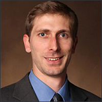
Brett Byram serves as Associate Professor of Biomedical Engineering at Vanderbilt University's School of Engineering and holds the Hoy Family Faculty Fellow distinction. He maintains dual affiliations with the Vanderbilt Institute for Surgery and Engineering (VISE) and the Vanderbilt University Institute of Imaging Science (VUIIS). Currently, he is the incoming Faculty Director of ACCRE—Vanderbilt’s High Performance Compute Center—and incoming chair for the NIH’s Image Technology Development (ITD) Panel, where he has served as a standing member. Educational Background: Ph.D. in Biomedical Engineering, Duke University (2011) B.S. in Biomedical Engineering and Mathematics, Vanderbilt University Dr. Byram's research centers on medical ultrasound with emphases in signal processing, elasticity imaging, and beamforming. His work spans beamforming/image formation, motion estimation, blood flow imaging, and hardware development—frequently integrating deep learning. The BEAM Laboratory, which he founded in 2013, develops ultrasound solutions for clinical challenges including transcranial imaging, placenta analysis, and kidney stone characterization. His methodology bridges advanced signal processing with tangible medical applications, particularly through AI-enhanced imaging techniques. Recent publications (2024-2025) reveal three dominant trends: deep learning integration for image reconstruction/stitching (e.g., SynStitch, LOTUS), clinical metric refinement (gCNR for lesion detectability), and specialized applications in obstetrics (placenta segmentation) and neuroimaging (transcranial ultrasound). These works consistently address data scarcity through synthetic training and domain adaptation while advancing quantitative assessment frameworks. Scientific Awards: Hoy Family Faculty Fellow Dr. Byram leads the BEAM Laboratory (Biomedical Elasticity and Acoustic Measurement), established in 2013 as part of Vanderbilt's Biomedical Engineering Department with VISE affiliation. His NIH ITD Panel leadership and ACCRE directorship reflect significant institutional trust. While specific grants aren't detailed, his 11+ years of continuous lab operation indicate sustained funding for projects spanning Siemens Healthcare collaborations, transcranial imaging development, and deep learning beamformer research—often translating academic innovations to clinical tools. The BEAM Lab functions as Vanderbilt's nexus for ultrasound innovation, maintaining strong VISE/VUIIS ties while pursuing hardware/software co-design. Current initiatives include real-time deep learning beamformers for echocardiography, transcranial functional ultrasound validation, and placenta segmentation systems. The lab's industry partnerships (notably with Siemens) and NIH panel involvement position it at the forefront of translating ultrasound research into clinical practice, with emerging work on diffusion models for 3D imaging and standardized biomechanical phantoms.