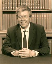
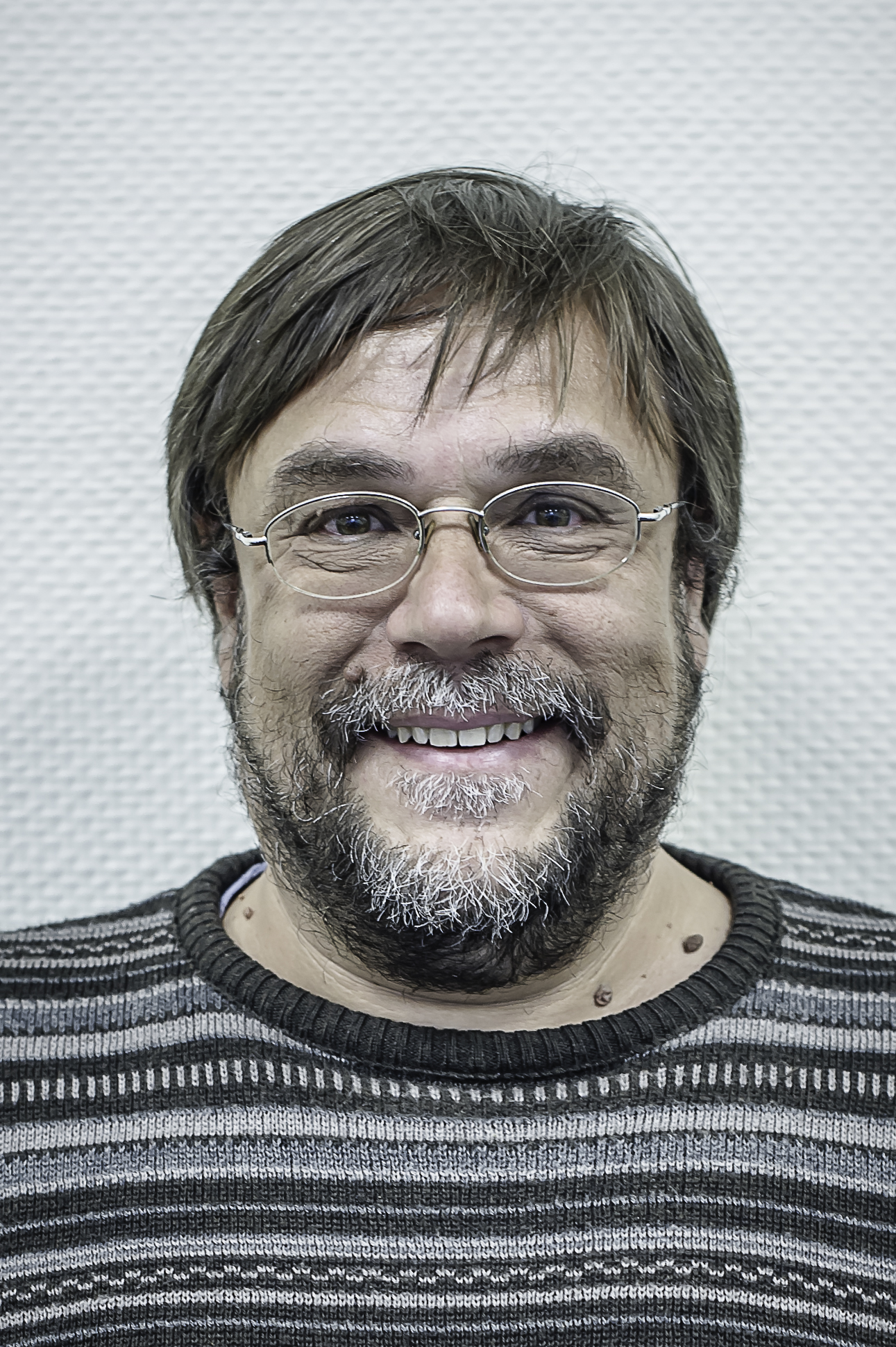
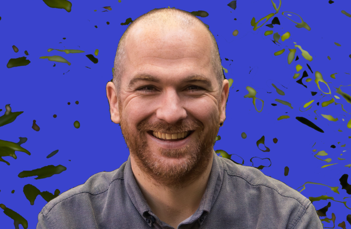
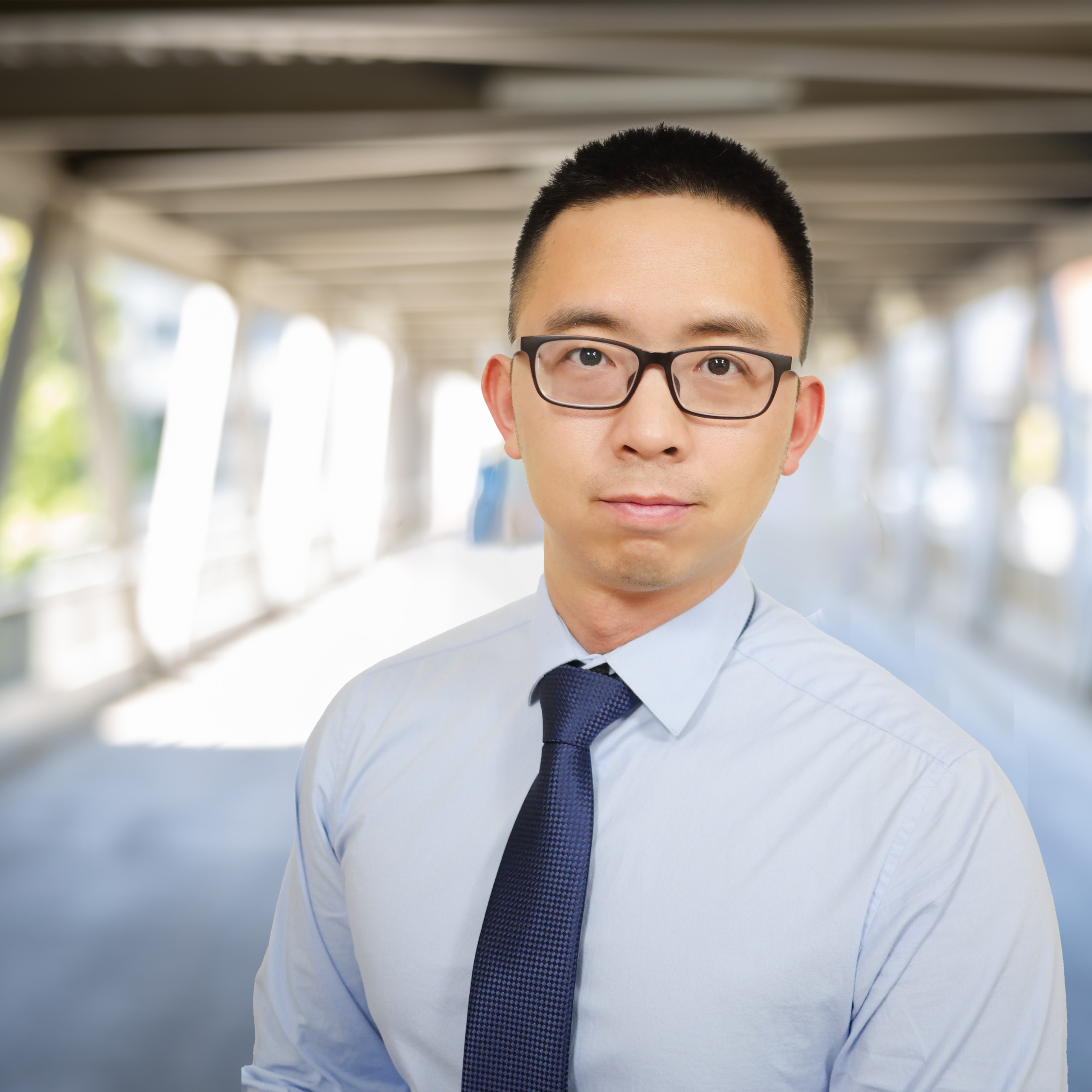
Mark Slifstein is a Professor of Psychiatry at Stony Brook University's Renaissance School of Medicine and Director of the PET Research Core. His career spans roles at Columbia University and the New York State Psychiatric Institute, where he held academic titles from Assistant to Associate Professor. PhD, Mathematics (1999), MS (1995), BA (1993) from New York University Summa cum laude graduate and Phi Beta Kappa member Dr. Slifstein's research focuses on pharmacokinetics , PET/SPECT neuroimaging , and dopaminergic/serotoninergic neurotransmission in schizophrenia. He specializes in multimodal imaging , data reduction methods , and image reconstruction techniques. Key research trends from his publications include neuroreceptor mapping , radioligand development , and quantitative PET analysis across schizophrenia and depression studies. Scientific Awards : Summa cum laude (1993), Phi Beta Kappa (1993) His work involves experimental design for PET studies, radiotracer production , and clinical imaging collaborations with institutions like the New York State Psychiatric Institute.
Louise Kauffmann is an Associate Professor at Grenoble Alpes University (UGA) in the Laboratory of Psychology and Neurocognition (LPNC), where she conducts research on visual perception and cognitive neuroscience. She is affiliated with the Faculty of Human and Social Sciences and has established herself as a leading researcher in visual perception, eye-tracking, and cognitive neurosciences. Education: PhD in Cognitive Psychology, LPNC (2012-2015) Postdoc at Max Planck Institute for Cognitive and Brain Science, Leipzig, Germany (2016-2018) Postdoc "NeuroCoG", GipsaLab/LPNC, UGA (2018-2019) Dr. Kauffmann's research focuses on understanding the mechanisms underlying visual perception and recognition of complex visual stimuli such as scenes, faces, and objects. She investigates how low-level characteristics of sensory inputs are used to achieve efficient recognition and how this process is modulated by prior knowledge and expectations about the visual environment. Her work employs complementary techniques including psychophysics, eye-tracking, and neuroimaging. Her primary research axes include the influence of predictive processes on subjective visual perception, the use of eye movements to investigate cognitive functioning, neurocognitive mechanisms underlying visual scene categorization, and mapping functional roles of visual subcortical structures. Her recent publications demonstrate a strong focus on predictive processing in visual perception, with particular attention to how contextual information and prior knowledge influence visual processing. Her work spans multiple domains including normal visual processing, visual impairments in aging, and neurological disorders such as autism spectrum disorders and developmental dyslexia. She frequently collaborates with researchers across France and internationally, particularly with institutions in Germany and Belgium. Dr. Kauffmann has received funding from prestigious organizations including the French National Research Agency (ANR-22-CE28-0021-01 "EXPER") and the CDP-NeuroCoG-IDEX of the University of Grenoble Alpes. She currently supervises several doctoral students including Clara Carrez-Corral (2023-2026), Pauline Rossel (2020-2023), and Chuyao Wang (2022-2026), often in co-supervision with other researchers at LPNC and collaborating institutions. Her research group is part of the LPNC laboratory, which is a CNRS UMR 5105 research unit focused on psychology and neurocognition. Dr. Kauffmann is actively involved in the academic community, serving as the Pedagogical coordinator of the Licence 3 of Psychology since 2022 and as Coordinator for International relations at UFR SHS since 2021. She is also a member of the Organizing committee for the "Semaine du cerveau" and an elected member of scientific boards at LPNC, UFR SHS, and CSPM H3S.
Foteini Liwicki is an Associate Professor at Luleå University of Technology, Department of Computer Science, Electrical and Space Engineering, leading the Embedded Intelligent Systems LAB and the Machine Learning Focus Group – Brain Analysis since 2022. Her work bridges Artificial Intelligence and Neuroscience with applications in communication disorders, neurodegenerative conditions, and mental health. Dr. Liwicki's research focuses on multimodal brain analysis, particularly inner speech mechanisms, neurodegenerative disorders like dementia, neurodevelopmental conditions such as ADHD, and the therapeutic effects of singing on mental health. She develops computational methods for EEG-fMRI integration and interpretable machine learning approaches to understand human communication across diverse populations. Her recent publications demonstrate a strong interdisciplinary approach, combining machine learning with neuroscience, geology, and educational technology. The research trends show increasing focus on multimodal data fusion, brain-computer interfaces, and practical applications of AI in healthcare and resource management. Scientific Awards: 2023-2025: Kompetensutveckling till professor, dnr LTU-154-2023 2022: Grants for Excellent Research Projects Proposals of SRT.ai 2022 2020-2021: Ansökan juniora lovande forskare, dnr LTU-4449-2019 Dr. Liwicki actively supervises multiple PhD and Master's students across diverse research areas including brain signal analysis, inner speech detection, geological data analysis, and AI applications in healthcare. She has received significant research funding including Kempestiftelserna grants for projects on inner speech, ADHD prediction, and singing therapy for psychiatric disorders. She leads the Machine Learning Focus Group – Brain Analysis, which develops computational methods for multimodal brain analysis, and collaborates extensively with international institutions including the University of Nantes, Kyushu University, and various European research centers.
Tiange Xiang is a Ph.D. student at Stanford University , affiliated with the Stanford AI Lab and Stanford Vision and Learning Lab. His research bridges generative models and AI for healthcare , focusing on 3D human reconstruction, medical imaging, and diffusion model applications. Education : Ph.D. and M.S. in Computer Science at Stanford University; B.S. in Computer Science and Technology (Advanced) at the University of Sydney. Advisors : Prof. Fei-Fei Li, Prof. Scott Delp, and Prof. Ehsan Adeli. His work includes foundational contributions to occluded human rendering (OccFusion, Wild2Avatar), medical anomaly detection (Exploiting Structural Consistency of Chest Anatomy, SQUID), and fMRI vision decoding (Seeing Beyond the Brain). He has pioneered 3D Gaussian splatting and score-distillation sampling techniques. Collaborators include leading researchers from Google DeepMind and Stanford. Scientific Awards : University Medal (University of Sydney), Stanford HAI Fellowship, Qualcomm Innovation Fellowship Finalist. He has served as a conference reviewer for CVPR, MICCAI, ICCV, and others, and as a teaching assistant for courses like CS 231n. His code repositories for BiO-Net and OccFusion are widely shared and implemented in Python.
Kimberley Phillips serves as Professor of Neuroscience at Trinity University's Center for the Sciences and Innovation, with dual affiliations in Psychology and Pre-Medical/Health Professions programs. Her research integrates neuroanatomy, primatology, and behavioral neuroscience to investigate primate cognitive evolution. Education: Ph.D. - The University of Georgia M.S. - The University of Georgia B.S. - Wofford College Dr. Phillips' research program centers on the neurological basis of skilled motor behaviors in capuchin monkeys, utilizing non-invasive MRI and DTI brain imaging alongside problem-solving task analysis. Her work bridges evolutionary biology and clinical neuroscience, particularly through Parkinson's disease modeling in marmosets and studies of age-related cognitive decline. Current investigations focus on loneliness impacts on neural aging through her NIH-funded $1.4M grant. Her publication record reveals consistent expertise in corpus callosum morphology, hemispheric asymmetry, and primate cognition across 15+ years. Recent work demonstrates increasing methodological sophistication through multimodal neuroimaging and cross-species comparisons spanning capuchins, marmosets, and chimpanzees. Scientific Recognition: NIH R01 Grant ($1.4 Million) for loneliness and cognitive decline research (2019) Trinity University Faculty IMPACT Award (2019) President of the American Society of Primatologists As principal investigator, Dr. Phillips directs a productive research program involving undergraduate and graduate students in both laboratory and field studies. Her leadership extends to developing best practices for primate neuroimaging through society-level initiatives. Current projects examine myelin degradation patterns in aging primates and their relationship to cognitive decline, with implications for human neurodegenerative disorders.
Karl Friston FMedSci FRSB FRS is a Wellcome Principal Research Fellow and Scientific Director at the Wellcome Trust Centre for Neuroimaging, and Professor at the Institute of Neurology, University College London. He also serves as an Honorary Consultant at The National Hospital for Neurology and Neurosurgery, UK. Friston is a theoretical neuroscientist and authority on brain imaging who has made seminal contributions to neuroscience methodology and theory. Friston's research interests span theoretical neurobiology, computational neuroscience, and computational psychiatry. He invented statistical parametric mapping (SPM), voxel-based morphometry (VBM), and dynamic causal modelling (DCM). His most significant theoretical contribution is the free-energy principle for action and perception (active inference), which provides a unified framework for understanding brain function. His work integrates predictive coding, Bayesian inference, and information theory to explain perception, action, and learning. His research has profound implications for understanding schizophrenia through the dysconnection hypothesis, which frames psychosis as a failure of hierarchical predictive coding. Friston's publication record demonstrates consistent thematic development across decades, with recent work focusing on active inference, free-energy minimization, and computational psychiatry. His articles reveal a progression from methodological innovations in neuroimaging to comprehensive theoretical frameworks that unify perception, action, and learning under Bayesian principles. The recurring themes include hierarchical generative models, precision weighting, and the role of prediction errors in neural processing. Young Investigators Award in Human Brain Mapping (1996) Fellow of the Academy of Medical Sciences (1999) President of the international Organization of Human Brain Mapping (2000) Minerva Golden Brain Award (2003) Fellow of the Royal Society (2006) Medal, College de France (2008) Weldon Memorial prize and Medal (2013) Charles Branch Award for unparalleled breakthroughs in Brain Research (2016) Glass Brain Award - lifetime achievement award in human brain mapping (2016) As Scientific Director of the Wellcome Trust Centre for Neuroimaging, Friston leads one of the world's premier neuroimaging research centers. His theoretical work has generated numerous research programs across multiple institutions investigating active inference in perception, action, psychiatry, and even developmental biology. His frameworks have been applied to diverse areas including robotics, machine learning, and philosophical questions about consciousness and agency. Friston's work continues to shape the theoretical foundations of cognitive neuroscience through both his mathematical innovations and conceptual frameworks that unify previously disparate phenomena.
François Cabestaing is a Professor in the Mechanical and Production Engineering department at the IUT of Lille, University of Lille, and leads the Brain-Computer Interfaces (BCI) team at the CRIStAL research center (UMR CNRS 9189). With administrative roles including President of CORTICO (French BCI association) since 2017, elected member of the CRIStAL Laboratory Council since 2015, and member of the IFRATH Board of Directors since 2006, he has established himself as a key figure in neurotechnology research. His research primarily focuses on brain-computer interfaces aimed at overcoming severe motor disabilities. Since 2004, he has specialized in BCI development, with earlier work (approximately 1994-2004) centered on image sequence processing and stereoscopic image analysis. His research spans neurotechnology, signal processing, human-computer interaction, and assistive technologies, with particular emphasis on applications for people with disabilities. Analysis of Cabestaing's publication history reveals a clear evolution from early work in image processing to a strong focus on brain-computer interfaces. Recent publications emphasize EEG-based BCIs, SSVEP and SSSEP techniques, sensory gating phenomena, and clinical applications for motor disability palliation. His work increasingly incorporates human factors considerations and explores applications in virtual reality and psychiatric treatment. Cabestaing has supervised numerous PhD students including Alban Duprès (defended 2016), Jimmy Petit (defended 2022), and Arne Van Den Kerchove (defended 2024). His current research team includes Gaël Van Der Lee working on neuromarkers in virtual reality and Maria Donantueno researching fMRI-based neurofeedback for schizophrenia. As head of the BCI team at CRIStAL since January 2015, Cabestaing oversees research on hybrid brain-machine interfaces, somatosensory filtering, and visual BCIs for eye-blind communication. His laboratory focuses on translating neurotechnology research into practical applications for people with severe motor disabilities, particularly through collaborations with medical institutions and patient organizations.
Chloe Cassandra Boyle serves as an Adjunct Assistant Professor in the Department of Psychiatry and Biobehavioral Sciences within the UCLA School of Medicine. Her research program focuses on the intersection of inflammation, mental health, and reward processing, with particular emphasis on how inflammatory processes contribute to depression, anhedonia, and altered reward mechanisms. Her primary research interests center on psychoneuroimmunology, examining the bidirectional relationships between the immune system and brain function. Dr. Boyle investigates how inflammatory challenges affect neural reward circuitry, with special attention to sex differences and vulnerable populations including breast cancer survivors and older adults. Her work employs experimental paradigms such as endotoxin administration and influenza vaccination to induce controlled inflammatory responses while measuring psychological and neural outcomes. Analysis of her publication record from 2016-2024 reveals consistent research productivity with a growing focus on sex differences, aging, and translational models connecting inflammatory processes with specific depression symptoms like anhedonia. Her work frequently employs multimodal approaches combining behavioral assessments, inflammatory biomarkers, and neuroimaging techniques to elucidate mechanisms linking inflammation with altered affect and cognition. Psychoneuroimmunology Inflammation-depression relationships Reward processing and anhedonia Sex differences in stress response Mindfulness interventions Older adult mental health Breast cancer survivorship Dr. Boyle maintains active collaborations with prominent researchers in the field including Michael Irwin, Steven Cole, and Patricia Ganz. Her work has received attention across multiple platforms with significant readership on Mendeley and coverage in news outlets, indicating impact within both academic and broader communities.
Kevin Whittingstall is a neuroscience researcher at the Centre de recherche du CHUS (Université de Sherbrooke) specializing in multimodal neuroimaging, with expertise in simultaneous EEG-fMRI integration and neurophysiological signal processing. His work bridges neuroscience, engineering, and clinical applications with a focus on visual system function and neurodegenerative processes. Education: Doctorate in Neuroscience, Dalhousie University (2005) Master's in Neuroscience, Dalhousie University (2002) Bachelor's in Psychology, Concordia University (1999) Research Focus: Dr. Whittingstall's work centers on neurovascular coupling and multimodal brain imaging , particularly the relationship between EEG oscillations and BOLD fMRI signals. His research explores visual system neurophysiology , white matter architecture , and metabolic changes in aging using advanced signal processing techniques. Key methodological contributions include tools for tractography visualization (Fiberweb) and EEG-fMRI coregistration. Publication Trends: Recent work (2016-2018) demonstrates a shift toward methodological innovation in brain connectivity mapping, with 60% of publications developing new neuroimaging techniques. Major themes include structural-functional coupling (35% of articles), visual system processing (25%), and aging/metabolism studies (20%), reflecting his dual focus on tool development and clinical neuroscience applications. Research Support: Natural Sciences and Engineering Research Council of Canada (NSERC) Grant (2018-2023) as Principal Investigator Focused Ultrasound Stimulation for Brain Research (2017-2018) as Co-applicant Multichannel Neural Signal Recording Instrumentation (2016-2017) as Co-applicant Professional Engagement: Dr. Whittingstall maintains active international collaboration, evidenced by 12 conference presentations (2013-2017) on neurovascular coupling across Canada, Europe, and North America. He served on the Organization Committee for Medical Image Computing and Computer Assisted Interventions (2017) and leads research within the Université de Sherbrooke's neuroimaging ecosystem.
Professor Albrecht Stroh is a Professor of Physiology and Director of the Institute of Physiology I at University Hospital Münster (UKM). He also serves as Research Group Leader and Head of the Mainz Animal Imaging Center (MAIC) at the Leibniz Institute for Resilience Research (LIR) in Mainz, Germany, and holds a Tenured Associate Professor position (W2) of Molecular Imaging and Optogenetics at the Institute of Pathophysiology, University Medical Center of the Johannes Gutenberg University Mainz. Professor Stroh's educational background includes: PhD studies (2002-2005) at Charité University Medicine Berlin Diploma in Biophysics (2001) from Humboldt University Berlin Studies in Biology (1994-1996) at Free University Berlin His research focuses on understanding the neural basis of resilient behavior in mouse models, particularly examining how neural networks adapt and maintain functionality despite pathological changes. His work primarily investigates the initial processing of sensory afferents in the cortex, local representation, and cortico-cortical processing in relation to resilient versus susceptible behavior. A key emphasis is placed on slow network oscillations and their role in resilience. His publications demonstrate a consistent focus on neural network dynamics, imaging techniques, and the relationship between molecular/cellular changes and functional outcomes. Professor Stroh has received numerous scientific awards and funding, including: The Kurt-Decker-Award from the German Society for Neuroradiology (DGNR) Significant DFG funding for advanced imaging equipment including a 9.4 T small animal MRI (2 M EUR) and two-photon microscopes (1.54 M EUR) A Young Investigators Award from the Roland-Ernst-Foundation A Certificate of Merit from ESMRMB 2009 His research group collaborates extensively with international partners including Stanford University, University of Washington, and Seattle Children's Research Institute. Current projects include 'Learning Resilience,' investigating neural excitability regulation, studying brain-wide resting networks in relation to resilience, and examining the role of spontaneous activity in mesoprefrontal circuits.
Ruud Hortensius is an Associate Professor in the Department of Social, Health and Organizational Psychology within the Social and Behavioural Sciences faculty at Utrecht University, where he leads the Human+ research team. He specializes in understanding social cognition during interactions with artificial intelligence, combining psychology, AI, and social neuroscience to investigate how AI shapes human social behavior at individual, group, and family levels. His educational background includes a PhD in Affective and Social Neuroscience (Cum Laude) from Tilburg University (2016), MSc in Neuroscience and Cognition from Utrecht University (2011), BSc in Psychology (Cum Laude) from Utrecht University (2008), and BSc in Social Work from University of Applied Sciences Utrecht (2005). Hortensius' research focuses on the neurocognition of real-world interactions with AI, moving beyond lab-based studies to measure social dynamics in natural settings. His work explores how long-term interactions with AI shape social behavior across individuals, groups, and families, with particular emphasis on the neural mechanisms underlying social cognition during human-AI interactions. He investigates whether AI agents function as tools, companions, or family members, and how these relationships impact social dynamics. His publications reveal a strong focus on social neuroscience methodologies applied to human-robot interaction, with recurring themes including neural network engagement during AI interactions, anthropomorphism, trust formation, and developmental aspects of human-AI relationships. The research spans multiple disciplines including psychology, neuroscience, and computer science, with increasing emphasis on family dynamics and long-term interaction patterns. 2025 NIAS-Lorentz Theme Group Fellow 2025 NWO Open Competition XS Grant 2024 NWO NGF AiNed XS Europe Grant 2023 ERC Starting Grant (F-AI-MILY project) 2023 Association for Psychological Science Rising Star award 2019 Bial Foundation Scientific Research Grant Hortensius actively supervises PhD students (with ius promovendi) and teaches courses in Social Neuroscience and Artificial Intelligence. His research is supported by significant grants including an ERC Starting Grant investigating AI's impact on family dynamics. He collaborates extensively across Utrecht University's Human-centered AI focus area, Human-AI alliance, and Embodied AI initiative, while also engaging in public outreach through the AI Helpdesk platform and artistic collaborations. He leads the Human+ research team within the Relationship Lab, developing innovative 'neurocognition-at-home' approaches to study social dynamics. Current projects include the F-AI-MILY ERC grant examining how AI integration affects family social dynamics at behavioral and neural levels, and the NIAS Lorentz Theme Group project 'Hybrid Families' investigating AI's role in family systems.
Ethan Meyers holds dual academic positions as a Visiting Professor in the Department of Statistics and Data Science at Yale University and an Associate Professor of Statistics at Hampshire College. He also maintains a research affiliation with the Center for Brains, Minds and Machines at MIT. His academic work bridges neuroscience, statistics, and computational methods. Dr. Meyers' research focuses on understanding how information is coded in neural activity, with particular emphasis on high-level visual and cognitive brain regions. His work combines experimental neuroscience with computational approaches, developing sophisticated tools to analyze high-dimensional neural recordings. His research spans neural population coding, working memory systems, face recognition mechanisms, and object perception. Analysis of his publication record from 2004-2018 reveals a consistent trajectory in computational neuroscience, with increasing focus on population-level neural dynamics. His work shows strong interdisciplinary connections between neuroscience, machine learning, and statistics, particularly in developing methods for decoding neural activity. Key themes include dynamic population coding, neural representations of visual information, and the development of computational tools for neural data analysis. Dr. Meyers has developed the Neural Decoding Toolbox, a significant contribution to the field that enables researchers to analyze neural population activity. His work with primate visual systems, particularly the face patch system, has provided insights into how neural information is processed and represented. His research demonstrates expertise in both experimental design and computational analysis of neural data.
Matt Sherwood is an Associate Professor in the Department of NeuroSci Cell Bio Physiology-SOM at Wright State University and serves as the Director of the Center of Neuroimaging and Neuro-Evaluation of Cognitive Technologies (CoNNECT). He holds a Ph.D. in Medical and Biological Systems Engineering, an M.S. in Biomedical Image and Signal Processing Engineering, and a B.S. in Biomedical Engineering, all from Wright State University. Research Focus Dr. Sherwood investigates brain function and neurological disorders using advanced neuroimaging techniques. His work includes: Real-time fMRI neurofeedback to enhance working memory and treat tinnitus. Neural correlates of visual object recognition and mirror imaging bias (linking Inferior Frontal Gyrus activity to MR spectroscopy biomarkers). Cognitive effects of transcranial direct current stimulation. Brain hemodynamics under hypoxic conditions. He collaborates with industry partners and led a Phase II Office of Naval Research project developing cognitive models for rapid training. Leadership As Director of CoNNECT, he oversees research in neuroimaging and cognitive technologies.
Dr. Ian Charest is an Associate Professor in the Department of Psychology at Université de Montréal, affiliated with the Faculty of Arts and Sciences. His research focuses on understanding how the human brain processes visual information through neuroimaging techniques like fMRI and MEG, combined with machine learning and psychophysical experiments. His work emphasizes individual representational idiosyncrasies and computational modeling of cognition. He leads the Charest Lab, which explores visual perception, consciousness, memory, and decision-making using advanced neuroimaging and computational tools. Key projects include analyzing natural scene semantics through machine learning (funded by NSERC), developing infrastructure for computational neuroscience (FCI grant), and investigating face recognition variability (SPIIE grant). Dr. Charest has supervised numerous graduate and undergraduate students, contributing to open-source tools like pyRSA, pyGLMdenoise, and pyslicetime. His research has been supported by prestigious agencies including the Fonds de recherche du Québec and the Canadian Institutes of Health Research.
Li-Ming Hsu is a Research Assistant Professor in the Department of Radiology at the UNC School of Medicine. His work focuses on neuroimaging techniques, particularly functional MRI (fMRI), to study brain networks and their roles in behavior, disease, and addiction. He specializes in translating rodent models to human applications, developing machine learning tools for neuroimaging analysis, and investigating the neurobiological mechanisms underlying brain functions. His research emphasizes understanding addiction mechanisms through rodent models, such as nicotine and cocaine studies, while advancing imaging methodologies like SORDINO and U-Net-based segmentation. He has pioneered techniques like optogenetic fMRI and chemogenetic stimulation to map therapeutic brain circuits. His work bridges fundamental neuroscience with clinical applications, aiming to improve diagnostic tools and therapies for neurological disorders like Alzheimer’s disease and depression. Key Areas: Neuroimaging, functional MRI, addiction models, network neuroscience, machine learning Awards: ISMRM Magna Cum Laude Merit Awards (2020, 2016) Methodologies: Optogenetics, deep learning, rodent-human translation, network redundancy analysis Publications reveal a focus on brain network dynamics in health and disease, with contributions to Alzheimer’s early detection, addiction circuitry, and fMRI methodological improvements. His work emphasizes translational potential, seeking to advance both basic science and clinical outcomes.