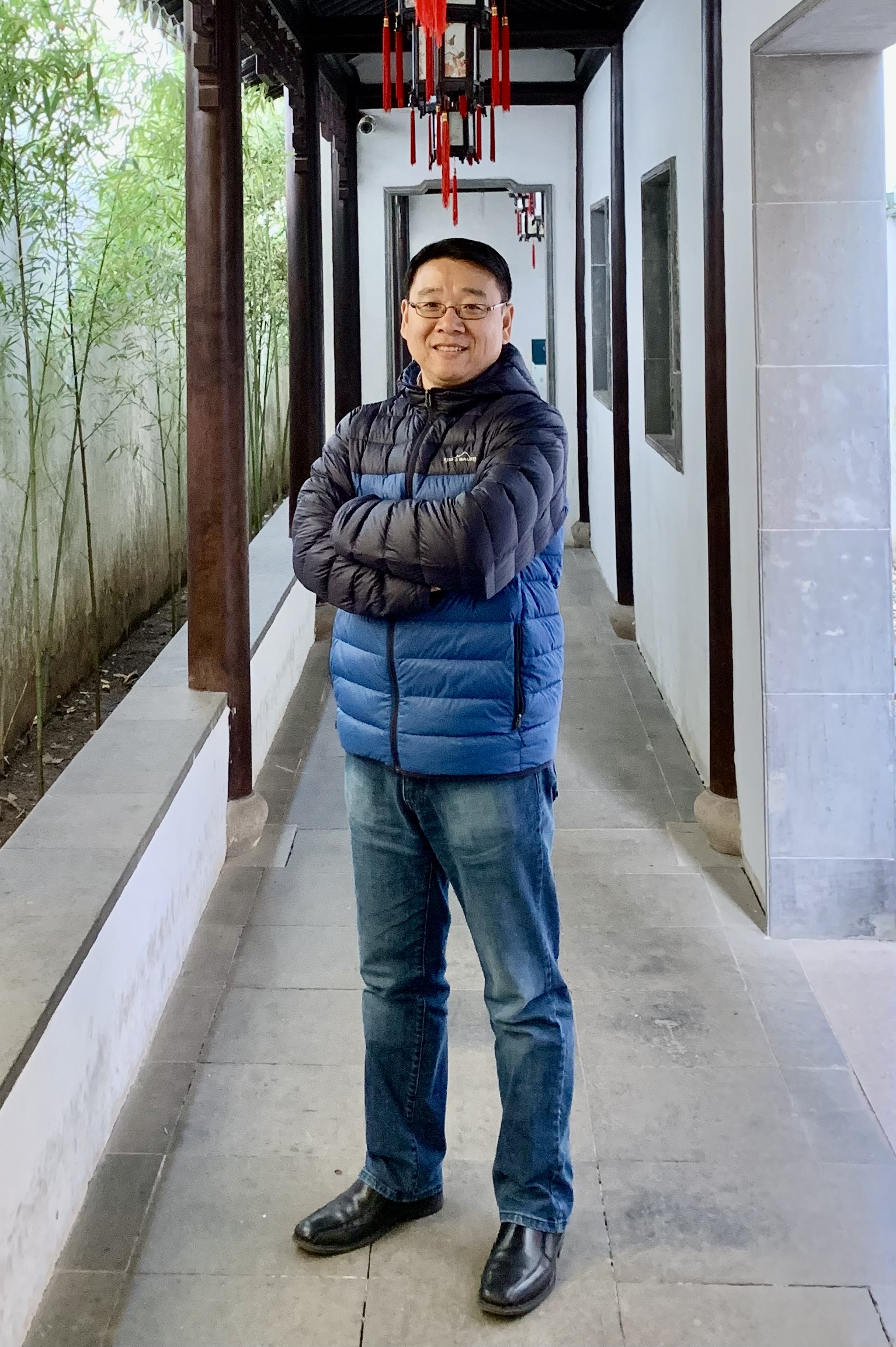
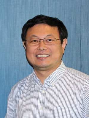
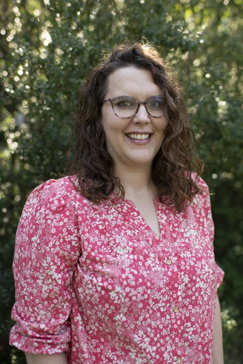
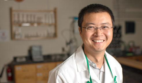
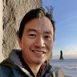
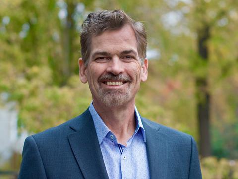
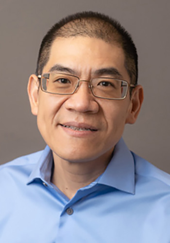
Weidong Yang is a Professor in the Department of Biology at Temple University's College of Science and Technology. He leads the Yang Laboratory, which focuses on developing advanced imaging techniques to address complex biological problems. His research emphasizes single-molecule biophysics, super-resolution microscopy, and molecular transport mechanisms. Developed techniques: SPEED microscopy, virtual 3D super-resolution imaging, sp-FRAP Current projects: Nuclear transport mechanisms and cytoplasm-to-primary-cilium trafficking His lab maintains an interdisciplinary approach, balancing technical innovation with biological applications to train future scientists. The lab's website can be found at www.yanglab.com .
Tong Ye, Ph.D., is an Associate Professor at Clemson University's College of Engineering, Computing and Applied Sciences. He directs the Nano and Functional Imaging Lab located in the Bioengineering Building, where his research focuses on advanced optical imaging techniques for biomedical applications. Dr. Ye received his doctorate from the Chinese Academy of Sciences in 1995. Research Focus Dr. Ye's research spans three interconnected domains: Optical Imaging Development : Creating novel microscopy platforms including superresolution fluorescence, nonlinear optical, and light sheet microscopy systems for deep tissue visualization. Structural-Functional Assessment : Developing label-free methods for analyzing cell metabolism, tissue viability, and microstructural properties using intrinsic optical signatures. Medical Device Innovation : Engineering clinical imaging tools such as compact nonlinear arthroscopes and laser speckle contrast devices for dermatological and orthopedic applications. Publication Trends Recent publications (2023-2025) demonstrate Dr. Ye's focus on integrating deep learning with advanced microscopy for quantitative tissue analysis. Key trends include: (1) Development of AI-enhanced imaging pipelines for automated assessment of cartilage viability and cardiac capillarization; (2) Innovation in super-resolution techniques (e.g., Phasor-FSTM) for live-cell imaging; and (3) Application of multimodal approaches in orthopedics, cardiology, and oncology. His work consistently addresses clinical challenges through label-free, noninvasive methodologies. Laboratory The Nano and Functional Imaging Lab specializes in pushing resolution boundaries across spatial scales – from nanoscopic cellular structures to whole-organ imaging. Current projects include developing deep learning-enabled diagnostic platforms for articular cartilage assessment, creating compact imaging devices for clinical use, and investigating microvascular changes in disease models using advanced optical techniques.
Jason McNeill is a Professor in the Department of Chemistry at Clemson University's College of Science. He obtained his Ph.D. in Chemical Physics from the University of California at Berkeley in 1999 and joined Clemson in 2002. His research focuses on laser spectroscopy, nanomaterials, and biological imaging, particularly using fluorescent nanoparticles for sensing and charge transport studies in organic semiconductors. Educational Background A.S., Chemistry, Sauk Valley Community College B.S., Chemistry, Northern Illinois University Ph.D., Chemical Physics, University of California at Berkeley His work spans super-resolution microscopy, single-molecule spectroscopy, and energy transfer dynamics in polymer nanoparticles. Recent publications highlight advancements in dual-mode imaging, charge carrier tracking, and biofunctionalized polymer dots. He received the NSF CAREER Award in 2006. Notable courses taught include Physical Chemistry I/II, Atomic and Molecular Structure, Quantum Chemistry, and Modern Laser Spectroscopy.
Brittany White-Mathieu is Assistant Professor of Chemistry at UNH's College of Engineering and Physical Sciences, developing innovative fluorescent probes for biological imaging. Her research integrates organic synthesis with cell biology to create tools for super-resolution microscopy. Key research areas include lipid expansion microscopy (LExM), nanohoop-based fluorophores, and clickable probes for cellular membrane studies. Current work focuses on macrocyclic structures with enhanced optical properties for live-cell imaging applications. Recent publications demonstrate advancement in super-resolution techniques, particularly for lipid membrane visualization. Synthetic methodologies for strained macrocycles represent foundational work enabling advanced probe development. Applications span neuroscience, cell biology, and diagnostic sensing.
Kanchana Vaishnavi Gandikota is a researcher at the Institute for Vision and Graphics at the University of Siegen. Her work focuses on image reconstruction, adversarial robustness, and generative models for computer vision applications. Research innovations: Reconstruction-free microscopy classification Text-guided super-resolution interfaces Transformer vulnerability analysis Recent articles address privacy-accuracy tradeoffs in medical imaging and diffusion model manipulation techniques. Develops computational methods for Fourier ptychographic microscopy.
Dr. Jixin Chen is Associate Professor of Chemistry at Ohio University specializing in single-molecule spectroscopy and surface chemistry. His research develops advanced microscopy techniques for studying molecular interactions and nanomaterial properties. Research areas: Super-resolution imaging of DNA-protein interactions Photophysics of perovskite nanomaterials Single-particle optical mapping Surface reaction kinetics His lab develops computational tools for analyzing single-molecule FRET data and stochastic adsorption processes. Funded by NIH and NSF, current projects focus on optical genome mapping and nanoscale heat transfer.
Vig Sundaresan is an Assistant Professor of Chemistry and Biochemistry at the University of Mississippi, affiliated with the College of Liberal Arts. He leads the Sundaresan Laboratory, focusing on developing high-throughput multimodal imaging techniques to study electrochemical and biological phenomena at the nanoscale and single-entity level. His research integrates super-resolution optical imaging with electrochemical methods to advance understanding in electrocatalysis, materials science, and biology. Dr. Sundaresan teaches undergraduate and graduate courses in analytical chemistry, electrochemistry, and instrumental analysis. Education: PhD in Chemistry from Temple University (2018), followed by postdoctoral work at the University of Notre Dame (2019–2021) and a role as Assistant Research Professor there (2021–2022). He has secured prestigious grants including the NSF EPSCoR Research Fellows Award and NSF CMI/EPSCoR RII Track-4 grants. His lab’s recent achievements include developing CLocK microscopy for nanoscale analysis and nanopore-based sensors. Research interests emphasize imaging electrochemical reactions at the nanoscale, with applications in designing sensitive chemical/biosensors. The lab employs correlated opto-electrochemical techniques to address fundamental questions in catalysis and biological systems. Current projects include NSF-funded studies on super-resolution imaging of nanoparticles and high-throughput electrocatalyst screening. Advising and collaborations involve mentoring graduate students and postdoctoral researchers. Notable lab members include Sachintha Illesinge, Nchumi Ndaleh, and Gintu Thomas (PhD students). Recent lab news highlights grants, student recruitment, and presentations at institutions like CECRI, India. The Sundaresan Lab operates within the Department of Chemistry & Biochemistry, offering interdisciplinary training. Their future work aims to bridge single-entity insights with ensemble-level system design for practical applications in energy and healthcare.
Yee-Hung (Mark) Chan is an Associate Professor in the Department of Biology at San Francisco State University (SFSU), affiliated with the Center for Cellular Construction. He earned a Ph.D. from Stanford University and completed postdoctoral research at UCSF. His lab focuses on understanding how cells regulate organelle size and distribution, using budding yeast vacuoles and fission yeast vacuoles as model systems. Research techniques include live-cell fluorescence microscopy, computational image analysis, and genetic/molecular biology approaches. Education: Ph.D., Stanford University; Postdoc, UCSF Lab Affiliation: Chan Lab at SFSU Key Research Areas: Organelle size scaling, vacuole inheritance, computational modeling of cellular structures Recent work explores vacuole localization in Schizosaccharomyces pombe and the role of microtubules in organelle dynamics. The lab actively trains undergraduates and graduate students in cell biology and microscopy techniques.
Matthew Dalva is a Professor of Cell and Molecular Biology at Tulane University, holding the Phyllis M Taylor Presidential Chair and serving as Director of the Tulane Brain Institute. He earned his Ph.D. in Neurobiology from Duke University in 1996. His research focuses on synaptic development and function, particularly mechanisms governing synapse formation, NMDA receptor regulation, and synaptic nano-architecture. The Dalva Lab employs advanced techniques like super-resolution microscopy (STED) and genetically encoded kinase activity reporters to study synapses in vivo. Research interests include synaptic plasticity, Eph/ephrin signaling pathways, and pain mechanisms. His team explores how synaptic structures adapt to activity and injury, with implications for neurological diseases like ALS and chronic pain. The lab is part of the School of Science & Engineering and collaborates on multidisciplinary projects combining molecular biology, biochemistry, and microscopy. Recent work highlights the role of EphB2 and ephrin-B2 in pain plasticity, synaptic module organization, and spinal cord neuroprotection. Open positions exist for postdoctoral researchers and lab managers skilled in molecular biology and imaging. Tulane Brain Institute affiliations enhance translational research efforts, emphasizing innovation in synaptic tools and technology.
Chaitanya Ullal is an Associate Professor in the Department of Materials Science and Engineering at Rensselaer Polytechnic Institute (RPI), affiliated with the School of Engineering. He leads the Ullal Lab, focusing on advanced materials science with expertise in polymers, optics, and nanolithography. His research emphasizes nanoscale structure-property relationships in hydrogels, polymer gels, and functional materials using super-resolution microscopy techniques like STED. Ullal earned a B.Tech from the Indian Institute of Technology Bombay and a Ph.D. from MIT. He joined RPI in 2013 and has received prestigious awards including the NSF CAREER Award (2017) for studying hydrogel structures and the American Chemical Society New Investigator Award (2016). His work bridges engineering and life sciences, addressing applications in biomedical materials, nanotechnology, and sustainable manufacturing. Key research thrusts include developing digital design frameworks for 3D printed materials, optimizing nanolithography processes, and understanding polymer dynamics at the molecular scale. He collaborates on projects like light-emitting diode enhancement and nanostructured template fabrication. Educational outreach includes STEM initiatives with Iridescent Learning.
Esther Wertz is an Associate Professor in the Department of Physics, Applied Physics & Astronomy at Rensselaer Polytechnic Institute. She obtained her B.S. in Physics from the University of Paris 7 in 2005 and a PhD in Physics from the same institution in 2010, where she pioneered work on polariton condensates in GaAs microcavities. After postdoctoral research at the University of Michigan with Dr. Julie Biteen, she joined RPI in 2015. Current Role: Associate Professor at Rensselaer Polytechnic Institute University: Rensselaer Polytechnic Institute Department: Physics, Applied Physics & Astronomy Research Center: Center for Materials, Devices, and Integrated Systems (CMDIS) Esther’s research focuses on nanoscale light-matter interactions, particularly in plasmonic nanostructures and perovskite materials. Her lab employs super-resolution microscopy to study quantum properties emerging from exciton-plasmon coupling. Key trends in her publications include: Investigations into chiral photon emission and ferroelectric perovskites Advancements in remote epitaxy for halide perovskites Development of plasmonic nanoantennas for single-molecule imaging Exploration of carrier lifetime enhancement and phononic effects She received the PicoQuant Young Investigator Award and secured an NSF CAREER grant in 2020. Her work bridges materials science , quantum optics , and nanophotonics , contributing to next-generation photonic and optoelectronic devices.
Wei-Chuan Shih is a Professor in the Electrical and Computer Engineering Department at the University of Houston's Cullen College of Engineering, with joint appointments in Biomedical Engineering, Materials Science & Engineering, and Chemistry. He leads the Nanobiophotonics Group and serves as Associate Editor for OSA Optics Express and SPIE Journal of Nanophotonics. Dr. Shih's work bridges fundamental nanophotonics with practical applications in healthcare diagnostics and environmental monitoring. Dr. Shih received his Ph.D. from Massachusetts Institute of Technology (MIT). His research centers on developing new technologies for biomedicine and portable chemical/biological sensing. His core expertise includes nanobiophotonics, plasmonics, surface-enhanced Raman spectroscopy (SERS), nanofabrication, optical imaging, microscopy, and spectroscopy. He works extensively on single nanoparticle analysis (exosomes, viruses), disease diagnosis through virtual biopsy, N/MEMS/microfluidics, lab-on-a-chip systems, and smartphone-based microscopy and sensing platforms. Dr. Shih's recent publications demonstrate a strong focus on plasmonic biosensing platforms, particularly using nanoporous gold structures. His work shows progression from fundamental plasmonic properties to practical applications in medical diagnostics and environmental monitoring. A significant trend is the development of smartphone-based sensing technologies for point-of-care applications, especially for heavy metal detection in water. His research increasingly integrates AI and machine learning with optical sensing techniques. Dr. Shih has received numerous prestigious awards and honors including: NSF CAREER Award (2012) NASA Early Career Faculty Award (2012) UH Award of Excellence in Research and Scholarship (2013) UH Cullen College of Engineering Research Award (2015) UH Cullen College of Engineering Rising Innovator Award (2019) Fellow, SPIE Senior Member, OSA/IEEE Senior Member (inaugural class), National Academy of Inventors MIT Martin Fellowship for Sustainability (2005) Dr. Shih has secured substantial research funding from multiple federal agencies including the NSF CAREER Award (2012-2018), NASA Early Career Faculty Award (2012-2016), NIH R21 (2014-2016), Department of Interior BSEE (2015-present), and NIH R01 EB-030623 (2021-2025). His research group has produced numerous PhD and Master's students who have co-authored many publications. Dr. Shih leads the Nanobiophotonics Group at the University of Houston, which maintains state-of-the-art facilities for nanofabrication, optical characterization, and biological testing. His team works at the intersection of nanotechnology, photonics, and biomedical engineering with strong collaborations across disciplines.
Peter McCourt is a Professor in the Department of Medical Biology at UiT The Arctic University of Norway. His research focuses on liver sinusoidal endothelial cells (LSECs), their role in scavenging pathogens and toxins, and the application of advanced microscopy techniques to study cellular ultrastructure. Key areas include aging-related changes in LSEC function, nanotherapeutic delivery systems targeting the liver, and the interplay between hemodynamics and liver architecture. Research interests span liver biology, endothelial cell biology, and biomedical imaging innovations. His work combines experimental and computational approaches to understand how LSECs regulate blood filtration, immune responses, and metabolic processes. Notable contributions include developing optical microscopy methods like transparent polymer waveguide chips for super-resolution imaging and investigating nanoparticle-mediated drug delivery to rejuvenate aged liver cells. Publications highlight advancements in correlative microscopy, fenestration dynamics in LSECs, and the role of stabilin receptors in scavenging oxidized proteins and viral particles. His research bridges basic science and translational medicine, addressing applications in aging populations and infectious diseases.
Dr. Song Wen is a Senior Lecturer in Biomedical Sciences at Middlesex University's Department of Natural Sciences. Her interdisciplinary research integrates biomedical science with nanotechnology and artificial intelligence to develop novel diagnostic and therapeutic approaches for cancer, particularly HPV-related cancers. Her research focuses on developing advanced drug delivery systems using liposomal nanotechnology, creating 3D tissue models for cancer research, and applying deep learning techniques to medical imaging for viral detection. She explores nanocarrier optimization for targeted cancer therapy, particularly using compounds like arsenic trioxide and baicalein. Dr. Wen's recent publications demonstrate expertise in developing deep learning algorithms for HPV detection in microscopy images, optimizing liposomal drug formulations for cervical cancer treatment, and evaluating natural compound synergies for leukemia therapy. Her work bridges computational approaches with experimental biomedical research. She contributes to educational initiatives in biomedical sciences and mentors students in interdisciplinary research methodologies that combine laboratory experimentation with computational analysis.
Jordan Beach is an Associate Professor of Cell & Molecular Physiology at Loyola University Chicago's Stritch School of Medicine. The Beach Lab investigates how cells generate and regulate contractile forces through myosin motor proteins, particularly during cell migration and division. Research employs advanced imaging techniques including: Super-resolution microscopy for myosin dynamics High-resolution live-cell imaging Quantitative analysis of cytoskeletal organization Current research themes include: Regulation of myosin filament assembly and amplification Integration of contractile units into functional networks Metabolic control of cellular contractility Tissue-specific contractile mechanisms Recent publications demonstrate innovations in understanding actomyosin organization and its implications for cellular mechanics and disease processes.