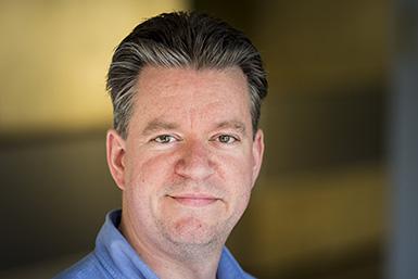
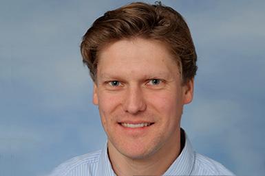
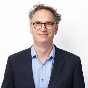
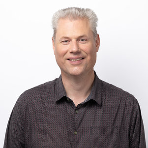
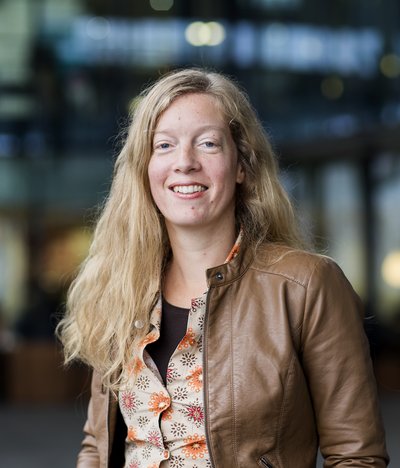
Michel Versluis is a Full Professor at the University of Twente, Netherlands, specializing in Physical and Medical Acoustics within the Physics of Fluids group. His work focuses on microbubbles and microdroplets for medical imaging and therapy, as well as microfluidic applications in medicine and nanotechnology. University of Twente, Physics of Fluids group His research bridges physics and biomedical engineering, with publications in high-impact journals like PNAS and IEEE Transactions. Recent work emphasizes ultrasound-driven microbubble dynamics, additive manufacturing of flow phantoms, and deep learning for super-resolution imaging. 2025 publications: vascular phantoms, PROTEUS simulator, acoustic microbubble control 2024 innovations: 3D-printed medical devices, immunogenic cell death optimization Contact: m.versluis@utwente.nl
Prof. Casper Hoogenraad is a full professor in Molecular Neuroscience at the Department of Cell Biology, Faculty of Science, Utrecht University. His research focuses on understanding how intracellular protein trafficking underlies neuronal development and function, with particular emphasis on the microtubule cytoskeleton, synaptic cargo trafficking, and synaptic plasticity. He leads an active research group within Utrecht University's Cell Biology department and collaborates extensively with other neuroscience research groups. Education: PhD, Erasmus University Rotterdam (1996-2001) Postdoc, Massachusetts Institute of Technology (2002-2005) Hoogenraad's research spans three main themes: cytoskeleton dynamics during neurodevelopment and synaptic plasticity, motor proteins and adaptors as regulators of synaptic transport, and psychiatric and neurologic disease disorders linked to intracellular transport. His work combines genetics, biochemistry, molecular, and cellular biology methods in in vitro (neuron cultures), ex vivo (brain slices), and in vivo (mice) systems, along with advanced microscopy techniques including immunofluorescent confocal microscopy, high-resolution live cell imaging, and photo-activated localization microscopy (PALM). Analysis of Hoogenraad's recent publications reveals a strong focus on microtubule organization, neuronal polarity, and the molecular mechanisms underlying synaptic function and dysfunction. His work frequently explores how disruptions in intracellular transport contribute to neurological disorders including Alzheimer's disease, schizophrenia, and autism spectrum disorders, with particular attention to the relationship between cytoskeletal organization and cargo transport in neuronal compartments. Scientific Awards and Memberships: ZonMW-VIDI (2004) European Young Investigators (EURYI) award (2005) NWO-ALW VICI (2011) ERC Consolidator grants (2013) FENS-Kavli Network of Excellence (2014) European Molecular Biology Organization (EMBO) (2015) Young Academy of Europe (YAE) (2015) IBRO Kemali Prize (2016) Hoogenraad leads a research group studying neuronal development and function, with a particular focus on how intracellular transport mechanisms contribute to both normal brain function and neurological disorders. His laboratory employs a multidisciplinary approach combining molecular, cellular, and systems neuroscience techniques to investigate the molecular basis of neuronal polarity, synaptic plasticity, and the pathogenesis of neurological disorders. He has secured significant research funding through prestigious grants including ERC Consolidator grants. The Hoogenraad lab operates within the Cell Biology department at Utrecht University, collaborating with other research groups focusing on cellular dynamics, biophysics, and neurobiology. The lab utilizes advanced microscopy techniques including immunofluorescent confocal microscopy, high-resolution live cell imaging (spinning disc microscopy and total internal reflection fluorescence microscopy), and quantitative analysis using advanced high-resolution microscopy (photo-activated localization microscopy). Current lab technicians include Phebe Wulf and Bart de Haan.
Peter Zijlstra is a Full Professor in the Department of Applied Physics at Eindhoven University of Technology (TU/e), leading the Molecular Plasmonics group. His research focuses on single-molecule sensing using plasmonic and nanophotonic approaches to study biomolecular interactions in complex environments. He is a core member of the Institute for Complex Molecular Systems at TU/e, collaborating across disciplines like chemistry, biomedical engineering, and mathematics. Education: MSc in Applied Physics, University of Twente (2005) PhD from Swinburne University of Technology (2009), studying plasmonic nanoparticles in optical data storage Postdoctoral fellowship at Leiden University under Prof. Michel Orrit Research Interests: Developing novel sensing concepts via nanophotonics and super-resolution microscopy. Key areas include plasmon-enhanced fluorescence, real-time biomolecular dynamics, and applications in cancer management. His work contributes to UN Sustainable Development Goals through advancements in biosensing technologies. Awards: 2013 NWO Vidi Award for research on plasmonic imaging of enzymes in living cells Teaching & Activities: Teaches courses like Advanced Optical Microscopy and Electromagnetism Supervised 32 academic works Contributed to conferences and editorial roles for journals like npj Biosensing Labs & Collaborations: Molecular Plasmonics group website: www.molecular-plasmonics.nl Marie Curie ITN SuperCol project: www.supercol.eu
John van Duynhoven is a Professor of Biophysics at Wageningen University and Research. His work bridges advanced imaging techniques and food science, focusing on lipid oxidation, protein processing, and emulsion structure. Affiliation: Wageningen University and Research Academic Rank: Professor Research Interests: He investigates food structure and stability using NMR spectroscopy, super-resolution microscopy, and magnetic resonance imaging. Key areas include lipid oxidation pathways, plant-protein extrusion, and granular flow dynamics. Scientific Contributions: Supervised multiple PhD projects on multiscale protein modeling, emulsion oxidation, and infant nutrition protein digestion. His recent articles highlight innovations in quantifying anisotropic food structures and oxidation products. Collaborations: Partnerships span imaging techniques, X-ray scattering, and food technology. Active projects include lipid oxidation mapping and protein extrusion modeling.
Johannes Hohlbein is an Associate Professor in Biophysics at Wageningen University & Research , with a focus on interdisciplinary research bridging food science, molecular biology, and advanced microscopy techniques. His work spans structural analysis of protein-based food materials and single-molecule studies of biomolecular processes. Interdisciplinary Research : Food Science, Biophysics, Molecular Biology Methodologies : Super-resolution microscopy, cryo-CLEM, Fourier transform analysis, single-particle tracking Scientific Contributions include: Structural anisotropy in soy protein extrudates Heterogeneity in food emulsions Lipid oxidation mechanisms DNA-targeting enzyme dynamics Statistical tools for nanoparticle tracking Supervised PhD Projects : Measurement and Modeling of Multiscale Protein Products (MP3) Biomolecule Localization in Food Matrices Food Oxidation Studies via Cryo-CLEM Single-Nanoparticle Diffusometry Plant Hormone Response at Single-Molecule Level
Prof. Dr. Lukas C. Kapitein is a leading researcher in Cell Biology, Neurobiology and Biophysics at the Faculty of Science, Utrecht University . His work bridges physics and neuroscience to understand how cells maintain their shape and intracellular organization, particularly in neurons. Academic Affiliation: Full Professor of Molecular and Cellular Biophysics since 2018 Key Collaborations: Co-manages the Gravitation project IMAGINE! with Anna Akhmanova Research Focus: The lab investigates the neuronal cytoskeleton , emphasizing microtubule organization and motor protein dynamics. They develop advanced optical methods to map cytoskeletal architecture and design intracellular assays to probe motor-cargo interactions, linking these to neurodegenerative disease mechanisms. Awards: ERC Consolidator Grant (2018), ERC Starting Grant (2013), NWO VIDI (2013), NWO ALW-VENI (2011), Erasmus MC Fellowship (2011). Students: PhD students include Albert Serweta, Thijs Makaske, Jasper Schelt, and Varsha Mahapatra. The lab also features postdocs and technical staff in microscopy and protein engineering. Methods: Combines protein engineering , super-resolution microscopy (STED, Localization, Expansion), and mathematical modeling to resolve microtubule polarity, transport rules, and dendritic spine dynamics.
Severine Le Gac is a Full Professor at the University of Twente, affiliated with the TechMed Centre and leading the Applied Microfluidics for BioEngineering Research group. Her work bridges engineering and biomedical sciences through innovative microfluidic platforms. Her research interests focus on the development and application of microfluidic systems for bioengineering, particularly organ-on-chip technologies. These platforms enable precise control over cellular microenvironments for applications in disease modeling, drug testing, and fundamental biological studies. Key areas include fibrosis-on-chip, lung-on-chip, and advanced cell imaging substrates. The recent publications reflect a strong trend toward developing physiologically relevant in vitro models using microfabrication and hydrogel technologies. Her work integrates engineering design with biological functionality, emphasizing applications in toxicology, regenerative medicine, and high-resolution imaging. The research spans from fundamental device development to translational applications in human health. She actively contributes to the scientific community through invited talks and editorial roles, demonstrating leadership in the lab-on-a-chip field. Although specific grant details are not listed, her sustained research output and invited presentations suggest successful funding and collaborative engagement. Le Gac leads a research team focused on advancing microfluidic bioengineering solutions, contributing significantly to the TechMed Centre's mission of translating medical technology to clinical practice. Her group develops novel devices that mimic human organ functions for improved experimentation and therapeutic development.
Bernd Rieger (born 1973) is a Professor at the Faculty of Applied Sciences of Delft University of Technology (TU Delft), Netherlands. He leads the Computational Imaging group within the Department of Imaging Physics (ImPhys). Appointed as Antoni van Leeuwenhoek full professor in 2017, he has established himself as a leading researcher in advanced microscopy techniques. His educational background includes an M.Sc. in physics from Technische Universität München (1999) and a Ph.D. in image processing and analysis from Delft University of Technology (2004). After completing his doctoral studies, he conducted postdoctoral research at the Max Planck Institute for Biophysical Chemistry in Göttingen, Germany. Rieger's research focuses on Computational Microscopy , which combines imaging physics and image processing specifically for light and electron microscopy applications in life sciences at the biomolecular level. His work spans optical nanoscopy, image processing algorithms, and electron microscopy techniques, with particular emphasis on improving resolution and developing novel imaging methodologies. The research has significant implications for understanding cellular structures and molecular interactions. Analysis of his recent publications reveals a strong focus on improving microscopy resolution techniques, developing image processing algorithms for microscopy data, and advancing super-resolution imaging methods. His work combines theoretical foundations with practical applications in biological imaging, particularly in the areas of structured illumination microscopy, DNA-PAINT, and Fourier analysis techniques for resolution assessment. ERC Consolidator Grant (2015) to investigate ways to further improve the resolution towards 1 nm in the field of optical nanoscopy Professor Rieger has successfully secured significant research funding, including the prestigious ERC Consolidator Grant. His research group collaborates extensively with other institutions and researchers across Europe. The group maintains strong connections with industry partners, particularly in the microscopy equipment sector, building on Rieger's earlier experience at FEI Electron Optics. The Rieger group operates advanced microscopy facilities at TU Delft, focusing on computational approaches to enhance imaging capabilities. The lab integrates physics-based modeling with sophisticated image processing techniques to push the boundaries of what's possible in optical and electron microscopy. Current research directions include 3D structured illumination microscopy, single-molecule localization techniques, and novel approaches to quantitative image analysis in biological contexts.
Sjoerd Stallinga is a full Professor in the Department of Imaging Physics within the Faculty of Applied Sciences at Delft University of Technology (TU Delft). He joined TU Delft in 2009 as an associate professor and was promoted to full professor in 2018. His academic journey began at the University of Nijmegen where he obtained both his graduate degree (1993) and PhD (1995) in theoretical liquid crystal physics. Stallinga's research focuses on computational optical imaging systems, with particular emphasis on biomedical applications. His primary research interests include computational imaging, super-resolution microscopy, digital pathology, optical nanoscopy, and general microscopy techniques. His work bridges theoretical physics, optical engineering, and biomedical applications, with a strong focus on developing novel imaging technologies for biological and medical research. His recent publications demonstrate a consistent focus on advancing imaging techniques, particularly in structured illumination microscopy, deconvolution algorithms, single-molecule localization microscopy, and image quality assessment. These works collectively contribute to pushing the boundaries of optical resolution and image processing in biological imaging. ERC Advanced Grant (2022) for making super detailed 3D images of proteins in living cells Zwaartekracht funding for living cells consortium (2022) Professor Stallinga has secured significant research funding including an ERC Advanced Grant and Zwaartekracht funding for a living cells consortium. His research group has developed innovative approaches in computational imaging and microscopy, with applications in biomedical research. The lab has produced numerous high-impact publications and has made significant contributions to the field of super-resolution microscopy and digital pathology. The ImPhys/Stallinga group at TU Delft focuses on the analysis, design, and realization of computational optical imaging systems. The team collaborates extensively with other researchers in the Netherlands and internationally, as evidenced by their numerous co-authored publications. Their work has practical applications in biomedical imaging, particularly in optical nanoscopy and digital pathology.
Ruud J.G. van Sloun is an Associate Professor in the Signal Processing Systems group within the Department of Electrical Engineering at Eindhoven University of Technology (TU/e). His research focuses on advanced sensing and signal processing algorithms, particularly deep learning methods for medical imaging applications such as ultrasound and MRI, as well as automotive radar. He holds a MSc and PhD in Electrical Engineering from TU/e, both awarded cum laude. He is affiliated with the Eindhoven MedTech Innovation Center and EAISI Health, collaborating with industry partners like Philips Research and Onera. Education: MSc (2014, cum laude) and PhD (2018, cum laude) in Electrical Engineering from TU/e. Research interests include deep learning for image reconstruction, probabilistic signal processing, and model-based approaches. His work contributes to UN Sustainable Development Goals related to healthcare innovation. Notable grants include ERC Starting Grant, NWO VIDI, and Google Faculty Research Award. He has supervised 17 research projects and authored over 200 publications.
Ivar de Vries is a Doctoral Candidate (PhD) at Eindhoven University of Technology, Department of Electrical Engineering, specializing in Biomedical Signal Processing. He holds the Dutch academic title 'ir.' (ingenieur), indicating a Master's degree in engineering. His research focuses on the intersection of biomedical engineering, obstetrics, and artificial intelligence, with particular emphasis on fetal monitoring and signal processing techniques. De Vries' primary research interests center around developing advanced signal processing algorithms for fetal monitoring, particularly in the areas of ECG denoising, predictive coding, and anomaly detection. His work combines machine learning techniques with clinical applications to improve fetal health assessment during pregnancy and labor. He has made significant contributions to understanding fetal physiology, particularly regarding the umbilical cord's role as a physiological buffer and developing AI-based approaches for cardiotocogram assessment. His publication record shows a clear trajectory of increasingly sophisticated applications of machine learning to fetal monitoring problems. Starting with foundational work on Kalman filtering for ECG denoising, he has progressed to developing contrastive predictive coding methods for fetal health assessment. His research spans both computational innovation and clinical application, with publications appearing in prestigious journals across biomedical engineering, obstetrics, and signal processing domains. De Vries is actively involved in the TKI-HTSM/23.0183 HASTA project (2022-2028), which focuses on healthy aging starting with a healthy start. This collaborative project involves multiple institutions and researchers working on maternal and fetal health. His research has gained notable attention, with publications being referenced in Wikipedia pages, picked up by news outlets, and shared on academic platforms like Mendeley.
Dr. Iddo Heller is an Assistant Professor in the Department of Physics and Astronomy at Vrije Universiteit Amsterdam, leading the Heller Lab focused on single-molecule biophysics. He holds a PhD in nanotechnology and quantum transport from Delft University of Technology and has pioneered methodologies combining optical tweezers with super-resolution microscopy. His work explores DNA-protein interactions, ice-binding proteins, and cryoprotection mechanisms. He has received NWO VENI and VIDI grants for his innovative research. Education: MSc in Applied Physics, Delft University of Technology PhD in Nanotechnology & Biosensing, Delft University of Technology Research Interests: Physics of DNA intercalation and structural transitions Mechanisms of ice-binding proteins in cryoprotection Single-molecule analysis of DNA repair/replication Development of advanced biophysical instrumentation Key Achievements: Pioneered super-resolution microscopy in optical tweezers (2011 VENI grant) Co-developed STED microscopy with optical tweezers for protein-DNA dynamics Recipient of 2017 NWO VIDI Grant for ice-binding protein research Scientific advisor to LUMICKS B.V., advancing nanomanipulation technologies Advising & Grants: Supervised 1 PhD thesis to date Active in interdisciplinary collaborations across physics, biology, and engineering Develops cutting-edge methodologies for biomolecular analysis Labs & Teams: Heller Lab at VU Amsterdam focuses on quantitative single-molecule biophysics Collaborates with LUMICKS on commercial instrumentation development
Ilja Voets is a Full Professor at Eindhoven University of Technology (TU/e) , holding positions in the Department of Chemical Engineering and Chemistry and the Institute for Complex Molecular Systems (ICMS) . Her research focuses on interdisciplinary studies of self-assembly processes in soft matter, particularly antifreeze proteins and smart materials. She leads the Self-Organizing Soft Matter group, collaborating with chemists, physicists, biologists, and engineers to bridge fundamental science and applied material design. Education: She earned a PhD cum laude in 2008 from Wageningen University (supervised by Arie de Keizer and Martien Cohen Stuart) and held a postdoc at the University of Fribourg (Switzerland). She joined TU/e in 2011 as an Assistant Professor, becoming a Full Professor in 2018. Research Interests: Her work spans colloidal self-assembly, polymer folding, protein biophysics, and ice-binding proteins. Key projects include designing antifreeze proteins for cryopreservation, light-responsive materials, and smart polymers for biomedical applications. She explores how environmental cues (e.g., light, temperature) can control self-assembly dynamics. Roles & Impact: She serves as an Associate Editor for ACS Nano and has contributed to Proceedings of the National Academy of Sciences and Angewandte Chemie . Her research bridges academia and industry, addressing challenges in food science, biomedical engineering, and materials innovation. Labs & Teams: Her group collaborates with TU/e’s ICMS to advance complex molecular systems research. Projects often involve correlative microscopy, computational modeling, and interdisciplinary design strategies.
Dr. Malina Iwanski is a Researcher at Utrecht University's Faculty of Science, affiliated with the Department of Cell Biology, Neurobiology and Biophysics. She completed her education at McGill University (B.Sc. Hons. in Physical Biology, 2013-2017; M.Eng. in Bioengineering, 2017-2019) and earned her PhD in Biophysics from Utrecht University in 2024 under Prof. Lukas Kapitein. Her research focuses on microtubule dynamics and cytoskeletal organization, with specialized interests in: Mechanisms of microtubule stability and orientation Advanced imaging techniques for 3D cytoskeleton mapping Kinesin motor protein functions Centrosomal and pericentriolar assembly mechanisms Recent publications demonstrate her work in developing novel microscopy methods (e.g., Lattice Light-Sheet Motor-PAINT) and investigating regulatory mechanisms in cell division. Her research consistently integrates biophysical approaches with cell biological questions, particularly regarding microtubule behavior in stem cells and interphase cellular organization.
Dr. Yuyang Wang is a Project Manager at Eindhoven University of Technology (TU/e) and an ICMS Core member. He leads the Advanced Microscopy Facility at ICMS, focusing on advancing biological and material imaging through cutting-edge microscopy techniques. His role bridges academic research and industrial applications, emphasizing interdisciplinary collaboration. Academic Background: PhD in Applied Physics (TU/e, 2020) - Research on plasmon-enhanced single-molecule fluorescence and enzymology. Postdoctoral research (2019-2021) at IBEC (Barcelona) and ICMS (TU/e), focusing on STED microscopy. Research Interests: Advanced microscopy techniques, nanoparticle dynamics, super-resolution imaging, polymer science, and bridging academia-industry innovation. His work contributes to UN Sustainable Development Goals through advancements in material science and bioengineering. Key Activities: Leadership in the Advanced Microscopy Facility, project management emphasizing communication and commercialization of scientific outcomes, and active participation in international collaborations.