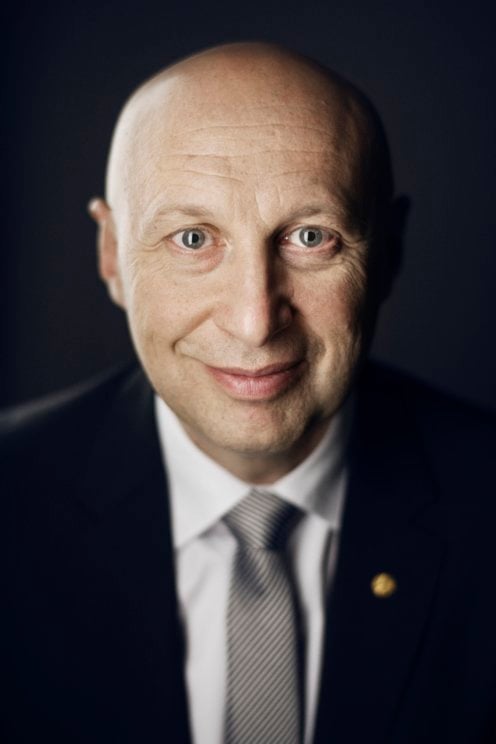
Stefan W. Hell is a Nobel laureate in Chemistry (2014) and a Research Professor at the Max Planck Institute for Biophysical Chemistry, Göttingen, Germany, and the German Cancer Research Center, Heidelberg, Germany. His work revolutionized optical microscopy by developing super-resolved fluorescence techniques such as STED microscopy and 4Pi microscopy , which circumvent the diffraction limit of light. Born: December 23, 1962 in Arad, Romania Education: Physics at the University of Heidelberg (PhD 1990); Habilitation in Physics at the University of Heidelberg (1996) Research Interests focus on super-resolution microscopy , optical physics , and biomedical imaging . His groundbreaking work includes concepts like STED (Stimulated Emission Depletion) and RESOLFT (Reversible Saturable Optically Linear Fluorescence Transitions), enabling nanoscale imaging of biological processes. By leveraging molecular state transitions, Hell’s techniques allow resolving structures previously limited by the diffraction of light. Scientific Awards include: Nobel Prize in Chemistry (2014) Kavli Prize in Nanoscience (2014) Advisees have contributed to advancing microscopy and biophysics, including Jan Wichmann, Franziska Meinecke, Thomas Klar, and Stefan Jakobs.
Professor Ramon Vilar Compte is Professor of Medicinal Inorganic Chemistry at Imperial College London 's Department of Chemistry within the Faculty of Natural Sciences . His multidisciplinary research group develops molecular tools for biological interrogation. Key affiliations include: CRUK Convergence Science Centre , Centre for Neurotechnology , Centre for Rapid Online Analysis of Reactions , and Grantham Institute Research focuses on: Medicinal Inorganic Chemistry of G-quadruplex DNA/RNA Metal complexes for cancer and antibacterial therapies Environmental remediation of toxic metals via molecular recognition Advanced optical imaging techniques using fluorescence/phosphorescence lifetime Recent publications demonstrate expertise in metal-based G-quadruplex probes , photocytotoxic antibiotics , and machine learning-driven drug discovery . Group members include 9 PhD students and 5 postdoctoral associates working across chemical biology and environmental science domains.
Charles Darr is an Assistant Professor at the University of Missouri, specializing in the Department of Chemical and Biomedical Engineering. His work focuses on nanomaterials, plasmonics, and biosensors for biomedical and chemical sensing applications. PhD in Chemical Engineering from University of Missouri MS in Chemical Engineering from University of Arkansas BS in Chemical Engineering from University of Arkansas Research interests span: Plasmonic-enhanced fluorescence for sensor development High-sensitivity detection of biomarkers (e.g., tuberculosis) Nanoparticle and nanostructured materials synthesis Antimicrobial surface modifications for medical devices Machine learning integration in biosensing systems Recent publications demonstrate expertise in creating advanced biosensing platforms using: Plasmonic gratings Fluorescent suprananoparticles Hybrid nanomaterials Single-molecule detection systems He actively manages undergraduate research and internship programs while teaching courses in: Bioengineering design Bioprocess analysis Materials characterization Professional development
Melike Lakadamyali is a Professor at the Perelman School of Medicine , University of Pennsylvania, with secondary appointments in Cell and Developmental Biology. Her research focuses on biophysics , super-resolution microscopy , and macromolecular assembly dynamics in cellular processes. Education: BS (2001) from University of Texas at Austin, PhD (2006) from Harvard University Her work aims to understand the spatiotemporal organization of cellular machinery through advanced single-molecule imaging and quantitative models . Key projects include vesicle transport , transcriptional regulation , and chromatin plasticity . Recent publications highlight methodological innovations like ECLiPSE classification and MiOS imaging-computational strategies. Articles span nucleosome dynamics , mitochondrial fission , and microtubule regulation , emphasizing biophysical quantification . Scientific Awards: EMBO Young Investigator (2013), Ramon y Cajal Fellowship (2014), Hans Fisher Junior Fellow (2016), La Vanguardia Finalist (2016) She advises a research team developing super-resolution tools for vesicular trafficking and chromatin studies , with advisory roles on Bruker/Vutara and BioQuant boards. Her lab employs quantitative approaches to study macromolecular complexes in cellular homeostasis and disease mechanisms .
Bappaditya Chandra is an Assistant Professor at the Department of Chemistry and Biochemistry , North Dakota State University. His research focuses on biomolecular condensates formed via liquid-liquid phase separation (LLPS), particularly their roles in cancer and disease mechanisms. Research Highlights : Transcription factor condensates, fusion oncoprotein dynamics, viral protein interactions, and synthetic bio-materials. Methodologies : Advanced microscopy, protein engineering, mass spectrometry, and computational modeling. Recent work in Nature Communications (2023) and Cancer Discovery (2022) demonstrates how LLPS drives oncogenic processes. His lab also investigates Amyloid-β aggregation in neurodegenerative diseases, applying fluorescence quenching and molecular simulations . Collaborative efforts include Annual Reviews of Cancer Biology (2023) and Journal of Molecular Biology (2018). Students : Dr. Monica Singh (postdoc), Jacob (PhD), and Ciara Reller (undergraduate). Contact : bappaditya.chandra@ndsu.edu
Cristina Ionica Øie is a Senior Researcher at the Department of Medical Biology, UiT The Arctic University of Norway, specializing in liver biology and advanced microscopy. She leads the Sugar Crush project (2018-2024), investigating metabolic mechanisms in non-alcoholic fatty liver disease (NAFLD) progression, funded by RCN FRIPRO. As Co-PI of InterNASH , she explores intercellular pathways for NASH treatment, supported by RCN FORNI, Novo Nordisk, and UiT Innovation. Her work bridges cell biology and engineering, using optical nanoscopy to study liver sinusoidal endothelial cell (LSEC) dysfunction. Research focuses: LSEC metabolism, NASH pathogenesis, and super-resolution microscopy. Key techniques: Structured illumination microscopy, photonic chip-based nanoscopy, and transcriptomics. She holds the 2018 Young Researcher Prize from UiT’s Faculty of Health Sciences. Her labs collaborate across disciplines, integrating nanoscopy with functional assays to dissect liver disease mechanisms. Current grants total ~9.6 MNOK, supporting projects like GenSEC (CRISPR screening in LSECs) and equipment acquisition (e.g., Seahorse metabolic analyzer). Publications emphasize LSEC morphology, bacterial phage interactions, and fenestration dynamics, with contributions to journals like npj Gut and Liver and Nature Photonics . Her work advances understanding of liver endothelial biology and therapeutic targets for metabolic liver diseases.
Lydia Kisley is the Ambrose Swasey Associate Professor of Physics in the Department of Physics at Case Western Reserve University's College of Arts and Sciences. Her research integrates physics, chemistry, and engineering to study molecular interactions in complex materials using advanced optical microscopy techniques. She leads an interdisciplinary lab focused on single-molecule spectroscopy and the development of novel microscopies. Her educational background includes a B.S. from Wittenberg University (2010) and a Ph.D. from Rice University (2015). B.S., Wittenberg University (2010) Ph.D., Rice University (2015) Dr. Kisley’s research interests center on understanding molecular behavior at bio/soft/metal interfaces. She aims to bridge molecular-scale phenomena with macroscopic material performance, particularly in industrial and medical applications. Her lab develops new microscopy and analysis methods to track molecular adsorption, diffusion, conformational changes, and reactivity across space, time, and temperature. Key research areas include separations (especially chiral and rare earth element extraction), corrosion (real-time, in situ monitoring), and extracellular matrix dynamics (protein transport in biological environments). Her work emphasizes quantitative, molecular-level insights to replace trial-and-error approaches in material design. The recent publications highlight a strong trend in single-molecule imaging applied to materials challenges. Her work spans from fundamental biophysics to applied engineering, with a growing emphasis on sustainability and green technologies. Articles focus on fluorophore development for corrosion detection, super-resolution imaging of biological environments, computational analysis of diffusion, and transformative approaches to separation science. A unifying theme is the development of methods to extract meaningful data from noisy or complex systems. She has received notable scientific recognition: U.S. Department of Energy Early Career Research Program Award (2024), $875,000 grant for rare earth element microscopy research Dr. Kisley actively mentors students and researchers at all levels. She advises graduate and undergraduate students in interdisciplinary projects, with several undergraduates contributing to publications and awards. She has secured significant external funding, including the DOE Early Career grant, to support her lab’s research on sustainable separation technologies. Her collaborations span departments at CWRU and include researchers at Lawrence Livermore National Laboratory. She encourages student involvement in high-impact science related to energy and sustainability. The Kisley Lab maintains active research facilities, including a microscopy laboratory in Rockefeller Hall and a wet chemistry lab in AW Smith Hall. The team includes postdoctoral researchers, graduate students, and undergraduates working on projects in biophysics, materials science, and chemical engineering. The lab fosters a collaborative environment with regular group meetings and opportunities for hands-on training in advanced instrumentation.
PD Dr. Basant Kumar Thakur is a researcher at the University Hospital Essen, affiliated with the Medical Faculty of the University of Duisburg-Essen. He leads the Cancer Exosome Research Lab within the Department of Pediatrics III, focusing on the role of extracellular vesicles (EVs) in cancer progression, particularly in pediatric acute myeloid leukemia (AML). His work bridges molecular oncology, liquid biopsy development, and cancer immunology. His research centers on three major themes: (i) identification and purification of cancer-specific EVs carrying dsDNA for clinical biomarker development; (ii) mechanisms of EV-DNA transfer into recipient cells; and (iii) functional consequences of this transfer on metastatic progression, including immune modulation via the cGAS/STING pathway. He has developed bead-based methods to isolate homogeneous EV populations and is analyzing large patient cohorts to establish EV-DNA as a tool for detecting minimal residual disease. The recent publications highlight a strong focus on EV biology, with applications in glioma therapy, plant-derived vesicles for drug delivery, and EV-DNA's role in immune activation and stem cell dysfunction. Keywords across articles include oncology, molecular biology, immunology, and nanomedicine, reflecting a multidisciplinary approach to cancer research. Scientific awards are not mentioned in the provided text. Dr. Thakur actively collaborates with Prof. Bernd Giebel (Transfusion Medicine, UK Essen) and contributes to EU and DFG-funded research programs. He is involved in PhD training through the ZMB (Centre for Medical Biotechnology) and participates in the European Liquid Biopsy Society. His lab investigates EV-based diagnostics and therapeutic strategies, particularly in pediatric cancers and solid tumors. The Cancer Exosome Research Lab is part of the ZMB and contributes to the Oncology research program. The team employs advanced techniques like single molecule localization microscopy and functional assays to study EV-DNA transfer and its impact on tumor microenvironment and metastasis.
Lingyan Shi is an Associate Professor in the Shu Chien-Gene Lay Department of Bioengineering at UC San Diego, with a faculty affiliation in Electrical and Computer Engineering. She leads the Shi Lab, pioneering multimodal super-resolution microscopy integrating stimulated Raman scattering (SRS), multiphoton fluorescence (MPF), and second harmonic generation (SHG) to study metabolic dynamics in aging, diseases, and immunology. Her research highlights include discovering the 'Golden Window' for deep-tissue imaging (1550–1870 nm), developing bioorthogonal metabolic imaging platforms (DO-SRS and STRIDE), and creating the Adam optimization-based Pointillism Deconvolution (A-PoD) for super-resolution SRS microscopy. She holds six patents and has received prestigious awards including the Blavatnik Regional Award (2018), Sloan Research Fellowship (2023), and Advancing Bioimaging Scialog Fellowships (2021–2023). Research Focus : Shi’s lab focuses on visualizing in situ metabolic activities in tissues, particularly lipid, protein, and nucleic acid synthesis. Applications target neurodegenerative diseases (e.g., Alzheimer’s), diabetes, cancer, and aging processes. Techniques emphasize sub-cellular resolution and real-time metabolic tracking. Publications & Impact : Over 50 peer-reviewed articles (2023–2025) explore topics like anti-aging effects of metformin, metabolic imaging in Drosophila brains, and exosome-based cancer classification. Her work bridges biosensor engineering , algorithm development , and clinical diagnostics . Awards & Mentorship : Recognized for mentoring underrepresented students and developing curricula in bioengineering. Teaches core courses in bioengineering at UCSD and actively promotes diversity in STEM.
Eugene Zubarev is an Associate Professor of Chemistry at Rice University, specializing in nanomaterial synthesis and self-assembly. He leads the Zubarev Research Group, focusing on colloidal metallic nanostructures, including gold nanorods, bimetallic nanoparticles, and their applications in catalysis, plasmonics, and biomedical engineering. His work spans from fundamental studies of self-assembly mechanisms to translational research in cancer therapy and nanomedicine. Education: B.S. in Chemistry (Moscow State University, 1993), Ph.D. in Chemistry (Russian Academy of Sciences, 1996). Postdoctoral research at University of Illinois and Northwestern University. Joined Rice University in 2005, promoted to tenured Associate Professor in 2009. Research interests include molecular self-assembly, nanocrystal superstructures, and functional hybrid materials. Key projects involve gold nanorod synthesis, anticancer functionalization, and catalytic bimetallic nanoparticles. His lab emphasizes scalable synthesis methods and nanomaterial characterization. Notable achievements include the Humboldt and Bessel Research Awards (2015). His work bridges materials science and biology, with over 150 publications since 2005. Current research explores tumor microenvironment interactions with nanorods for targeted radiosensitization and drug delivery.
Xiaogang Liu is a Research Fellow at the National University of Singapore, specializing in supramolecular coordination chemistry and optoelectronic materials. With a Ph.D. and M.Sc. in Chemistry from Northwestern University and East Carolina University respectively, and a B.Eng. from Beijing Technology and Business University, his work bridges inorganic and organic materials for advanced technological applications. Education Ph.D. in Chemistry, Northwestern University, U.S.A. M.Sc. in Chemistry, East Carolina University, U.S.A. B.Eng., Beijing Technology and Business University, China Research Focus Liu’s research explores metal-organic complexes for optoelectronic devices, including light-emitting diodes, solar cells, and biosensors. He investigates photon upconversion, X-ray photonics, and mechanoluminescence, emphasizing molecular-level engineering and interdisciplinary applications in medical imaging and wearable technology. Publication Trends His recent publications highlight innovations in lanthanide-doped nanoparticles, perovskite nanocrystals, and X-ray detection technologies. Themes include real-time sensing, super-resolution microscopy, and energy transfer mechanisms, with contributions to journals like Nature , Nature Materials , and Nature Photonics . Scientific Honors RSC Centenary Prize (2024) President’s Science Award (2016) Additional Contributions Liu’s work enables low-cost optoelectronic devices and sensors, with implications for healthcare, renewable energy, and advanced imaging. His research integrates chemical synthesis, nanotechnology, and photonics, positioning him as a leading figure in materials science.
Prof. Pieter van der Zaag is a Professor in the Molecular Biophysics department at the University of Groningen's Zernike Institute for Advanced Materials. He also holds a position at the Faculty of Medical Sciences, University Medical Center Groningen (UMCG). His research focuses on bionanotechnology, advanced optical imaging techniques for 3D tissue analysis, and studying cancer drug interactions at cellular levels. He leads the van der Zaag group and the Optical Molecular Imaging Groningen (OMIG) group, collaborating with international teams in physics, medicine, and biomolecular sciences. Research interests include developing microscopy technologies (e.g., Light Sheet, confocal, and super-resolution techniques like STED) to visualize cancer drug distribution and tumor growth dynamics. His work bridges nanotechnology and medical applications, such as improving tissue adhesive materials and diagnostic imaging in surgery. He chairs the NWO Physics for Technology and Instrumentation advisory committee and co-chairs the Intra-Operative Imaging study group of the European Society for Molecular Imaging (ESMI). Prof. van der Zaag has advised numerous PhD and master’s students and holds patents in medical imaging, microfluidics, and diagnostic devices. His labs focus on interdisciplinary projects, combining physics, engineering, and biomedicine to advance clinical and research tools.
Dr. Siegfried Musser is a Professor in the Department of Cell Biology and Genetics at Texas A&M University's School of Medicine. He holds a PhD from Caltech and joined Texas A&M in 2001 after postdoctoral work at UC Davis and Brandeis University. His research focuses on biophysical mechanisms of protein translocation and biomolecular condensates, using advanced microscopy techniques like single-molecule fluorescence and super-resolution imaging. Key projects include studying nuclear pore complexes, the bacterial Tat machinery, and phase-separated FUS condensates linked to neurodegenerative diseases. Education: AB in Biochemistry from UC Berkeley (1990), PhD in Chemistry from Caltech (1996), postdoctoral training at UC Davis (1996-1999) and Brandeis University (1999-2001). Research Interests: Protein translocation systems (Nuclear Pore Complex, Tat machinery), biomolecular condensates, ALS/FTD pathogenesis, and single-molecule imaging technologies. Current work explores transport pathways in nuclear pores, Tat translocation energetics, and FUS phase maturation using MINFLUX and polarization-PALM approaches. Awards: Junior Faculty Research Excellence Award (Texas A&M Health Science Center). Lab Roles: Mentors graduate students (e.g., Thom Weidman) and postdocs (e.g., Abhishek Sau). Ongoing funding includes grants from the Edward Mallinckrodt Foundation. The lab seeks postdocs, graduate students, and undergraduates for biophysics and cell biology projects. Labs/Teams: Musser Lab at Texas A&M Health Science Center, specializing in single-molecule biophysics and cellular transport mechanisms.
Yuling Yan serves as Professor and Founding Chair of the Department of Bioengineering at Santa Clara University's School of Engineering, holding the Phil and Bobbie Sanfilippo Professorship. She maintains an affiliation as Consulting Faculty at Stanford University's Department of Otolaryngology. Her academic journey includes faculty positions at the University of Hawaii-Manoa (2002-2005), University of Wisconsin-Madison, and University of the Ryukyus (1997-1999), with postdoctoral training at McGill University and Max Planck Institute for Biochemistry. Professor Yan's research focuses on AI-driven medical diagnostics, specializing in machine learning applications for early disease detection including vocal pathologies, cardiac arrhythmia, skin cancer, brain aneurysms, and breast cancer. Her work integrates biomedical imaging (MRA, ultrasound, CT), biosignal analysis, and fluorescence microscopy with molecular switch probes. Current projects involve collaboration with radiologists at Santa Clara Valley Hospital to develop deep learning systems for medical image analysis. Her publication trends reveal an evolution from foundational work in vocal fold dynamics and optical probe development (2011-2015) toward AI/ML applications in medical diagnostics (2017-present), with increasing emphasis on deep neural networks for cancer detection and cardiac monitoring. Key contributions include novel methodologies for high-speed laryngeal imaging analysis and optical lock-in detection microscopy. Active research funding includes NIH Multi-Investigator Awards for high-contrast single-molecule imaging and NSF grants for laryngeal imaging tools development. She teaches graduate courses including Machine Learning for Biomedical Applications and Deep Learning for Medical Image Analysis, alongside undergraduate bioengineering fundamentals.
Marco Salerno is a Researcher at the Department of Physics (DIFI) of the University of Genoa (UniGe). His work focuses on the application of advanced physics techniques to life sciences, environmental studies, and cultural heritage conservation. He teaches courses such as Physics 2 for Management Engineering and Physics Laboratory for Electrical Engineering, Biotechnology, and Cultural Heritage Conservation programs. Researcher, Department of Physics (DIFI), University of Genoa Teaches interdisciplinary physics courses across multiple degree programs Email: marco.salerno@unige.it Salerno's research bridges physics with biomedical and environmental applications, utilizing cutting-edge imaging technologies like confocal fluorescence microscopy and MINFLUX nanoscopy. His work explores nanoparticle-cell interactions, chromatin pattern analysis, and innovative nanomaterial synthesis processes. Recent publications highlight his expertise in nanoparticle dynamics, cellular imaging, and environmental engineering. While no formal awards are listed, his contributions to interdisciplinary physics research suggest ongoing recognition in his field. Studies gold nanoparticle uptake in HeLa cells Develops closed-loop nanoparticle synthesis methods Investigates carbon nanotubes' impact on fibroblast growth Implements confocal microscopy for nuclear chromatin analysis Focuses on physics applications for cultural heritage conservation Pioneers MINFLUX nanoscopy techniques He is involved in laboratory-based instruction and research that connects physical methodologies with biomedical and environmental challenges, demonstrating a multifaceted approach to applied physics.