Ulrich Vogt is a Professor in Applied Physics at Kungliga Tekniska Högskolan (KTH) and leads the X-ray Optics and Nanoimaging group within the Bio-Opto-Nano unit. He serves as Vice-head of the Applied Physics department for undergraduate education. His research focuses on developing advanced X-ray microscopy techniques, particularly at synchrotron facilities like MAX IV’s NanoMAX beamline. He specializes in X-ray optics, nanoimaging, and diffractive optical elements for applications in materials science, biology, and medicine. Key contributions include the design of the NanoMAX beamline, optimization of X-ray zone plates via metal-assisted chemical etching, and advancements in multi-beam ptychography. Vogt has pioneered compact X-ray microscopy systems using laser-plasma sources and liquid-jet targets. His work integrates nanofabrication, computational imaging, and synchrotron instrumentation to achieve sub-100 nm resolution in hard and soft X-ray regimes. Teaching responsibilities include courses on experimental physics, photonics, and X-ray applications. His lab collaborates internationally on projects like the European XFEL, emphasizing high-brightness sources and radiation-resistant optics. Recent innovations include adaptive multi-beam ptychography and stereo X-ray imaging for 3D nanoscale visualization. Research highlights span over 100 peer-reviewed articles, with a focus on coherence characterization, beamline instrumentation, and nanostructured materials. Vogt’s grants include a Röntgen-Ångström Cluster award supporting multi-beam ptychography and cryo-microscopy advancements.
Max Planck Institute for Solid State ResearchGermany
Dr. Manish Garg is the Head of the Research Group 'Quantum Microscopy and Dynamics' at the Max Planck Institute for Solid State Research in Stuttgart, Germany. He holds a Group Leader (W2) position since 2024, having previously served as a Research Group Leader from 2020-2023 in the Department of Nanoscale Science. His research integrates attosecond science, scanning tunneling microscopy, and ultrafast Raman spectroscopy to develop a four-dimensional quantum microscope capable of capturing electrons and atoms in action at fundamental space-time limits. PhD (Dr. rer. nat.) in Physics from LMU Munich, Germany (2012-2017) Integrated B.S.-M.S. from Indian Institute of Science Education and Research, Kolkata, India (2007-2012) Dr. Garg's research focuses on developing techniques to observe and control quantum phenomena at the atomic scale. His group has pioneered methods to visualize electron dynamics with attosecond temporal resolution and sub-Ångström spatial resolution, enabling unprecedented insights into molecular processes. Recent breakthroughs include selective excitation of molecular vibrations, real-time tracking of electron oscillations in nanodevices, and imaging coherent phonon wave packets in graphene nanoribbons. His publication record shows a consistent trajectory of high-impact research in leading journals including Nature, Science, and Nature Communications. The work demonstrates expertise across multiple disciplines including attosecond physics, nanoscale imaging, quantum dynamics, and ultrafast spectroscopy, with applications ranging from fundamental quantum mechanics to potential light-wave electronics. Rudolf-Kaiser Preis (2023) Max-Auwärter Award (2022) IMPRS Advanced Photon Science Graduate Fellowship (2012-2016) INSPIRE Fellowship, DST India (2007-2012) Dr. Garg leads a research group comprising postdocs and PhD students working on cutting-edge quantum microscopy techniques. His group has secured significant recognition for developing methods to directly measure electron oscillations in quantum nanodevices and visualize atomic motion in single molecules. Current research focuses on advancing four-dimensional quantum microscopy to capture electronic and atomic dynamics in molecules, two-dimensional materials, and superconductors at fundamental space-time limits. The Quantum Microscopy and Dynamics group operates within specialized facilities at the Max Planck Institute for Solid State Research, utilizing low-temperature scanning tunneling microscopes operating in ultrahigh-vacuum conditions. Their work on picocavities and nonlinear optical spectroscopy at atomic length scales represents the frontier of quantum measurement science.
Wilson Ho is the Donald Bren Professor and Distinguished Professor of Physics and Astronomy at the University of California, Irvine (UCI). He holds a B.S. and M.S. from Caltech (1975) and a Ph.D. in Physics from the University of Pennsylvania (1979). Prior to UCI, he was on Cornell University's faculty and worked at AT&T Bell Labs. His research focuses on nanoscale phenomena using Scanning Tunneling Microscopy (STM) and Inelastic Electron Tunneling Spectroscopy (IETS). Key areas include probing atomic/molecular behavior, quantum tunneling, single-molecule chemistry, and surface magnetism. Notable achievements include observing single hydrogen atom tunneling and developing STM-based quantum sensing techniques. Recent work includes quantum stochastic rectification in molecules and THz-driven molecular sensors. Over 20 students have graduated under his mentorship, many advancing to prestigious postdocs and industry roles. Awards include the Heinrich Rohrer Medal and National Academy of Sciences membership. Lab activities include the Quantum Superposition Microscope, THz-STM integration, and catalytic studies on surfaces. Collaborations span institutions globally, with a focus on advancing nanoscale measurement science and molecular-level understanding.
Kayla Nguyen is an Assistant Professor in the Department of Physics at the University of Oregon, affiliated with the College of Arts and Sciences. Her research focuses on advancing electron microscopy techniques, particularly ptychography, to study magnetism, topology, and material properties at the angstrom scale. She leads the kxnlab , which develops innovative imaging methods for topological materials, magnetic textures, and nanoscale systems. Key research interests include high-resolution imaging beyond diffraction limits, cryo-electron microscopy for biological specimens, and quantitative analysis of magnetic materials using 4D-STEM and Lorentz microscopy. Her work integrates computational approaches with experimental techniques to address challenges in material characterization and structural analysis. Nguyen’s publications highlight advancements in ptychography, sub-angstrom resolution, and imaging chiral magnetic systems. Her research often bridges fundamental physics with applications in nanotechnology, materials science, and art conservation (e.g., analyzing pigments in historical artworks). Advising and grants: While specific grant details are not listed, her research aligns with cutting-edge microscopy and materials science initiatives. She mentors graduate students in experimental and theoretical projects related to her lab’s focus areas. Labs/teams: The kxnlab collaborates with interdisciplinary groups to pioneer microscopy innovations, including air-compatible electron microscopy and pixel array detector technologies.
Dr. Yaroslav Gerasimenko serves as Group Leader of the Lightwave-STM research group within the Huber group at the University of Regensburg, Germany, where he directs the ERC-funded Orbital Cinema project. His position represents an independent faculty-level research role focused on ultrafast quantum material dynamics. His academic training includes a PhD in Condensed-Matter Physics (2008-2014) from the P. N. Lebedev Physical Institute of the Russian Academy of Sciences and undergraduate studies in Physics and Microelectronics (2002-2008) at the Moscow Institute of Electronic Technology, Russia. Postdoctoral experience spans the University of Regensburg (2020-2022), Jozef Stefan Institute in Slovenia (2016-2020), and P. N. Lebedev Institute (2015). Gerasimenko pioneers lightwave-controlled scanning tunneling microscopy to achieve simultaneous atomic spatial and subcycle temporal resolution. His research centers on quantum materials including transition metal dichalcogenides (e.g., 1T-TaS 2 ) and metal halide perovskites, with emphasis on charge density waves , Mott physics , and non-equilibrium phase transitions . Key innovations involve terahertz plasmonics in graphene and quantum jamming transitions, often leveraging light-induced metastable states. Recent publications (2023-2025) demonstrate breakthrough capabilities in atomic-scale ultrafast imaging , including Nature cover stories on subcycle microscopy and Nature Photonics cover stories on atomic-scale spectroscopy. The work establishes new paradigms for visualizing electron dynamics at fundamental spatiotemporal limits, with applications spanning quantum computing components and next-generation photovoltaics. Scientific recognition includes: ERC Starting Grant for Orbital Cinema project (2023) As principal investigator of the ERC project, Gerasimenko leads instrumentation development for lightwave-STM systems. His research program involves collaborations with the Jozef Stefan Institute (Slovenia), P. N. Lebedev Institute (Russia), and international quantum material consortia. Current efforts focus on extending ultrafast nanoscopy to topological materials and quantum annealers. The Lightwave-STM laboratory at University of Regensburg houses custom cryogenic scanning probe microscopes integrated with multi-terahertz laser systems, enabling experiments at 0.1-atom spatial and 1-femtosecond temporal resolutions under extreme conditions.
University of California, Los AngelesUnited States
Tamir Gonen is a Professor of Biological Chemistry and Physiology at the David Geffen School of Medicine, University of California, Los Angeles (UCLA) , and an Investigator at the Howard Hughes Medical Institute (HHMI) . He is a pioneer in microcrystal electron diffraction (MicroED) , a transformative cryo-EM method for atomic-resolution structure determination. Education: Doctor of Science (DSc), University of Auckland (2025) PhD in Structural Biology, Harvard Medical School (2005) PhD in Biochemistry, University of Auckland (2002) BSc (Hons) in Inorganic Chemistry and Biochemistry, University of Auckland (1998) His research focuses on membrane protein structure and function , particularly those in the blood-brain barrier , using MicroED, X-ray crystallography, NMR, and molecular dynamics. He has determined structures of ion channels, transporters, and drug compounds at resolutions better than 1 Å, advancing drug discovery and understanding disease mechanisms. Recent work includes high-throughput MicroED for ion channel dynamics, energy filtering to enhance resolution, and polymorphic drug characterization . His lab also develops protocols for suspended drop crystallization and focused ion-beam milling of samples. Scientific Awards: Fellow, American Crystallographic Association (2025) Doctor of Science, University of Auckland (2025) Carl Branden Award, The Protein Society (2024) Investigator, HHMI (2017) Member, Royal Society of New Zealand (2017) American Diabetes Association Career Development Award (2009) He leads the Gonen Lab , which emphasizes multidisciplinary approaches and method development in structural biology. His trainees have become faculty at top global institutions, extending his impact on the field.
Dr. Tatiana Latychevskaia is a Principal Investigator at the Paul Scherrer Institute (PSI) and holds an academic position at the University of Zurich's Department of Physics. She leads the Laboratory for Multiscale Bioimaging, focusing on developing advanced imaging techniques. Her research integrates theoretical and experimental approaches to push the boundaries of nanoscale visualization. Research Interests: Her group innovates in coherent high-resolution 3D imaging, including holography, coherent diffraction imaging, and convergent beam electron diffraction. Key areas are algorithm development for wavefront propagation, iterative phase retrieval, low-energy electron imaging (20–300 eV), and wavefront modulation for beam shaping. Applications span 2D materials, biological specimens, and nanostructured systems. Publication Trends: Recent work emphasizes algorithmic advances in phase retrieval and holographic reconstruction (2025), 2D material characterization using electron diffraction (2023–2025), and novel lensless imaging techniques (2024). Her publications demonstrate consistent leadership in quantitative electron microscopy and computational imaging. Teaching & Mentoring: She lectures on Electron Microscopy (PHY427) and Modern Microscopy (PHY425) at the University of Zurich. Actively supervises a diverse team including Postdocs, PhD students, and undergraduate researchers. Current advisees include Dylan Brault (Postdoc), Piet Fang (PhD), and Zhiyong Zhuang (Master's).
Max Planck Institute for the Science of LightGermany
Hisham Mazal is a Research Fellow at the Max Planck Institute for the Science of Light (MPL), part of the group led by Prof. Vahid Sandoghdar. His research focuses on advancing cryogenic super-resolution fluorescence microscopy to study protein structures in native environments. Key objectives include developing workflows for correlative light-electron microscopy using vitrified samples and enhancing detection sensitivity for small proteins via machine learning. Education: BSc in Biotechnology Engineering (ORT Braude College, 2010-2013), MSc in Chemical and Biological Physics (Weizmann Institute, 2013-2015), PhD in Single-Molecule Protein Dynamics (Weizmann Institute, 2016-2020). Joined MPL as a postdoc in 2020. Research interests span cryogenic microscopy innovations, protein dynamics, membrane protein analysis, and machine learning applications in imaging. His work bridges structural biology and biophysics, with recent contributions to PIEZO1 channel studies, α-Synuclein aggregation, and sub-10kDa protein detection. Notable collaborations include work on AAA+ protein machines and enzymatic activity modulation, leveraging single-molecule FRET and advanced microscopy techniques. His lab integrates interdisciplinary approaches to uncover functional protein mechanisms at atomic scales.
University of Illinois Urbana-ChampaignUnited States
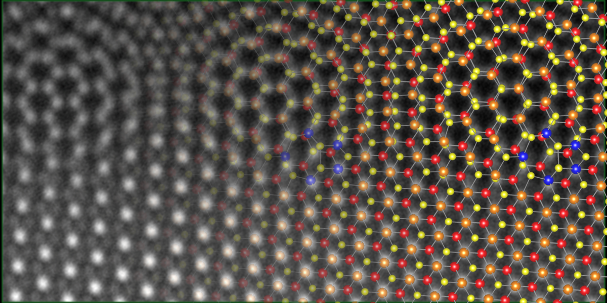
Pinshane Huang is an Associate Professor and Ivan Racheff Faculty Scholar in the Department of Materials Science and Engineering at the University of Illinois Urbana-Champaign. She holds a B.A. in Physics from Carleton College and Ph.D. in Applied Physics from Cornell University. Her research focuses on transmission electron microscopy and spectroscopy of 2D materials, soft-hard interfaces, and atomic-scale defects. Huang leads the Huang Research Group, headquartered at the Materials Research Laboratory. Education: PhD, Cornell University, Applied and Engineering Physics MS, Cornell University, Applied and Engineering Physics BA, Carleton College, Physics (with Distinction) Research Interests: Huang’s work bridges physics, chemistry, and materials science. Her group develops advanced electron microscopy techniques (e.g., ptychography) to image atomic structures in 2D materials like graphene and transition metal dichalcogenides. Key areas include defect dynamics, strain engineering, and material interfaces. Recent breakthroughs include sub-angstrom resolution imaging in uncorrected microscopes and atom-by-atom visualization of silica glass defects. Key Contributions: Pioneered high-resolution ptychography methods Discovered atomic-scale dynamics in 2D materials Developed machine learning tools for microscopy data analysis Awards & Recognition: Presidential Early Career Award for Scientists and Engineers (2019) David and Lucile Packard Fellowship (2017) Sloan Research Fellowship (2018) Multiple teaching awards including Grainger College of Engineering Teaching Excellence Award (2021) Labs & Collaborations: Huang’s lab is part of the Materials Research Laboratory (MRL) at UIUC. Current projects are funded by NSF, AFOSR, 3M, and the Packard Foundation. Collaborations span academia and industry, focusing on quantum materials, nanoelectronics, and energy applications.
David Smith is Regents' Professor in the Department of Physics at Arizona State University and concurrently a Distinguished Global Futures Scientist within the Global Futures Scientists and Scholars initiative. Since 1984 he has directed ASU’s Center for High Resolution Electron Microscopy and served as Principal Investigator for the NSF National User Facility for High Resolution Electron Microscopy. His leadership roles also include Associate Chair for Space & Infrastructure in Physics (2013–present), Director of the John M. Cowley Center (1991–2006) and President of the Microscopy Society of America (2009). Education D.Sc., University of Melbourne, Australia (1988) Ph.D., University of Melbourne, Australia (1978) B.Sc. (Hons.), University of Melbourne, Australia (1970) Research Overview David Smith’s research is devoted to the development and application of atomic-resolution electron microscopy . Over four decades he has pioneered instrumentation and techniques that allow direct visualization of atomic arrangements in solids, enabling breakthrough insights into semiconductor heterostructures , nanostructures , oxide/semiconductor interfaces , magnetic multilayers and quantum-confined systems . Recent emphasis targets wide-band-gap nitrides for optoelectronics, two-dimensional electron gases at oxide interfaces, and nanoscale dopant distributions critical for next-generation electronic devices. His work integrates aberration-corrected TEM , off-axis electron holography , in-situ microscopy and quantitative image simulation to correlate structure with functional properties at the sub-ångström scale. Selected Scientific Awards & Honors Helmholtz International Fellowship Award, Helmholtz Foundation (2014) Distinguished Physical Scientist Award, Microscopy Society of America (2014) Harold Rose Distinguished Lectureship Award, German Microscopy Society (2019) Fellow, Materials Research Society Fellow, American Physical Society Fellow, Microscopy Society of America Fellow, Institute of Physics (U.K.) Grants & Doctoral Advising Smith has served as PI or co-PI on numerous federal and industry grants exceeding tens of millions of dollars. Current and recent funding includes: DOD-AFOSR – “Modulation-Doped Heterovalent Structures for High-Speed Electronic Device Applications” (2015–2018) UT-Austin – “Charge Transfer at Metal Dielectric Interfaces under Extreme Environments” (2014–2017) WYLE LABS – “Advanced Nanostructural Techniques for Nitride Device Operation” (2013–2016) NSF-MPS-PHY – “SusChEM: FRG: Molecular routes to new classes of polar/non-polar alloy semiconductors” (2013–2016) DOD-ARMY-ARO – “Advanced Microscopy and Analytical Studies for Hg-based Infrared Detector Materials and Substrates” (2013–2016) He routinely supervises Ph.D. dissertations and M.S. theses; course offerings include PHY 799 Dissertation , MSE 554/555 Electron Microscopy II & Lab , and PHY 792 Research . Laboratories & Teams Smith directs operations within the John M. Cowley Center for High Resolution Electron Microscopy , one of the premiere university-based microscopy facilities in the United States, housing multiple aberration-corrected TEMs, environmental TEMs, and dedicated specimen-preparation suites. The center supports interdisciplinary teams spanning physics, materials science, chemistry, electrical engineering and geoscience, and hosts national and international visiting researchers via the NSF user facility program.
Andre Mkhoyan is a Professor in the Department of Chemical Engineering and Materials Science at the University of Minnesota Twin Cities, operating within the College of Science and Engineering. He leads the Analytical Electron Microscopy Lab , focusing on atomic-scale materials characterization. Research Focus: Sub-Angstrom resolution transmission electron microscopy (TEM/STEM), quantitative spectroscopy, nanomaterials, and electron beam channeling phenomena Key Facilities: Access to aberration-corrected FEI Titan and Thermo Fisher TALOS FX200 transmission electron microscopes His research group has produced groundbreaking work in Science , Nature , and Nature Materials publications, covering topics like spintronic device degradation , 2D material interfacial states , and metal-organic framework stability . Recent scientific recognition includes Fellowship in the Microscopy Society of America (2024). Notable achievements include: $1M+ in research funding from NSF, SRC, and University initiatives Development of novel TEM sample preparation techniques Mentoring 15+ graduate students including Supriya Ghosh and Hwanhui Yun The lab's technological impact spans semiconductor manufacturing (through SRC partnership), materials discovery (graphene oxide for filtration), and fundamental understanding of crystal defect behavior at nanoscale dimensions.
University of Illinois Urbana-ChampaignUnited States
Bryan K Clark is an Associate Professor in the Department of Physics at the University of Illinois, specializing in Computational Condensed Matter Physics and Quantum Computing. He leads the Clark Research Group focused on developing computational tools to understand many-body physics and advance quantum computing capabilities. His research spans the intersection of quantum information, condensed matter physics, machine learning, and computing. Clark's group develops computational methods including neural network backflow approaches, quantum Monte Carlo techniques, tensor networks, and machine learning algorithms for quantum systems. His work addresses fundamental questions in quantum many-body physics, quantum algorithms, and the quantum-classical boundary. His recent publications (2024-2025) demonstrate expertise across quantum simulation, quantum error mitigation, neural network quantum states, and applications of machine learning to physics problems. These works show a strong trend toward integrating machine learning with quantum computing and developing novel approaches for quantum state preparation and characterization. Ranked Excellent Teacher by students multiple times (Spring 2024, Spring 2022, Spring 2020, Fall 2018, Fall 2017) Recipient of DOE grants for quantum information science research Secured NSF funding for machine learning applications in nano-photonics manufacturing IBM funding as part of the IIDAI institute for shadow tomography and error mitigation Clark actively advises PhD students, with recent successful defenses by Faisal Alam, Matt Thibodeau, Chad Germany, James Allen, and Abid. His pedagogical contributions include developing computational physics curriculum at Illinois, notably Physics 246 (Intro to Computational Physics) and Physics 446 (Modern Computational Physics), where students build quantum computing simulators, implement numerical renormalization, and explore connections between Ising models and machine learning.
Francisco Lagunas Vargas is an Assistant Professor in the Department of Mechanical Engineering & Materials Science at Washington University in St. Louis's McKelvey School of Engineering. He will join in June 2025. His research focuses on atomic-scale analysis of materials' properties using advanced Scanning Transmission Electron Microscopy (STEM). Education: PhD in Materials Science, University of Illinois Chicago (2023) BS in Materials Science, Southern Illinois University Edwardsville (2018) Research Interests: Developing structure-property relationships in functional materials for energy and environmental applications. Specializes in real-time observations of nanoscale interactions in 2D/1D materials and quantum/photonic devices. Utilizes sub-angstrom resolution STEM for atomic-scale analysis. Awards: Outstanding Dissertation Award (PhD) Access to Excellence Fellowship James Kouvel Fellowship Future Work: Aims to optimize materials for renewable energy systems and next-generation sensors through atomic-resolution imaging. Active in lab筹建 and interdisciplinary collaborations.
Prof. Stefan Hell is a renowned physicist and director at the Max Planck Institute for Multidisciplinary Sciences, leading the Department of NanoBiophotonics. He holds positions as Hon. Prof. at the University of Göttingen's Faculty of Physics and has directed multiple research departments since 2002. His work pioneered breakthroughs in super-resolution microscopy, including STED, RESOLFT, and MINFLUX nanoscopy, enabling nanometer-scale imaging in biology and materials science. Education: He earned a Physics Diploma (1987) and Doctorate (1990, summa cum laude) from Heidelberg University, followed by postdoctoral work at EMBL (1991–1993). He habilitated in Physics at Heidelberg in 1996. Research focuses on developing fluorescence nanoscopy techniques that surpass the diffraction limit, with applications in cellular and molecular imaging. His group's innovations have revolutionized biological research by enabling visualization of sub-cellular structures at unprecedented resolutions. Key awards include the Nobel Prize in Chemistry (2014) and the Kavli Prize in Nanoscience (2014). His work has been published in top journals like Science, Nature, and Proceedings of the National Academy of Sciences. He advises researchers in his lab and collaborates internationally. His research groups are affiliated with the Max Planck Institutes in Göttingen and Heidelberg, focusing on advancing optical nanoscopy and its biomedical applications. Labs/Teams: Head of the NanoBiophotonics department and Optical Nanoscopy division, advancing MINFLUX-based technologies and stochastic single-molecule imaging.
David Anthony Muller is the Samuel B. Eckert Professor of Engineering in the School of Applied and Engineering Physics at Cornell University, and co-director of the Kavli Institute at Cornell for Nanoscale Science. His research focuses on atomic-scale characterization and control of matter for energy storage, conversion, and quantum materials. Education: B.S. and M.S. in Physics from University of Sydney; Ph.D. in Physics from Cornell University (1996) Using high-resolution electron microscopes in specialized environments, his group explores chemistry, electronic structure, and bonding in systems ranging from transistors to turbine blades. Key areas include 2D materials, superconductors, and nanoscale device physics where quantum effects dominate. His recent work on electron ptychography has pushed imaging resolution to atomic-scale limits, enabling precise lattice vibration analysis and sub-picometer strain mapping. This research has significant implications for semiconductor technology, energy systems, and quantum computing materials. Scientific Awards: Joseph F. Keithley Award (2024), John Cowley Medal (2023), AAAS Fellow (2022), Ernst Ruska Prize (2021), Peter Duncumb Award (2016), Fellow of Microscopy Society of America (2013), Fellow of American Physical Society (2011) As director of the Electron Microscopy Facility at PARADIM, Muller leads development of advanced imaging tools recognized by Guinness World Records for highest resolution microscope technology. His work bridges fundamental physics and applied engineering through industry collaborations.