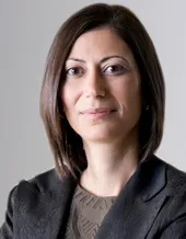
Melike Lakadamyali is an Assistant Professor of Physiology at the University of Pennsylvania’s Perelman School of Medicine (since 2017). She holds a PhD from Harvard University (2006) and has held roles including Senior Group Leader at ICFO-The Institute of Photonic Sciences (Spain). Her research integrates advanced microscopy techniques (super-resolution nanoscopy, single-molecule biophysics) to study protein spatial-temporal organization in cells, particularly in neurons. She focuses on nuclear organization, synaptic architecture, and intracellular trafficking dynamics. Her methods combine optical innovations with molecular tools like microfluidics and biophysical modeling. At TUM, she was a Hans Fischer Fellow (2016) collaborating with Thomas Misgeld on the Focus Group ‘Subcellular Dynamics in Neurons.’ Key awards include the EMBO Young Investigator Award (2013) and ERC Starting Grant. Lakadamyali’s work has been published in Cell, Nature Methods, and PNAS, advancing quantitative imaging and cellular mechanics understanding. Her research emphasizes bridging microscopy innovation with biological questions, such as how protein localization impacts function. Collaborations include advisory roles at BioQuant (Heidelberg) and editorial work for Scientific Reports.
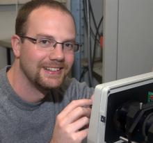
Sören Doose serves as a Privatdozent (equivalent to Researcher) at the Chair of Biotechnology and Biophysics at the University of Würzburg. His work focuses on advanced imaging techniques with particular expertise in super-resolution microscopy. He maintains an active research program with numerous high-impact publications in leading scientific journals including Science, Nature Methods, and Advanced Materials. Dr. Doose's research interests center on pushing the boundaries of fluorescence imaging technology, particularly in the sub-10nm range. His work spans multiple disciplines including biophysics, molecular imaging, and protein engineering. He has made significant contributions to developing novel microscopy techniques such as photoswitching fingerprint analysis and protein-based nanorulers that enable unprecedented resolution in biological imaging. His research has applications across immunology, neuroscience, and cancer research, demonstrating the interdisciplinary nature of his work. Analysis of Dr. Doose's recent publications reveals a consistent focus on overcoming technical limitations in super-resolution microscopy. His research demonstrates expertise in genetic code expansion, click chemistry labeling, and advanced image analysis techniques. A notable trend across his work is the development of tools that enable molecular-level visualization of biological processes, particularly in challenging environments like confined synaptic clefts or cellular membranes. His research has significant implications for understanding disease mechanisms at the molecular level. Dr. Doose maintains an extensive collaborative network, frequently working with Markus Sauer (appearing as co-author on nearly all publications) and researchers across multiple institutions in Germany and internationally. His work spans multiple disease contexts including cancer immunotherapy, neurodegenerative disorders, and viral infections, demonstrating the broad applicability of his imaging techniques. His laboratory appears to focus on developing and applying cutting-edge microscopy techniques to answer fundamental biological questions. The research group likely maintains specialized equipment for super-resolution microscopy including dSTORM, SIM, and lattice lightsheet systems as referenced on the department website. Their work bridges physics, biology, and chemistry to develop novel approaches for visualizing molecular processes in living systems.
Dr. Frank Garwe is a Scientist at the Leibniz Institute of Photonic Technology (Leibniz-IPHT) in Jena, Germany, working within the Research Department of Nanobiophotonics and the Molecular Plasmonics work group. His research focuses on the intersection of nanotechnology, photonics, and molecular systems, with particular expertise in plasmonic phenomena and their applications in sensing and nanofabrication. Dr. Garwe's primary research interests center around nanophotonics and plasmonics , with specific focus on graphene-based optical sensors, molecular plasmonics, and DNA nanowire technologies. His work explores how light interacts with matter at the nanoscale, particularly examining surface plasmon resonances in metal nanoparticles and their applications in sensing, imaging, and energy transfer. His research spans multiple disciplines including materials science , optical physics , and nanotechnology , with applications in biomedical sensing, catalysis, and terahertz technology. Analysis of Dr. Garwe's publication record reveals a consistent research trajectory focused on plasmonic phenomena and their applications. His work demonstrates expertise in utilizing various metallic nanoparticles (particularly gold and silver) for sensing applications, energy transfer along molecular structures, and nanofabrication techniques. A notable trend in his research is the exploration of long-range excitation transfer along DNA nanowires, which has implications for molecular communication systems. More recently, his work has expanded into graphene-based plasmonic structures and their applications in terahertz sensing. Dr. Garwe has actively contributed to the field through numerous publications in high-impact journals including Nano Letters, ACS Nano, and Applied Physics B. His collaborative work spans multiple institutions and demonstrates strong interdisciplinary connections between physics, materials science, and biotechnology. Within the Leibniz Institute of Photonic Technology, Dr. Garwe is a key member of the Molecular Plasmonics work group in the Nanobiophotonics department. This research group focuses on the fundamental interactions between light and matter at the nanoscale, particularly examining how plasmonic effects in metal nanostructures can be harnessed for applications in sensing, imaging, and nanofabrication. The group's work bridges the gap between fundamental physics and practical applications in biotechnology and materials science.
Professor Marc Bramkamp is a leading microbiologist at the Institute of General Microbiology , Christian-Albrechts-University Kiel , specializing in bacterial cell biology, chromosome organization, and membrane dynamics. He previously held faculty roles at LMU Munich and the University of Cologne. His research explores the spatio-temporal organization of bacterial cells, focusing on cell division mechanisms (e.g., Min system, ParABS in Corynebacterium), membrane fluidity (flotillins), and phage interactions . Key projects include studying Bacillus subtilis and Corynebacterium glutamicum as models for polar growth and division. Article trends highlight his work on membrane fusion (DynA), chromosome segregation (ParB-CTPase), phage resistance (e.g., prophage-encoded actin-like proteins), and bacterial lipid domains. His publications span biochemistry , cellular microbiology , and antibiotic mechanisms . He leads the Central Imaging Facility at Kiel, advancing super-resolution microscopy (e.g., PALM) for bacterial studies. Collaborations involve DFG-funded projects and interdisciplinary networks in synthetic biology.
Jan Vogelsang is a researcher at the University of Regensburg within the Faculty of Physics and the Institute of Experimental and Applied Physics . He has held academic positions at the Ludwig-Maximilians-University of Munich and the University of Texas at Austin, with a focus on single-molecule spectroscopy and super-resolution microscopy. Education: PhD in Physics from the University of Bielefeld (2007), Ludwig-Maximilians-University Munich (2009). Research Interests: Single-molecule photophysics, super-resolution microscopy, conjugated polymer aggregates, exciton dynamics, and fluorescence correlation spectroscopy. His work bridges single-molecule spectroscopy with bulk measurements to understand electronic coupling in π-conjugated systems. Recent studies explore exciton localization , photoredox catalysis , and chiral electronic structures . Publications span journals like Nature , ACS Nano , and Angewandte Chemie , often addressing challenges in organic optoelectronics and nanoscale photonics . His methodological expertise includes fluorescence lifetime measurements , solvent vapor annealing , and alternating laser excitation (ALEX) . Collaborations with Prof. J. M. Lupton’s group at Regensburg and Prof. P. Tinnefeld’s team in Munich have been central to his career. No specific awards or students are mentioned in available records.
PD Dr. Marcel Müller is a Senior Lecturer in the Faculty of Physics at Bielefeld University , affiliated with the AG Biomolekulare Photonik (Biomolecular Photonics Research Group). His academic work focuses on advanced optical imaging technologies and their applications in biomedical research. University: Bielefeld University College: Faculty of Physics Research Group: AG Biomolekulare Photonik Academic Rank: Senior Lecturer Dr. Müller's research explores cutting-edge developments in super-resolution microscopy , particularly through structured illumination microscopy (SIM) and optical fluctuation imaging . His recent work addresses challenges in improving spatial resolution , image denoising using machine learning, and cost-effective imaging solutions for live cell analysis. He specializes in optimizing camera technologies and real-time reconstruction algorithms to enhance microscopy capabilities. The 15 most recent publications highlight his contributions to optical nanoscopy , with particular emphasis on structured illumination , deep learning denoising , multiplane imaging , and noise-controlled reconstructions . His work spans from hardware development (e.g., image-splitting prisms , DMD devices ) to software innovations (e.g., real-time reconstruction algorithms ), all aimed at making super-resolution microscopy more accessible and efficient for biological applications. Key Research Areas: Structured Illumination Microscopy (SIM) Super-resolution Optical Imaging Deep Learning for Image Denoising Cost-effective Microscopy Systems Live Cell Imaging Camera Technology Optimization Dr. Müller's laboratory, the AG Biomolekulare Photonik , develops innovative optical imaging tools that bridge physics and biology. His team focuses on modular microscope designs and open-source reconstruction frameworks to democratize access to advanced microscopy techniques. Current projects include optimizing video-rate imaging and exploring nonfluorescent nanoparticle probes for cellular dynamics studies.
Professor Dietmar Schmucker leads the Department of Neural Circuit Development at the LIMES Institute (Life and Medical Sciences Institute) at the University of Bonn. Previously, he was a Professor at University Leuven (KUL) in Belgium and an Associate Professor at Harvard Medical School. His research focuses on understanding the molecular mechanisms underlying neural circuit development, particularly neuronal wiring specificity and synaptogenesis. Dr. Schmucker's research interests center on how complex neural networks develop from a limited genetic repertoire. His lab investigates molecular diversity of membrane receptors, particularly the Dscam family, and their roles in neuronal self-avoidance, axon branching, and synapse formation. Using model organisms like Drosophila and Xenopus tropicalis , his team employs genetic, biochemical, and imaging approaches to study neural development and regeneration. His research has revealed how molecular diversity of the Dscam receptor provides tens of thousands of isoforms essential for neuronal self-recognition and circuit assembly. Recent work focuses on phosphatase Prl-1's role in spatially restricted synaptogenesis and Wnk kinase's evolutionary conserved function in protecting neurons from degeneration. His publications span top journals including Cell, Nature, Science, and Neuron. Alexander von Humboldt Professorship (2019) ERC Synergy Grants Professor Schmucker's lab maintains active research programs in neuronal wiring mechanisms and regeneration. His team combines reverse genetics with high-resolution imaging to investigate synapse formation at single-synapse resolution. Current projects include studying the molecular organization of vertebrate DSCAM and clustered protocadherins in Xenopus tropicalis , and novel mechanisms of axonal branching specificity.
Dr. Mark Schüttpelz is a researcher at the Faculty of Physics, Universität Bielefeld . He is actively involved in the development and application of advanced optical imaging techniques, particularly in the fields of super-resolution microscopy and single-molecule detection . His research program integrates key areas such as Biophysics , Nanoscopy , and Fluorescence Microscopy , with a focus on cellular nanostructures , DNA imaging , and optical waveguide technologies . The recent publications highlight significant contributions to multiscale biomedical imaging , photoswitching mechanisms , and cost-efficient super-resolution systems . Dr. Schüttpelz has developed innovative approaches in single-molecule localization microscopy and deep-learning image reconstruction . His work spans both fundamental biophysical questions and practical applications in hepatology and plant molecular biology , demonstrating a strong interdisciplinary orientation within the university's Transcending Boundaries research framework.
Professor Philip Tinnefeld is a leading academic at Ludwig-Maximilians-Universität München (LMU Munich), serving as Dean of the Faculty of Chemistry and Pharmacy and head of the Tinnefeld Lab. His research integrates molecular biology, physical chemistry, biophysics, and nanophotonics to advance single-molecule detection, super-resolution microscopy, and DNA nanotechnology. Notable contributions include the development of dSTORM and DNA-PAINT techniques, graphene energy transfer (GET), and MXene-based biosensors. He coordinates the EU-funded BioHYBRITE doctoral program and leads interdisciplinary projects at the Center for NanoScience (CeNS). Research Focus: Single-molecule dynamics, DNA origami-based tools, graphene/MXene nanomaterials, and super-resolution microscopy innovations. Recent work includes studies on DNA nanoantennas, force spectroscopy, and MINFLUX-based imaging. Lab Activities: The lab actively publishes in top journals (e.g., Nature Methods, Nature Nanotechnology), hosts international collaborations, and trains PhD students in advanced techniques. Key students include Cindy (PhD 2025), Fiona (2025), and Renukka (2024). Awards include CeNS Publication Awards and funding from EU Marie Skłodowska-Curie Actions.
Thorben Dammeyer is a Researcher at the Institute of Semiconductor Technology , part of the Faculty of Electrical Engineering, Information Technology, and Physics at the Technical University of Braunschweig . His work bridges molecular biology and nanotechnology, focusing on protein engineering, fluorescence imaging, and microbial systems. Primary Affiliation : Institute of Semiconductor Technology, Technical University of Braunschweig Email : t.dammeyer@tu-braunschweig.de Research Interests : Dr. Dammeyer’s research spans biochemistry, protein engineering, and microbial biotechnology. Key contributions include advancing inclusion body refolding protocols, developing DNA origami-based fluorescence standards, and elucidating protein complex roles in bacterial motility. His work often integrates structural biology with practical applications in biopharma. Publications highlight trends in super-resolution microscopy , recombinant protein production , and marine microbial metabolism . He has pioneered tools for quantitative imaging and explored pigment biosynthesis in cyanobacteria and viral enzymes.
Erin Schuman is a Director at the Max Planck Institute for Brain Research in Frankfurt, leading the Department of Synaptic Plasticity. She holds a BA in Psychology from the University of Southern California (1985) and a PhD in Neuroscience from Princeton University (1990). Her research focuses on the molecular and cellular mechanisms underlying synaptic function, emphasizing protein synthesis and degradation in neurons. Key contributions include discovering local protein synthesis in dendrites (1996), developing BONCAT/FUNCAT techniques for tracking protein synthesis, and studying synaptic diversity via proteomics. Schuman has been recognized with prestigious awards, including the Körber Prize (2024) and Brain Prize (2023). She has mentored numerous students and postdocs, advancing understanding of neurodevelopmental disorders and neural circuitry. Her lab uses zebrafish models to explore social behavior and employs advanced imaging and proteomic methods. Education: BA Psychology (USC, 1985), PhD Neuroscience (Princeton, 1990) Key Roles: Director at Max Planck (2009–), HHMI Investigator (2004–2010) Techniques: BONCAT/FUNCAT, single-cell sequencing, super-resolution microscopy
Richard Taylor is a Research Fellow at the Max Planck Institute for the Science of Light, specializing in nanophotonics and interferometric scattering microscopy (iSCAT). He holds a Master's (1st Class Honors) in Physics from the University of Birmingham (2009) and a PhD from the University of Cambridge (2013), where his doctoral work focused on plasmonic nanoparticles and led to a patent for plasmonic junctions in surface-enhanced spectroscopy. Since 2013, he has worked in Prof. Vahid Sandoghdar's group, developing iSCAT microscopy for live-cell applications and became a Humboldt Postdoctoral Fellow in 2015. His research interests include visualizing nanoscale biological dynamics, particularly membrane organization and cellular function. He has pioneered tools like nanofluidic delivery systems for precise molecular manipulation in live cells. His work bridges plasmonics, microscopy innovation, and single-molecule biophysics. Education: BSc (Hons), Physics, University of Birmingham, 2009 PhD in Nanophotonics, University of Cambridge, 2013 Key research contributions include high-precision protein tracking, iSCAT microscopy development, and SERS-based analysis of nanoparticle clusters. He co-organized the inaugural international workshop on iSCAT microscopy in 2020, advancing scientific outreach and collaboration. Awards: Humboldt Postdoctoral Fellowship (2015) Grants/Advising: Not explicitly listed; focus on experimental and theoretical nanoparticle systems. His lab develops microscopy tools for studying biological systems at unprecedented spatiotemporal resolution, with applications in biophysics, nanomedicine, and materials science.
Sylvia Speller is a Professor of Physics at the University of Rostock, where she leads research in nanoscale physical, molecular, and biological processes. She is affiliated with the Institute of Physics and has previously held a professorship at Radboud University Nijmegen, where she directed NanoLab Nijmegen, fostering academic-industry collaboration in nanoscience and technology. PhD in Physics, University of Osnabrück (1995) Habilitation in Physics (2002) Postdoctoral research in Eindhoven and Leuven Full Professor, Radboud University Nijmegen (2001–2012) Professor of Physics, University of Rostock (2012–present) Her research focuses on understanding and controlling nanoscale processes through the development and application of scanning probe microscopy methods. Key areas include energy transfer, chemical reactions, and biophysical interactions at surfaces involving metal nanoparticles, molecular aggregates, and proteins. She investigates systems under complex environments, adapting SPM techniques for specific experimental conditions. Analysis of her recent publications reveals a strong emphasis on scanning probe microscopy , particularly STM and AFM, applied to porphyrin assemblies , quantum dots , biomolecular interactions , and surface nanostructures . Her work bridges physics, chemistry, and biology, with applications in nanotechnology and materials science. She has contributed significantly to understanding molecular self-assembly, mechano-catalysis, and single-molecule force spectroscopy. Her scientific contributions have been published in high-impact journals such as Nature Nanotechnology , Physical Review B , Nano Letters , and Langmuir . While no specific awards are listed in the provided text, her sustained publication record and leadership roles indicate significant recognition in the field. Sylvia Speller teaches advanced physics courses including Experimental Physics VI (Nuclei and Particles), Surfaces and Nanostructures, and a Seminar on Scanning Probe Microscopy. She has advised multiple students and collaborators on projects involving molecular imaging, surface dynamics, and nanoscale characterization. Her research has been supported by interdisciplinary grants, particularly in nanoscience and biomaterials, though specific funding sources are not detailed here. She leads a research group focused on scanning probe techniques and has been instrumental in initiatives like the Advanced Scanning Probes for Innovative Nanoscience and Technology (ASPRINT). Her lab emphasizes method development and real-time imaging of dynamic processes at solid-liquid interfaces, contributing to both fundamental science and technological applications.
Tatjana Tchumatchenko is a Group Leader at the Max Planck Institute for Brain Research in Frankfurt and affiliated with the University of Bonn Medical Center. She leads the Theory of Neural Dynamics group, focusing on computational models of neural coding, synaptic plasticity, and dendritic computation. Her work integrates mathematics, physics, and computer science to understand how neurons and networks process information. Institution: Max Planck Institute for Brain Research, Frankfurt Secondary Affiliation: University of Bonn Medical Center Group: Theory of Neural Dynamics Research Focus: Computational Neuroscience, Neural Coding, Synaptic and Dendritic Dynamics Her research spans from molecular-level processes like mRNA and protein distribution in dendrites to network-level phenomena such as information transmission, oscillations, and learning. She develops theoretical models and computational tools to analyze neural data and predict novel effects testable by experiments. Her interdisciplinary approach bridges theoretical neuroscience with experimental biology, often in close collaboration with experimental groups worldwide. The 15 most recent publications highlight a strong trend toward integrating molecular, structural, and functional aspects of synaptic and dendritic computation. Key themes include competitive synaptic plasticity, energy constraints on molecular localization, astrocyte involvement in learning, and the development of novel analytical methods for imaging and electrophysiology data. Her work increasingly connects computational principles with biological realism, influencing both neuroscience and artificial intelligence. Heinz Maier-Leibnitz-Prize (2016) ERC Starting Grant (2020) Boehringer Ingelheim FENS Research Award (2022) Young Academy of Europe Fellow (2019) Focus Magazine: 25 Young Innovators Shaping Germany’s Future (2017) Tchumatchenko has mentored over thirty students and postdocs, many of whom have received prestigious fellowships. Her research is supported by the Max Planck Society, DFG, and Hessian funding agencies. She actively contributes to the neuroscience community through organizing workshops, serving on program committees (Bernstein Conference, CNS, FENS), and promoting women in science. She currently chairs the Bonn Center for Neuroscience and co-organizes international workshops on dendritic computation and synaptic plasticity. Her lab operates at the intersection of theoretical modeling and experimental collaboration, with members shared across scientific groups. She emphasizes training the next generation of computational neuroscientists and fostering interdisciplinary dialogue.
Katharina Kaiser is an Assistant Professor at the Fourth Physical Institute of the University of Göttingen. Her research focuses on atomic-scale imaging and manipulation of single molecules using advanced techniques such as scanning tunneling microscopy (STM) and atomic force microscopy (AFM). She investigates quantum phenomena in molecular systems, molecular electronics, and soot formation mechanisms, bridging physics, chemistry, and nanotechnology. Recent Research Trends: Her publications (2019–2025) emphasize quantum technologies, molecular engineering, and environmental chemistry. Key areas include plasmonic-enhanced fluorescence, molecular conductance tuning for neuromorphic computing, and characterization of soot precursors through π-radical localization. Her work also explores synthesis of novel carbon allotropes like cyclo[18]carbon and nonbenzenoid polycyclic hydrocarbons. Technical Expertise: She specializes in Combined AFM/STM imaging Single-molecule spectroscopy Quantum photon emission Nanoscale charge dynamics