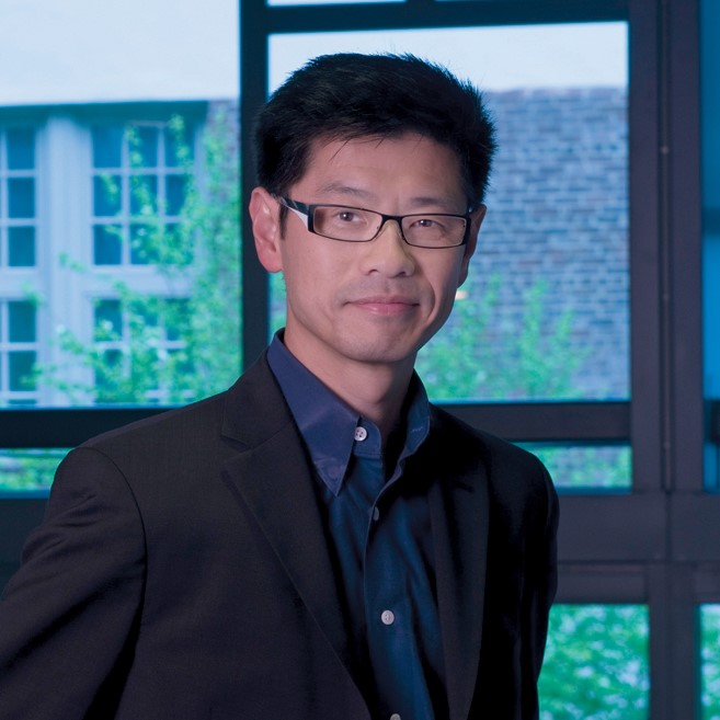
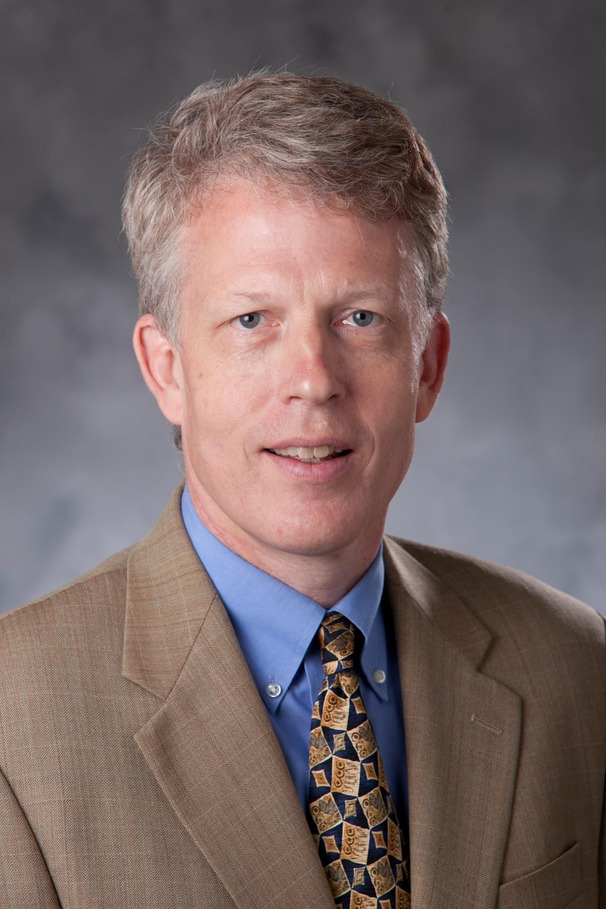
Chi Liu is a Professor of Radiology & Biomedical Imaging at Yale School of Medicine . He serves as Associate Director of Biomedical Imaging Technology at the Yale Biomedical Imaging Institute and Director for Research Faculty Affairs in the Radiology & Biomedical Imaging department. Education : PhD from Johns Hopkins University (2008) Postdoctoral Training : University of Washington (2010) Certification : American Board of Science in Nuclear Medicine (Nuclear Medicine Physics and Instrumentation) His research focuses on quantitative cardiac and oncological PET/CT and SPECT/CT imaging , emphasizing deep learning algorithms , reconstruction algorithms , data correction , and dynamic imaging . Key clinical applications include early detection of chemotherapy-induced cardiotoxicity , multimodality imaging of heart failure , and motion variability elimination in therapy response assessment . The 15 most recent publications reveal a strong emphasis on deep learning techniques for low-dose imaging , motion correction , and cross-tracer generalizability in PET/SPECT systems. These works span applications in cardiac imaging , neuroscience , oncology , and theranostics . Scientific Award : Bruce Hasegawa Young Investigator Medical Imaging Science Award (2012) Contact: chi.liu@yale.edu | ORCID 0000-0002-7007-1037
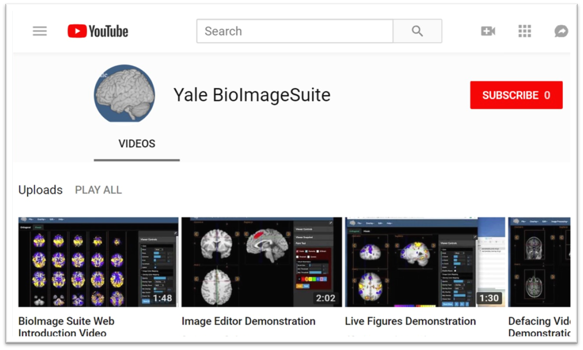
Xenophon Papademetris is a Professor of Biomedical Informatics & Data Science and Radiology & Biomedical Imaging at Yale School of Medicine. He serves as Associate Director of Biomedical Imaging Data Sciences at Yale Biomedical Imaging Institute and directs the Medical Software and Medical Artificial Intelligence Certificate Program. PhD in Electrical and Information Sciences from Yale University (2000) BA from Cambridge University (1994) Postdoctoral Fellowship at Yale University (2002) His research focuses on medical image analysis, machine learning, and biomedical software development. He has developed tools like BioImage Suite Web and contributed to standards committees at the Association for the Advancement of Medical Instrumentation (AAMI). His work spans modalities including MRI, CT, PET, and optical imaging. Recent publications emphasize neuroimaging analysis, explainable AI in healthcare, and multimodal data integration across species. He leads NIH-funded research under the BRAIN Initiative (R24 MH114805) and has authored a textbook on Medical Software published by Cambridge University Press. IEEE Senior Member Yale Brown-Coxe Postdoctoral Fellowship Harding Bliss Prize for Excellence in Engineering He directs the BioImage Suite Project, creating web-based image analysis tools using JavaScript and WebAssembly. His teaching includes both academic courses and a Coursera program on Medical Software with over 14,000 enrollments.
Steven Meikle is a Professor of Medical Imaging Physics and Head of the Imaging Physics Laboratory at the Brain and Mind Centre, University of Sydney. He also serves as Deputy Director (Preclinical) of Sydney Imaging and Deputy Director of the National Imaging Facility's Sydney node. His expertise spans advanced imaging technologies, with a focus on PET/SPECT instrumentation and molecular imaging. He holds a B.App.Sc.(Hons) from the University of Technology Sydney and a PhD from the University of New South Wales. Research focuses include developing novel PET systems like Open-field PET (for freely moving rodents) and Total Body PET, which enhance imaging sensitivity and enable real-time behavioral studies alongside brain function analysis. Collaborations include Tsinghua University (China) and UC Davis (USA). He leads projects on motion correction, quantitative imaging, and AI-driven analysis. Key achievements include over 180 peer-reviewed publications, editorial roles in Physics in Medicine and Biology , and leadership in professional societies. Awards include IEEE Senior Membership and Australian Institute of Physics Fellowship. Current student projects explore Total Body PET applications, motion correction, and radiopharmaceutical evaluation. Teaching roles include medical physics courses in diagnostic radiography and medical physics programs. He advises on imaging ethics, facility implementation, and translational research bridging basic science and clinical applications.
Professor Jerome Liang is a distinguished faculty member at Stony Brook University's Renaissance School of Medicine, holding professorships in Radiology, Biomedical Engineering, Electrical and Computer Engineering, and Computer Science. He serves as Co-Director of Radiology Research and has established himself as a leading expert in medical imaging reconstruction techniques. Dr. Liang's educational background includes a Ph.D. in Physics from City University of New York, postdoctoral training at Duke University, and fellowship at Albert Einstein College of Medicine. His undergraduate degree in Modern Physics was obtained from Lanzhou University in China. His primary research interests focus on advanced medical imaging techniques, particularly low-dose computed tomography image reconstruction, quantitative SPECT reconstruction, high-resolution PET imaging, tissue segmentation from multi-spectral images, computer-aided diagnosis systems, and virtual colonoscopy development. His work bridges engineering principles with clinical applications to improve diagnostic imaging capabilities while reducing radiation exposure. Analysis of his recent publications reveals a strong focus on machine learning applications in medical imaging, particularly in polyp classification, dual-energy CT spectral analysis, and virtual endoscopy. His research consistently aims to enhance diagnostic accuracy while optimizing radiation dose and improving visualization techniques for various medical conditions. 1981 China-US Physics Examination and Application Program (CUSPEA) Winner (Top 25 among 250,000 candidates) 1990 NIH First Investigator Award 1996 American Heart Association Established Investigator Award 1996 Radiological Society of North America Certificate of Merit Award 2002 SUNY Chancellor's Entrepreneur Award 2007 IEEE Society Fellow 2011-2013 SBU, BNL and CSHL Certificates of Excellence in Research and Invention 2013 Stony Brook School of Medicine Award for Excellence in Translational Research Dr. Liang has secured significant research funding including NIH/NCI R01 grants for "Advanced Virtual Colonoscopy for Early Cancer Screening" and "Radiogenomics of Colorectal Polyps." He currently leads active protocols including IRB 93995-MODCR005 focused on integrating virtual and optical colonoscopies with pathological analysis. His laboratory (IRIS - Imaging Research and Informatics) continues to advance medical imaging technology while mentoring the next generation of researchers in this critical field.
James C. Gee is a Professor of Radiologic Science in Radiology at the University of Pennsylvania's Perelman School of Medicine. He serves as Director of the Penn Image Computing and Science Laboratory and Co-Director of the Translational Biomedical Imaging Center , with affiliations in Bioengineering and Applied Mathematics graduate groups. His research focuses on biomedical image analysis, specialization in segmentation, registration, and morphometry applied to neurodegenerative diseases and multi-organ systems. Education : B.S. in Computer Science/Electrical Engineering (University of Washington, 1987), Ph.D. in Computer and Information Science (University of Pennsylvania, 1996) Research : Quantitative medical imaging methods, brain connectomics, neurodegeneration mapping, and translational imaging technologies Publications : 15+ recent works on AI-driven image analysis for Alzheimer's disease, cardiac amyloidosis, and radiomics applications Leadership : Directs MSE-DS Online Degree Program, co-chairs Radiology DCOAP Committee, and founded RISE (Radiology Initiative to Support Inclusive Excellence) His laboratory develops advanced computational tools like ITK-SNAP for biomedical imaging, with applications in both in vivo clinical imaging and ex vivo histology . The work spans cross-disciplinary collaborations in computer science, neuroscience, and clinical medicine.
Associate Professor Andre Kyme is an academic staff member in the School of Biomedical Engineering at The University of Sydney. His research focuses on developing enabling technologies for biomedical imaging, including motion compensation in MRI/PET, robotic platforms for image-guided therapy, and cross-disciplinary applications like plant salt uptake analysis using PET. He collaborates with institutions globally and advises students on projects like lameness detection in horses and AI-based motion correction. Research Interests: Kyme's work spans motion correction in medical imaging modalities, medical robotics integration with imaging systems, and innovative applications of imaging technologies in non-traditional fields. His team emphasizes leveraging advancements in computer vision, machine learning, and instrumentation to improve imaging performance and accessibility. Recent Projects: Current research includes MRI-compatible robotic platforms for therapy applications, AI-driven lameness detection in horses, and pediatric neuroimaging improvements. He leads the BREEZE initiative to enhance MRI accessibility for children with cerebral palsy through eye-gaze communication technology. Publications: His work spans 20+ years with over 50 peer-reviewed publications in journals like Physics in Medicine and Biology and IEEE Transactions. Key areas include PET/SPECT/CT motion correction algorithms, robotic systems for medical imaging, and novel imaging applications in plant science. Teaching: Kyme instructs core biomedical engineering courses including thesis supervision and capstone projects at both undergraduate and postgraduate levels. Labs/Teams: Active in the Brain and Mind Centre and Biomedical Imaging, Visualisation and Information Technologies groups at Sydney. Collaborates with industry partners like TeleMedVet and academic institutions including University of California Davis and Chinese University of Hong Kong.
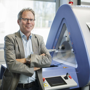
Prof. Freek J. Beekman is a Full Professor and head of the Biomedical Imaging section within the Department of Radiation Science & Technology at Delft University of Technology (TU Delft), Faculty of Applied Sciences. He is a leading figure in biomedical imaging, with extensive contributions to nuclear imaging technologies, including SPECT, PET, and CT. His research spans detector development, image reconstruction algorithms, hybrid photonic imaging, and the application of artificial intelligence in medical imaging. Research Interests: His work focuses on advancing imaging modalities through innovations in hardware (e.g., multi-pinhole collimators) and software (e.g., deep learning for attenuation correction). He has pioneered ultra-high-resolution imaging systems, particularly for preclinical and clinical SPECT, and has developed integrated platforms like U-SPECT-BioFluo. His recent research explores glymphatic delivery of nanoparticles, infection imaging, and AI-driven reconstruction techniques, reflecting a strong translational focus. Publication Trends: His most recent publications (2021–2023) emphasize deep learning in SPECT, multi-isotope imaging, high-resolution ex vivo systems, and applications in neuroimaging and oncology. The articles demonstrate a consistent focus on improving image quality, resolution, and clinical utility through physics-informed and AI-enhanced methods. Scientific Awards: NWO Physics Valorization Prize Innovation of the Year Award by the World Molecular Imaging Society (2015, 2018) Edward Hoffman Memorial Award (2017) Bruce Hasegawa Memorial Award (2021) FOM Valorization Award (2013) TU Delft Entrepreneurial Award (2010) Advising and Grants: While specific student names are not listed, his leadership in large collaborative projects and supervision of numerous publications suggests active mentoring. He has secured significant funding through national and international grants, evidenced by his invention of over 20 patent families and successful technology transfer. His founding and leadership of MILabs BV (sold to Rigaku) highlights his impact on commercialization and industry-academia collaboration. Labs and Teams: He leads the Biomedical Imaging research group at TU Delft, which develops cutting-edge imaging systems such as VECTor (SPECT-PET) and EXIRAD-HE. His teams have produced technologies used globally in academic and pharmaceutical research, contributing to tracer development and therapeutic innovation.
Dale Bailey is a Professor in the Faculty of Medicine & Health at the University of Sydney, with clinical appointments at Royal North Shore Hospital's Department of Nuclear Medicine. He previously directed the Sydney Vital Northern Translational Cancer Research Centre and is a member of the Centre for Drug Discovery Innovation, the University of Sydney Nano Institute, and the Charles Perkins Centre. Nuclear Medicine Physicist Theranostics Researcher Clinical Imaging Expert His research focuses on quantitative functional imaging using SPECT and PET, hybrid imaging modalities, radiation dosimetry for personalized cancer therapy, and the biological effects of radiation. He pioneered combined structure/function imaging and advanced radionuclide therapy applications. Recent publications emphasize PET/CT with novel radionuclides, metabolic tumor volume as a prognostic biomarker, and dosimetry validation methods . Key themes include cancer, nuclear medicine, and translational research. PhD in Physics Fellow of the Institute of Physics and Engineering in Medicine (FIPEM) Chartered Scientist (CSci) in the UK International Consensus Guidelines Contributor He supervises research students in radiotheranostics and collaborates on clinical trials for neuroendocrine tumors and glioblastoma. Grants and projects involve hybrid imaging optimization, radiation safety, and personalized treatment planning.
Anas Alani, MD is an Assistant Professor in both Medicine and Radiology at the School of Medicine, University of California, Los Angeles. His academic career spans cardiovascular medicine with a strong emphasis on advanced imaging techniques and coronary pathophysiology. Dr. Alani's research interests focus on: Cardiovascular imaging using computed tomography Coronary artery disease and atherosclerosis Diabetes-related cardiovascular complications Valvular heart disease and interventions Cardiovascular risk assessment in special populations Extra-coronary calcification patterns His scholarly work demonstrates expertise in applying advanced imaging modalities to understand cardiovascular pathophysiology and improve diagnostic accuracy. Dr. Alani has contributed significantly to the field of coronary plaque characterization, positive remodeling in smokers, and the application of CT angiography in various clinical scenarios including diabetes, HIV, and metabolic syndrome. Dr. Alani's publication record shows consistent productivity since 2013, with 24 publications through 2024. His work frequently appears in high-impact cardiology and radiology journals including the Journal of the American College of Cardiology, American Journal of Cardiology, and JACC: Cardiovascular Interventions. His research demonstrates methodological rigor with extensive use of CT angiography, intravascular ultrasound, and other advanced imaging techniques to quantify coronary plaque characteristics. Dr. Alani is currently serving as Principal Investigator for a clinical trial titled "Effect of Tirzepatide on progression of coronary atherosclerosis using MDCT" sponsored by Eli Lilly & Co. (2025-2026), indicating ongoing active research in diabetes and cardiovascular disease. His work frequently collaborates with Dr. Matthew Budoff and other cardiovascular imaging experts, contributing to multicenter studies including the DCCT/EDIC Research Group and Multicenter AIDS Cohort Study.
Walter Noordzij is an Assistant Professor at the University of Groningen within the Faculty of Medical Sciences , affiliated with the Basic and Translational Research and Imaging Methodology Development in Groningen (BRIDGE) group. His research focuses on nuclear medicine imaging technologies , particularly PET/CT applications in oncology, cardiology, and healthcare economics. He has contributed to studies on cardiac sympathetic innervation in amyloidosis, financial literacy of medical residents regarding imaging costs, and prostate cancer management using PSMA PET/CT. Recent work also addresses physician workload trends in Dutch nuclear medicine and innovative parametric imaging techniques. Research interests include positron emission tomography-computed tomography (PET/CT) , fluorodeoxyglucose (FDG) imaging , and multidisciplinary approaches to prostate cancer diagnosis . He actively collaborates internationally, evidenced by his participation in the 19th International Conference on Radiation Therapy and the EANM Focus Meeting on Prostate Cancer . No awards are explicitly listed, but his involvement in high-impact studies suggests active recognition in the field. He has supervised 3 students but their names are not disclosed. His work spans clinical imaging methodology, translational research, and healthcare system analysis, reflecting a commitment to bridging basic science and clinical practice.
Weihua Zhou is a Tenured Associate Professor at Michigan Technological University in the College of Computing, with affiliations in Applied Computing, Biomedical Engineering, Computer Science, Electrical and Computer Engineering, and Mathematical Sciences. He holds a PhD from Southern Illinois University Carbondale, an MS and B.Eng. from Wuhan University, and completed postdoctoral research at Emory University. Academic Positions : Tenured Associate Professor (2025–present), Tenure-Track Assistant Professor (2019–2025) at Michigan Tech; Nina Bell Suggs Endowed Professorship (2015–2019) at the University of Southern Mississippi. Research Interests : Focuses on medical imaging and health informatics , particularly machine learning applications in cardiovascular diagnosis , osteoporosis risk stratification , and senile dementia early detection . Additional work includes radiomics for COVID-19 severity assessment , federated learning in medical segmentation , and deep learning for proximal femur strength prediction . Scientific Awards : USM Nina Bell Suggs Endowed Professorship, USM College of Arts and Sciences Scholarly Research Award, American Heart Association Research Leaders Academy (2017, 2018), USM Butch Oustalet Distinguished Professorship Research Award. Lab & Tools : Leads the Medical Imaging & Informatics Lab (MIILab-MTU) and contributes to the NSF/MRI GPU Cluster. Open-sourced tools include KD4COVID19 for radiomics analysis and ECGTools for ECG classification.
Daniel Appelbaum, MD, is a Professor of Radiology at the University of Chicago, specializing in nuclear medicine and molecular imaging. He serves as a radiologist and nuclear medicine physician at the University of Chicago Medicine, where he leads a section involved in multiple oncology PET/CT clinical trials. Dr. Appelbaum chairs the University of Chicago Radioactive Drug Research Advisory Committee (RADRAC) and is an active member of the national Society of Nuclear Medicine Committee on Education. Mallinckrodt Institute of Radiology at Washington University, St. Louis, MO (2000) - Fellowship in Nuclear Medicine University of Chicago, Chicago, IL (1999) - Residency in Diagnostic Radiology Mount Sinai School of Medicine, New York, NY (1995) - MD Cornell University, Ithaca, NY (1991) - BA in English Literature American Board of Radiology Certification (1999) American Board of Nuclear Medicine Certification (2000) Dr. Appelbaum's research focuses on nuclear and molecular imaging in both basic science and clinical settings. His current work includes developing a novel brain imaging agent for multiple sclerosis and a metabolic catalyst to enhance tumor detection with PET imaging. He has pioneered new strategies for computer-aided diagnosis (CAD) in nuclear medicine, for which he holds an AI patent. His research spans oncologic, neurologic, and cardiac applications of PET and PET/CT imaging, with special emphasis on metabolic tumor volume analysis, molecular targeted therapies, and quantitative imaging biomarkers. His laboratory investigates applications of artificial intelligence in radiographic image interpretation, particularly for cancer detection and monitoring treatment response. Analysis of Dr. Appelbaum's recent publications (2015-2025) reveals a strong focus on quantitative nuclear medicine imaging, particularly PET/CT applications in oncology. His work consistently explores metabolic tumor volume as a prognostic marker across various cancers including lung cancer, lymphoma, and neuroendocrine tumors. There is a clear trajectory toward integrating artificial intelligence with nuclear medicine imaging, as evidenced by his research on computer-aided diagnosis systems and quantitative image analysis. His publications span multiple disciplines including oncology, neurology, cardiology, and hematology, demonstrating the broad applicability of nuclear imaging techniques across medical specialties. America's Top Doctor (Castle Connolly, 2014-present) Chicago's Top Doctor (Chicago Magazine, 2012-present) Marc Tetalman Teacher of the Year Award (University of Chicago, 2006-2007) As an educator, Dr. Appelbaum has made significant contributions to nuclear medicine education, serving on the national Society of Nuclear Medicine Committee on Education for many years. He authored the textbook Nuclear Medicine RadCases as a comprehensive review resource for the field. He frequently accepts lecture invitations on nuclear and PET imaging at medical institutions and conferences worldwide. His research program has secured multiple clinical trials funding, particularly in oncology PET/CT applications for monitoring therapy response. Dr. Appelbaum's AI-related patent in computer-aided diagnosis represents a significant translational research achievement with potential clinical impact. Dr. Appelbaum leads a research section at the University of Chicago focused on nuclear and molecular imaging. His team collaborates extensively with oncologists, neurologists, and cardiologists on clinical trials and translational research projects. The group is particularly active in developing novel imaging biomarkers and quantitative analysis methods for cancer imaging. They maintain strong collaborations with computer science researchers for AI development in medical imaging. The section participates in multiple multi-center clinical trials evaluating PET/CT for cancer staging, treatment response assessment, and prognostication across various malignancies.
Niek Prakken is affiliated with the Faculty of Medical Sciences at the University of Groningen's University Medical Center Groningen (UMCG), where he is part of the Vascular Medicine department and the Basic and Translational Research and Imaging Methodology Development in Groningen (BRIDGE) group. His research focuses on cardiovascular imaging techniques such as Cardiac Magnetic Resonance Imaging (MRI) and Positron Emission Tomography (PET), with a particular emphasis on applications in athletes' heart conditions, cardiomyopathies, and innovative imaging methodologies. Awarded prizes include the Conacyt-ATTP Validation Award (2022), Heartfoundation Dekker Grant (2016), and the Frederik Philips Prize (2010). Serves as an editor for Frontiers in Cardiovascular Medicine since 2023 and actively contributes to academic organizations like the Society for Cardiovascular Magnetic Resonance (SCMR). Collaborates internationally, with recent work involving institutions across multiple countries. His research interests span sports cardiology, cardiac imaging advancements, and translational imaging methodologies. He has published over 88 peer-reviewed articles and actively participates in academic activities such as editorial work, presentations, and advisory roles.
Dr. Catherine Farrow is a Senior Lecturer at the Sydney Medical School, University of Sydney, specializing in respiratory physiology and advanced pulmonary imaging techniques. Her research investigates ventilation distribution, airway function, and pulmonary pathophysiology in conditions including asthma, COPD, and COVID-19. Research interests include: Ventilation heterogeneity in obstructive lung diseases SPECT/CT imaging for pulmonary function assessment Respiratory sequelae of COVID-19 infection Impact of obesity on ventilation patterns Peripheral airway function in aging and disease Clinical applications of oscillometry and nitrogen washout techniques Her recent publications (2017-2025) demonstrate a strong focus on pulmonary imaging innovations and COVID-19 respiratory pathophysiology. Research themes include ventilation-perfusion abnormalities in post-COVID recovery, AI-assisted radiological assessment, asthma phenotyping using SPECT/CT, and pulmonary function testing standards during pandemics. Longitudinal work shows consistent emphasis on translating imaging biomarkers to clinical applications. Collaborations include multidisciplinary teams across institutions globally, evidenced by multi-center publications on COVID-19 pneumonitis and position statements for respiratory societies.
Timothy Garvey Turkington is an Associate Professor in the Department of Radiology at Duke University School of Medicine and a Faculty Network Member of the Duke Institute for Brain Sciences. Holding a Ph.D. from Duke University (1989), his academic career spans over three decades with continuous contributions to nuclear medicine physics. His research focuses on PET imaging physics, including instrumentation development, reconstruction algorithms, and image processing techniques. Key research thrusts include improving quantitative accuracy in PET, reducing scan times and radiation doses in PET/CT systems, developing novel imaging devices for PET and SPECT applications, and expanding PET's clinical utility in oncology, neurology, and cardiology. His work bridges theoretical physics with practical clinical implementation, particularly in breast imaging and quantitative biomarker development. Analysis of his 15 most recent publications reveals a strong emphasis on instrumentation optimization (particularly for breast imaging), quantification accuracy in clinical settings, and the development of standardized protocols for PET/CT systems. His work increasingly focuses on translational applications where physics innovations directly impact clinical decision-making, especially in cancer imaging and therapy response assessment. Dr. Turkington teaches graduate courses including PHYSICS 523: Modern Medical Diagnostic Imaging System, MEDPHY 782: Advanced Practicum for Clinical Development in Medical Physics, and MEDPHY 530: Modern Medical Diagnostic Imaging System, training the next generation of medical physicists. His research has been supported by significant grants including the RSNA 2016 Project (2014-2018), Simultaneous Emission and Transmission Mammotomography (2002-2014), and the Harmonized PET Reconstructions for Cancer Clinical Trails (2013-2014), demonstrating sustained funding for his innovative work in medical imaging physics.