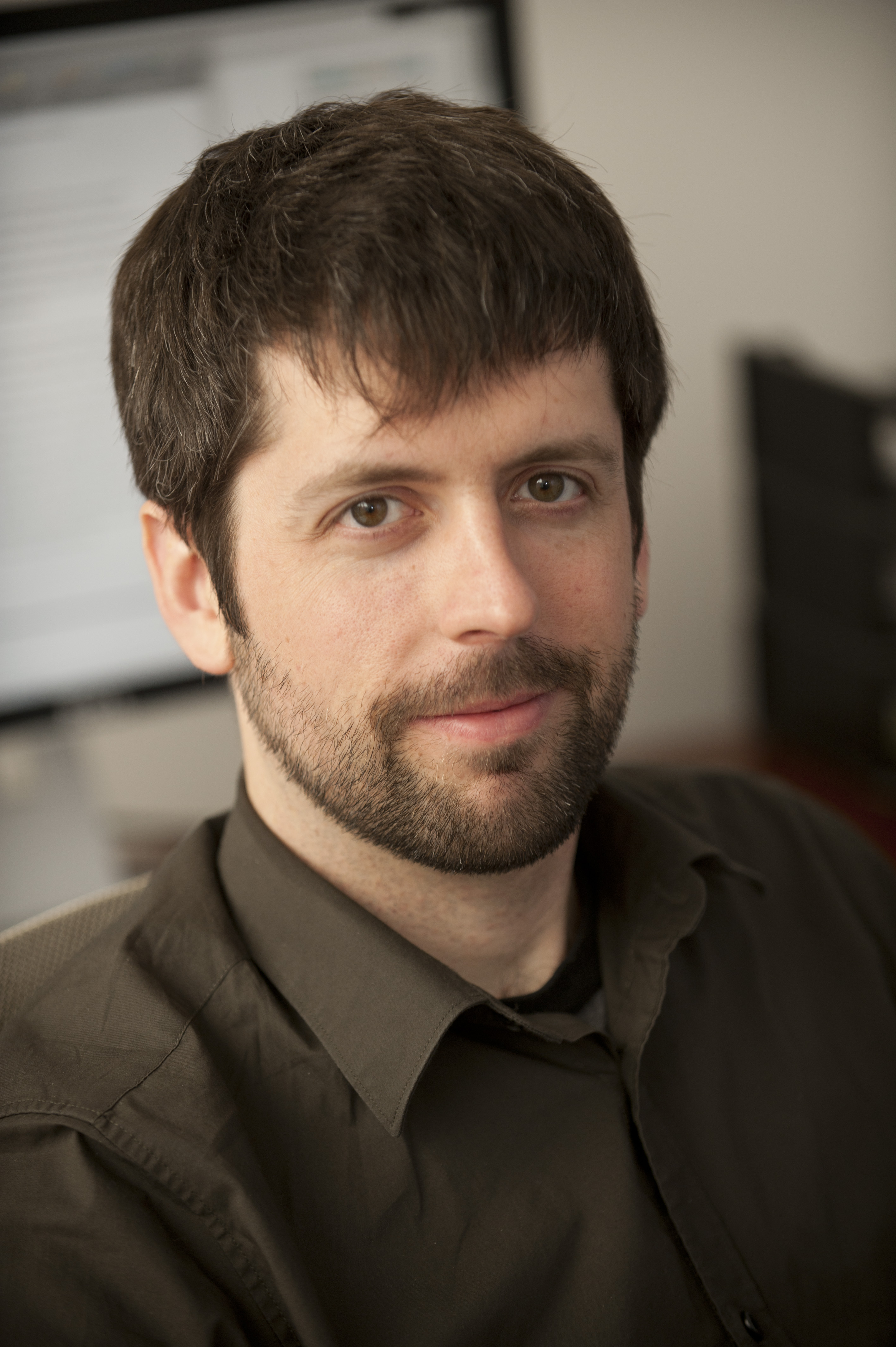
Mark Kramer is a Professor in the Department of Mathematics & Statistics at Boston University. He belongs to the Applied Mathematics research group, focusing on mathematical, statistical, and machine learning approaches to characterize brain activity. His work bridges data-driven neuroscience with computational methods, exploring topics like biophysical models of neurons, field models of neural populations in epilepsy, and theoretical questions about brain rhythms. His research interests include: Biophysical modeling of single-neuron dynamics Neural population activity in pathological states Machine learning for detecting abnormal brain rhythms Analysis of cross-frequency coupling and coherence Kramer has developed educational resources like Case Studies in Neural Data Analysis using both MATLAB and Python. These materials teach practical data analysis techniques for spike trains and field data, emphasizing hands-on implementation over theoretical mathematics. He has received funding from NIH and NSF for computational neuroscience projects. His recent publications focus on epilepsy research, sleep spindle analysis, and neural signal processing. The work spans from developing statistical frameworks to understanding network dynamics in seizure termination and exploring phase consistency in neural data. Notably, his coherence studies revealed non-intuitive coupling patterns between brain regions, demonstrating that low-amplitude rhythms can be more informative than dominant ones.
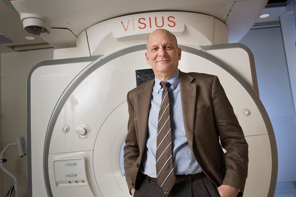
Keith D. Paulsen is the MacLean Professor of Engineering at Dartmouth College’s Thayer School of Engineering and holds the title of Professor of Radiology & Surgery at the Geisel School of Medicine. He serves as Scientific Director of the Center for Surgical Innovation at Dartmouth-Hitchcock Medical Center and Co-Director of the Translational Engineering in Cancer Research Program at the Norris Cotton Cancer Center. His roles emphasize interdisciplinary collaboration between engineering, medicine, and oncology. Paulsen earned a BSc in Biomedical Engineering from Duke University (1981), followed by MS (1984) and PhD (1986) degrees in Engineering Sciences from Dartmouth College. His research focuses on biomedical imaging, cancer therapeutics, and image-guided surgery, with particular expertise in optical and electromagnetic methodologies. He has pioneered technologies such as fluorescence-guided surgery, quantitative scatter imaging, and non-linear image reconstruction techniques, aiming to enhance surgical precision and cancer diagnosis. His awards include fellowships from OSA, SPIE, AIMBE, IEEE, and the National Academy of Inventors. Paulsen’s work has led to startups like CairnSurgical (where he serves as CTO) and InSight Surgical Technologies, translating research into clinical tools. Key projects include intraoperative imaging systems for brain and spine surgery, microwave imaging for breast cancer, and optical molecular imaging for real-time surgical guidance. Paulsen teaches advanced computational methods (ENGS 205, 105) and courses on medical device innovation (ENGM 189.1/2). His lab, part of Dartmouth’s Optics in Medicine cluster, collaborates with radiology, surgery, and oncology departments to develop clinical technologies funded by NIH, NCI, and DoD grants.
Cristian Tomasetti serves as Professor and Director of the Early Detection and Prevention Division at City of Hope's Beckman Research Institute, where he leads the Division of Mathematics for Cancer Evolution and Early Detection within the Department of Computational and Quantitative Medicine. His work bridges mathematical modeling with cancer biology to develop novel approaches for cancer detection and prevention. Dr. Tomasetti's research focuses on the mathematical foundations of cancer etiology, evolution, and early detection. His laboratory has pioneered algorithms for early cancer detection using cell-free DNA sequencing and protein data, developing some of the first multi-analyte blood tests capable of identifying cancer at its earliest stages. His work on cancer evolution has produced the most comprehensive mathematical models of tumorigenesis currently available, providing critical insights into mutation rates, driver gene requirements, and fitness advantages in cancer development. His influential research on the relationship between stem cell divisions and cancer risk has reshaped understanding of cancer etiology. The analysis of Dr. Tomasetti's recent publications reveals a consistent focus on liquid biopsy technologies, particularly circulating tumor DNA analysis for early cancer detection and monitoring. His work spans multiple cancer types including colorectal, pancreatic, ovarian, and bladder cancers, with a strong emphasis on translating mathematical models into clinically applicable diagnostic tools. The research demonstrates an evolution from theoretical models of cancer development to practical applications in cancer screening, risk assessment, and treatment monitoring. Dr. Tomasetti has established himself as a leader in the field of mathematical oncology, with his laboratory at the forefront of developing computational approaches to cancer prevention and early detection. His work on cell-free DNA testing has significant potential to transform cancer screening paradigms, moving toward less invasive and more effective methods for detecting cancer before symptoms appear.
Dr. Uwe Grünefeld is a Visiting Professor at the Faculty of Computer Science , Institute for Computer Science and Business Information Systems (ICB) of the University of Duisburg-Essen. He has been actively contributing to Human-Computer Interaction research through multiple publications in 2025-2022 focusing on Virtual Reality , Augmented Reality , and Robotics . Research Interests span across immersive technology applications for health behavior change (situated artifacts, weight visualization mirrors), haptic feedback systems (EMS for weight perception, vibrotactile directional cues), and behavioral biometrics (hand tracking identification, gaze-based user recognition). His work addresses cross-reality system design , collaborative robotics , and human-in-the-loop simulation methodologies . Key Publications demonstrate significant contributions to VR/AR user engagement, with particular focus on Physical activity promotion through situated artifacts Advanced haptic feedback techniques for immersive environments Behavioral biometric identification systems Robot motion intent communication Cross-reality transition visualization His research often employs mixed-method approaches combining technical implementations with user studies involving quantitative and qualitative data collection.
Professor Kylie Tucker is a distinguished academic at the University of Queensland, serving as Professor and School Director of Teaching and Learning in the School of Biomedical Sciences within the Faculty of Health, Medicine and Behavioural Sciences. She is also an Affiliate of the Centre for Innovation in Pain and Health Research (CIPHeR) and currently serves as President of the International Society of Electrophysiology and Kinesiology (ISEK) for the term 2024-2026. Professor Tucker leads a dynamic research environment focused on advancing knowledge about muscles and movement control, with significant contributions to understanding how pain impacts movement, methods for estimating muscle forces, and assessment of childhood movement control and adolescent skeletal maturity. Professor Tucker earned her Bachelor of Arts, Bachelor of Science, and Doctor of Philosophy from the University of Adelaide. Her academic journey has positioned her as a leader in neuromuscular research, particularly in the areas of motor control and pain adaptation. Within the School of Biomedical Sciences, she has held significant leadership roles including Deputy Director of Teaching and Learning (2018-2020), inaugural chair of the REMEDE committee (2021-2023), and Director of Teaching and Learning (2024-2025). She also co-facilitates UQ's flagship Career Progression for Women program. Her research interests span motor control, pain research, biomechanics, electromyography, neuromuscular control, pediatric movement, scoliosis, and muscle physiology. Professor Tucker's work has transformed understanding of pain's impact on movement and advanced assessment methods for childhood movement control and skeletal maturity. She has recently proposed new insights into scoliosis progression, identifying unique muscle features that can be non-invasively detected early in curve progression. Approximately 3-7% of children worldwide develop adolescent idiopathic scoliosis, often requiring surgical intervention when conservative treatments fail. Analysis of Professor Tucker's recent publications reveals a strong focus on neuromuscular control mechanisms, particularly in relation to pain, scoliosis, and pediatric movement disorders. Her work integrates advanced methodologies including electromyography, shear wave elastography, and biomechanical modeling to investigate muscle function across diverse populations. A notable trend is her leadership in consensus projects (CEDE) establishing standardized methodologies for electromyography research, reflecting her commitment to methodological rigor in the field. Professor Tucker actively mentors the next generation of researchers, supervising numerous PhD students across projects related to scoliosis, knee osteoarthritis, pain research, and pediatric movement disorders. Her research is supported by significant funding including NHMRC MRFF EPCDR grants for chronic musculoskeletal conditions in children and the SRS Research Grant for novel insights into adolescent idiopathic scoliosis. She leads the Motor Control and Pain Research Lab, a collaborative environment bringing together basic science and clinical researchers. The lab focuses on two main research streams: Motor Control and Pain Research and Child and Adolescent Neuromotor Control Research. Professor Tucker teaches across 10 UQ programs with class sizes ranging from 70-1400 students, demonstrating her commitment to education alongside her research leadership.
Dr. Caroline Paquette is an Associate Professor in the Department of Kinesiology & Physical Education at McGill University's Faculty of Education, where she serves as Associate Dean, Administration. She directs the Human Brain Control of Locomotion (HBCL) Laboratory, focusing on the neural mechanisms of balance and locomotion using biomechanics, neuroimaging, and non-invasive brain stimulation. Her research aims to improve mobility in older adults and neurological patients, particularly those with Parkinson's disease and post-stroke impairments. PhD, Rehabilitation Science, McGill University MSc, Kinesiology, Laval University BSc, Kinesiology, Laval University Postdoctoral Fellowships: Neurology (Lady Davis Institute) and Neuroscience (Oregon Health and Science University) Her research spans motor control, neuroimaging, and non-invasive brain stimulation, with applications in Parkinson's disease, stroke rehabilitation, aging, and locomotor adaptation. She has published in high-impact journals like Neuroimage, Parkinsonism & Related Disorders, and Neurorehabilitation and Neural Repair. Dr. Paquette's Google Scholar publications reveal expertise in: Freezing of gait in Parkinson's disease Neuroplasticity in stroke and aging Functional connectivity analysis Exercise interventions for neurodegenerative disorders Robotic compensation in PET imaging Non-invasive brain stimulation applications She supervises graduate students at the HBCL Laboratory, located at the Education Building and Currie Gymnasium, Montreal, Canada.
Susann M. Brady-Kalnay, PhD , is the Sally S. Morley Designated Professor of Brain Tumor Research at the Case Western Reserve University (CWRU) School of Medicine. She holds professorships in the Departments of Molecular Biology and Microbiology, Neurosciences, and Pathology, and is a member of the Cancer Imaging Program at the Case Comprehensive Cancer Center. Her work bridges molecular biology, cancer imaging, and targeted therapy. Research Interests : Dr. Brady-Kalnay specializes in receptor protein tyrosine phosphatases (PTPs) in cancer progression cell adhesion dynamics in brain tumors signal transduction mechanisms molecular imaging agents for tumor detection novel therapeutics targeting proteolyzed PTPmu fragments Her lab's landmark discovery of PTPmu proteolysis in glioblastoma multiforme (GBM) has led to platform technologies for "see(k) and destroy" cancer strategies. Imaging Innovations : The lab develops fluorescent peptides (e.g., SBK2) for real-time surgical imaging SBK2-Gd MRI agents with superior tumor contrast radioisotope-conjugated PET agents for targeted radiotherapy nanoparticle diagnostics for invasive gliomas ultrasound nanobubble contrast agents These tools address critical gaps in imaging tumor boundaries and microenvironment interactions. Scientific Awards : Elected Senior Member, National Academy of Inventors (2022) Equalize National Woman Academic Inventor Finalist (2021) Mather Spotlight Prize for Women of Achievement, CWRU (2019) Distinguished Faculty Researcher, CWRU (2018) Special Achievement Award, University of Dayton National Alumni Association (2011)
Dr. Patrick Reinard is an Assistant Professor in the Department III - Papyrology at the University of Trier. His research focuses on Papyrology, Epigraphy, and socio-economic history of Greco-Roman Egypt, with specific expertise in Jewish communities, economic strategies, and material culture analysis. Co-editor of Oeconomica and Muziris series Principal investigator in Roman economic behavior and documentary evidence Specializes in papyrus letters as economic data sources His recent publications analyze market dynamics, trust mechanisms, and sustainability in Roman trade. Collaborative work includes hyperspectral papyri imaging projects and digital pandemic-era pedagogy initiatives. He contributes extensively to academic conferences and editorial boards, with particular interest in Greco-Roman epistemology and its contemporary reception.
Joakim Lindblad is a Professor at the Department of Information Technology, Uppsala University , and holds affiliated roles as Senior Research Associate at the Mathematical Institute of the Serbian Academy of Sciences and Arts, and Head of Research at Topgolf Sweden AB. With over two decades of expertise in image analysis and machine learning , his work bridges computational methods with biomedical applications. Key affiliations: Uppsala University, Serbian Academy of Sciences, Topgolf Sweden Specializations: Deep Learning, Multimodal Image Registration, Quantitative Microscopy His research focuses on reliable image processing frameworks that integrate intensity and spatial information , particularly for biomedical applications . Recent publications highlight innovations in autofluorescence-based cancer detection , self-supervised one-class learning for sparse instance identification, and rotation-equivariant CNNs for robust analysis of cytology images. Recent article trends demonstrate expertise in multimodal image analysis (2024: 3 papers), oral cancer detection (2025: 2 papers), and multiscale biomedical imaging . His 2025 work on the Uppsala Storytelling Dataset introduces novel frameworks for multimodal dataset creation in AI research. While no scientific awards are explicitly mentioned, his extensive publication record (2000-2025) across top venues like Pattern Recognition , PLOS ONE , and IEEE Transactions indicates significant academic impact. His methodological contributions span stochastic distance transforms , fuzzy set defuzzification , and multimodal image registration techniques. Collaborative work with researchers like Nataša Sladoje and interdisciplinary teams has produced innovations in automated cytology analysis , TEM image enhancement , and AI-driven medical diagnostics . His 2021-2022 projects introduced contrastive learning approaches for multimodal image registration and explainable AI frameworks for infant engagement analysis.
Dr. Yian Yin is an Assistant Professor of Information Science at Cornell University within the Cornell Bowers College of Computing and Information Science. His research focuses on computational social science, data science, and network science to study scientific progress, innovation, and complex social processes. He has affiliations with Northwestern University’s Northwestern Institute on Complex Systems (NICO) and Kellogg Center for Science of Science and Innovation (CSSI). Education: Ph.D. in Industrial Engineering and Management Sciences from Northwestern University Bachelor's degrees in Statistics and Economics from Peking University Research Interests: Computational tools for understanding scientific collaboration and innovation Analysis of policy impacts on scientific output during crises Quantitative frameworks for studying failure in scientific endeavors Cross-disciplinary applications in art, economics, and public health Awards: Forbes 30 Under 30 in Science (2023) Emerging Researcher Award from Complex Systems Society (2023) Advancement Activities: Organized Summer Institute in Computational Social Science (SICSS) Chicago Program Co-Chair for International Conference on Science of Science and Innovation Labs/Teams: Leads computational social science research groups at Cornell, focusing on science-of-science methodologies and innovation ecosystems.
Delphine Périé-Curnier is a Full Professor in the Department of Mechanical Engineering at Polytechnique Montréal and Director of Graduate Studies. Her research focuses on developing quantitative MRI techniques for non-invasive characterization of living tissue mechanical properties, particularly in cardiotoxicity detection and musculoskeletal mechanobiology . She leads the Bioperformance Analysis and Innovation Laboratory (LIAB) and contributes to the Institute of Biomedical Engineering. Education: Ph.D. from Paul Sabatier University, Toulouse, France Her work bridges medical imaging , biomechanical modeling , and finite element analysis to predict disease progression through pathomechanism understanding. Key projects include exercise-induced cardiac changes in childhood cancer survivors and spinal biomechanics in scoliosis. Recent publications (2023-2024) emphasize cardiovascular MRI for childhood cancer survivorship and hemodynamic modeling in left ventricle analysis. She supervises 26 graduate students, with completed theses spanning topics like doxorubicin cardiotoxicity , knee replacement stability , and spatial cardiac MRI protocols . Teaching includes graduate courses in biomedical design , advanced biomechanics , and modeling techniques .
Dr. William M Holmes is a Senior Research Fellow and Senior MRI Physicist at the University of Glasgow's School of Psychology & Neuroscience, affiliated with the Glasgow Experimental MRI Centre. He holds a PhD in Physical Chemistry from the University of Nottingham and specializes in advancing MRI techniques for biomedical and physical sciences. His work bridges physics, neuroscience, and clinical applications, with a focus on cerebral blood flow imaging, glymphatic system dynamics, and disease modeling in rodents. Key roles: MRI method development, neuroimaging biomarkers, porous media analysis Leadership: Glasgow Experimental MRI Centre Research interests emphasize novel MRI applications in stroke, neurological disorders, and material science. Over 80 peer-reviewed publications demonstrate contributions to perfusion imaging, biofilm dynamics, and translational MRI techniques. Recent projects include: Quantitative arterial spin labeling methods Glymphatic system's role in multiple sclerosis Non-invasive rodent disease modeling
Silvia S. Martins, MD, PhD, is Professor of Epidemiology at Columbia University Mailman School of Public Health, where she directs the Substance Use Epidemiology Unit and co-directs the NIDA T32 Substance Abuse Epidemiology Training Program. She serves as Vice Dean for Faculty and holds affiliations with Columbia's Global Health Initiative, Injury Science Center, and Population Research Center. Her education includes an MD from UFPR-Brazil (1998) and a PhD from the University of São Paulo (2003). Dr. Martins' research examines substance use epidemiology through interdisciplinary lenses, focusing on: Policy impacts of cannabis/opioid regulations on overdose rates and usage patterns Machine learning applications for analyzing drug policy effectiveness Child/adolescent psychiatric epidemiology and global substance use disparities Urban health interventions and community-based substance abuse prevention Her work spans national contexts including the U.S., Brazil, and Uruguay. Her publications demonstrate strong thematic coherence, with recent work emphasizing: Quantitative analysis of cannabis/opioid policy impacts using longitudinal data Innovative methods (e.g., machine learning, geospatial modeling) in epidemiological research Health equity dimensions of substance policies across racial/ethnic groups Maternal health and polysubstance use during public health crises Awards & Honors: Tow Leadership Scholar (2021-2023) Columbia University Irving Medical Center Mentor of the Year (2021) Calderone Health Equity Award (2021) National Hispanic Science Network Mentoring Award (2021) Dean's Award for Excellence in Mentoring (2017) Joseph Ciarrocchi Award for Youth Gambling Research (2011) As principal investigator on multiple NIH grants since 2006, she leads projects examining: Synergistic effects of opioid/cannabis policies in the U.S. Drug overdose trends in Latin America (Brazil, Colombia, Mexico) Unemployment insurance effects on drug outcomes during COVID-19 She mentors early-career researchers through the NIDA T32 program, with >90 mentee-led publications. She directs the Substance Use Epidemiology Unit and co-leads the PHIOS interdisciplinary group. Additional affiliations include Columbia’s Center for Injury Science and Prevention, Institute of Latin American Studies, and Global Mental Health Program.
Jie Deng, Ph.D., is a Professor in the Department of Radiation Oncology at UT Southwestern Medical Center, where she serves as faculty in the Division of Medical Physics & Engineering. She is a certified MRI and MRI for radiation therapy medical physicist by the American Board of Medical Physics and holds a leadership role as a magnetic resonance safety officer. Dr. Deng is actively involved in both clinical and research aspects of medical imaging and radiotherapy, with a strong emphasis on integrating advanced imaging technologies into therapeutic workflows. Dr. Deng earned her academic degrees from prestigious institutions: a Bachelor of Science in Biomedical Engineering from Southeast University in China, a Master’s in Bioengineering from the University of Illinois at Chicago, and a Ph.D. in Biomedical Engineering from Northwestern University. She further enhanced her expertise by obtaining a Master of Science in Law from the Northwestern Pritzker School of Law, reflecting a multidisciplinary approach to her scientific work. Her research interests center on MRI physics , quantitative imaging , oncological imaging , and the application of artificial intelligence in medical imaging. She has pioneered work in MRI-guided radiation therapy, imaging biomarkers for therapeutic response, and AI-driven image reconstruction and artifact reduction. Her recent publications demonstrate a consistent focus on improving imaging accuracy, speed, and clinical utility, particularly in liver, pediatric, and oncological applications. The analysis of her 15 most recent articles reveals a strong trend toward deep learning-based image reconstruction , quantitative MRI biomarkers , and synthetic image generation for radiotherapy planning. Topics such as 4D-MRI, synthetic CT, motion artifact reduction, and AI fusion models dominate her scholarly output, indicating a forward-looking research trajectory centered on intelligent, fast, and precise imaging for personalized cancer therapy. Dr. Deng actively contributes to the scientific community through presentations at major conferences including the International Society for Magnetic Resonance in Medicine (ISMRM) and the American Association of Physics in Medicine (AAPM), where she shares innovations in MRI, adaptive radiotherapy, and AI integration. As an educator, Dr. Deng mentors medical physics residents and graduate students, delivering lectures on MR-only simulation, MR-linear accelerator practices, and medical imaging fundamentals. While no specific grants are mentioned in the text, her extensive publication record in high-impact journals suggests active research funding and collaborative projects. She is affiliated with key professional organizations and serves on UT Southwestern’s MRI Safety Committee, ensuring safe and effective use of MRI in clinical and research settings. Her work bridges the gap between engineering innovation and clinical application, making significant contributions to the field of radiation oncology and medical physics.
Professor Mikko Nissi holds the Chair of Medical Physics and Engineering at the University of Eastern Finland’s Department of Technical Physics, Faculty of Science, Forestry and Technology. He leads research on quantitative magnetic resonance imaging (qMRI), focusing on musculoskeletal diseases and methodological advancements in ultra-short echo time techniques. His work integrates inverse problems research and machine learning to predict tissue properties unobservable via conventional MRI. Education: PhD in Physics (University of Kuopio, 2008), Adjunct Professor since 2015. Key roles include Associate Professor (2020–2024) and Academy Research Fellow (2015–2020). Active in research groups like the Academy of Finland Flagship (FAiME) and UEF’s Musculoskeletal Diseases (MSKD) community. Research interests span qMRI relaxometry, T1ρ/T2 anisotropy, and AI-driven virtual histology. Notable projects include EU-funded microMRI infrastructure for industrial applications and grants from the Research Council of Finland for AI-assisted MRI methods. Teaching: Course on MRI principles and applications. Advises on musculoskeletal biomechanics and finite element modeling. Recognized for contributions to medical physics, including societal roles in the Finnish Society for Medical Physics and International Society for Magnetic Resonance in Medicine. Hobbies: 3D printing for research tooling and handicrafts. Publications (>100 articles) address cartilage degeneration, myocardial perfusion, and MRI applications in agriculture/forestry.