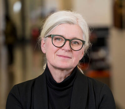
Dr. Charlotte Brassey is a Senior Lecturer in Zoology at Manchester Metropolitan University, specializing in functional morphology, comparative anatomy, and 3D imaging techniques. Her research explores mammalian genitalia evolution and animal kinematics using advanced imaging technologies. She serves as Associate Editor for Proceedings of the Royal Society B and is a grant reviewer for major UK research councils. Her research spans: Evolution of mammalian baculum and clitoris 3D shape quantification methods AI-based animal tracking Biomechanical modeling Her publications focus on comparative anatomy across species, employing CT scanning and morphometric analysis to understand functional adaptations in vertebrates and arthropods. The research shows consistent application of 3D imaging to evolutionary questions across diverse taxa. Dr. Brassey supervises PhD students investigating reproductive biomechanics in mammals and predatory structures in arthropods. She serves on governance boards including the EPSRC National X-Ray Computed Tomography Facility and NERC Peer Review College.
Professor Enrico Dall'Ara is a leading figure in musculoskeletal biomechanics at the University of Sheffield , where he holds the Professorship of Musculoskeletal Biomechanics since 2024. He directs the Skelet.Al Laboratories , serves as Research Director for Insigneo's Computational Modelling in Medicine theme, and leads the Non-Clinical Task Force in the Avicenna Alliance. Education: PhD in Biomechanics (Vienna), MSc Mechanical Engineering (Bologna) Current Roles: President of European Society of Biomechanics, Deputy Head of IMSB Research Group Research Focus: Subject-specific computational models for bone strength prediction, fracture risk assessment, and mechanobiological studies in preclinical/clinical settings. Key applications include osteoporosis , osteoarthritis , and vertebral metastases , integrating imaging , experimental , and computational methods . Recent Article Trends: His computational work spans finite element modeling , digital volume correlation , machine learning in segmentation , and multi-scale adaptation studies , validated through microCT , synchrotron tomography , and MR imaging across mouse models and human specimens . Scientific Awards & Leadership: President, European Society of Biomechanics (2024) Director, Skelet.Al Laboratories (2020) Research Director, Insigneo (2021) Council Member, Avicenna Alliance (2023) Editorial roles in Journal of Mechanical Behaviour and Frontiers in Bioengineering Advising & Grants: Mentors 14+ PhD students across international institutions. Secured £2.6M+ in grants including EPSRC Virtual Human Twin (£1.74M), Horizon Europe METASTRA (£773k), and EUROSTARS projects. Collaborates with EPSRC, Horizon2020, and AO Spine.
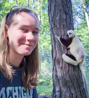
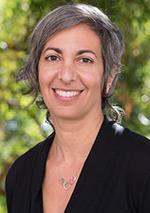
Neysa Grider-Potter is an Assistant Professor of Anatomy and Neurobiology at Northeast Ohio Medical University (NEOMED). Her research focuses on comparative primate locomotion and musculoskeletal functional morphology, with current projects exploring head stability during locomotion, spine function, and locomotor energetics in lemurs. She holds a Ph.D. in Anthropology from Arizona State University (2019), an M.A. in Anthropology from Arizona State University, and dual B.S. degrees in Anthropology and Zoology from Michigan State University. Her work integrates 3D imaging techniques, biomechanical modeling, and evolutionary perspectives to understand mammalian head-neck function and its influence on craniovertebral form. Education: Ph.D. in Anthropology, Arizona State University (2019) M.A. in Anthropology, Arizona State University B.S. in Anthropology, Michigan State University B.S. in Zoology, Michigan State University Research Interests: Comparative primate locomotion and musculoskeletal adaptations Evolution of orthograde posture and bipedalism Functional morphology of the primate spine and neck muscles 3D anatomical analysis using microCT and fascicle-tracking algorithms Her articles highlight interdisciplinary approaches to studying locomotion energetics, cervical vertebrae morphology, and muscle activity patterns across primates and other mammals. While no formal awards are listed, her publications demonstrate significant contributions to understanding evolutionary biomechanics. Laboratory and Team: Her lab at NEOMED investigates craniovertebral form-function relationships, emphasizing how locomotion influences head-neck anatomy. Collaborations with institutions like Osaka University and Rocky Vista University reflect her international research network.
Dr. Florian Witzmann serves as Curator for Fossil Fishes and Amphibians at the Museum of Natural History Leibniz Institute for Evolution and Biodiversity Research in Berlin, Germany, a position he has held continuously since June 2016 (and previously from April 2009 to November 2014). He also functions as Editor-in-Chief of 'Fossil Record,' the museum's paleontological open-access journal. His academic credentials include a habilitation completed in November 2014 at Humboldt University of Berlin with the thesis 'Physiological aspects of the fish-to-tetrapod transition - skin structure, breathing and feeding in early tetrapods,' supervised by Prof. Johannes Müller, and a PhD earned in September 2004 from the same institution focusing on the Permo-Carboniferous temnospondyl amphibian Archegosaurus decheni. His research program centers on three primary interconnected domains: (1) physiological adaptations during the fish-to-tetrapod transition in the late Paleozoic, particularly examining morphological and histological changes in dermal bones and scales alongside restructuring of the gill skeleton related to feeding and respiration mechanisms; (2) evolution of cranial muscles in early tetrapods and amphibians; and (3) paleopathology, with special emphasis on spinal diseases in fossil amphibians and reptiles and the evolutionary history of bone healing mechanisms. His work frequently employs advanced imaging techniques including microCT, FIB-SEM tomography, and phylogenetic disease bracketing to analyze both modern and fossil specimens. Dr. Witzmann's scholarly impact is reflected in publications across prestigious interdisciplinary journals including Nature, Science Advances, Current Biology, The Lancet Diabetes & Endocrinology, and JAMA Oncology. His research has identified ancient diseases in the fossil record such as the oldest known osteopetrosis case in human skeletons, cancer in a 240-million-year-old stem-turtle, and metabolic bone diseases in Permian amniotes. His methodological approach bridges paleontology, evolutionary developmental biology, and clinical medicine to understand deep-time patterns of disease and physiological adaptation. Feodor Lynen-Research Fellow of the Alexander von Humboldt-Foundation (2014-2016) at Brown University's Department of Ecology and Evolutionary Biology Endeavour Leadership Award recipient (2019-2020) supporting research at the Queensland Museum, Brisbane His academic trajectory demonstrates consistent progression from doctoral research on temnospondyl amphibians through postdoctoral work on dermal bone histology to his current leadership role in paleopathology and evolutionary morphology. Dr. Witzmann maintains active collaborations with researchers across Europe, North America, and Australia, contributing significantly to our understanding of vertebrate evolution and the deep history of physiological adaptations.
Associate Professor Brian Anderson is a glaciologist at the Antarctic Research Centre , Victoria University of Wellington, New Zealand. He specializes in glacier dynamics, mass balance, and climate-glacier interactions using mathematical modeling. BSc(Hons) and PhD in Geology from University of Canterbury Supervises PhD and Masters students in Antarctic and Southern Alps glacier studies Active in climate change research, leading grants from 2011–2025 His research interests span: Glacier modeling under past/future climate scenarios Snow property analysis on Antarctic landfast ice Impact of debris cover and lake calving on ablation Holocene climate reconstruction from glacier records Paleoclimatology of subantarctic islands Sea level rise projections from land ice loss Recent publication trends (2018–2025) show focus on: Antarctic ice sheet response to warming Extreme glacier mass loss attribution Paraglacial sediment dynamics 2D glacier modeling for paleoclimate Equilibrium line altitude (ELA) analysis High-resolution snow/ice core studies Notable collaborations: A. Mackintosh, H. Purdie, R. Dadic, J. Oerlemans
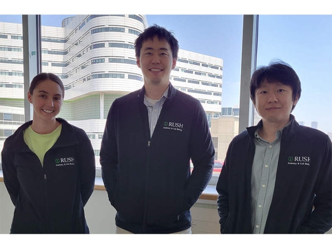
Frank C. Ko, PhD is an Assistant Professor in the Department of Anatomy & Cell Biology at Rush Medical College, Rush University , with secondary appointments in the Department of Orthopaedic Surgery. His research focuses on molecular mechanisms of intramembranous bone regeneration and musculoskeletal homeostasis , particularly examining periostin-expressing cells and circadian rhythm interactions with bone metabolism. PhD in Musculoskeletal Biomechanics from Cornell University Postdoctoral training at Massachusetts General Hospital and Beth Israel Deaconess Medical Center Research spans bone regeneration , implant fixation , and disease models (osteoporosis, osteoarthritis, sarcopenia). Key projects include: Periostin cell lineage tracing in marrow ablation recovery Wnt/β-catenin activation for accelerated healing Phosphate-mTORC1-BMAT interactions Gut-bone axis through colonic circadian disruption Translational preterm pig skeletal modeling Current funding includes NIH R01 and R21 grants. Scientific accolades feature the Searle Innovator Award . His lab collaborates with orthopedic surgeons and computational biomechanists to develop novel therapeutic strategies for musculoskeletal repair and implant integration.
Federico Bernardini is a Researcher in Archaeological Research Methodologies at Ca' Foscari University of Venice's Department of Humanities. His research focuses on archaeometry, digital archaeology, and landscape studies in northeastern Italy and the Adriatic region. He employs interdisciplinary approaches including geochemical analysis, LiDAR, microCT, and remote sensing to investigate prehistoric to Roman-era material culture, trade networks, and settlement patterns. Bernardini teaches courses in the Master's program in Digital and Public Humanities, including Digital Archaeology , Digital Archaeology and Heritage, Ethics and Politics , and Virtual Archaeology and Web Technologies . His publications emphasize technological innovation in archaeology, with recent work exploring pottery provenance, lithic analysis, and landscape reconstruction through advanced imaging techniques. His scholarly output demonstrates consistent collaboration with international teams and application of cutting-edge methodologies. Research trends include: 1) Integration of scientific analyses (geochemistry, tomography) in artifact studies; 2) Digital reconstruction of archaeological landscapes; 3) Trans-Adriatic cultural connections from Neolithic to Roman periods; and 4) Development of non-destructive analytical protocols for cultural heritage materials.
Soban Umar serves as a Professor-in-residence within the Department of Anesthesiology at the David Geffen School of Medicine at the University of California Los Angeles (UCLA). His research program bridges anesthesiology, cardiology, and pulmonary medicine with a specialized focus on pulmonary hypertension and right ventricular failure mechanisms. Dr. Umar's work is characterized by its translational approach, moving from basic molecular mechanisms to potential clinical applications. Dr. Umar's research interests encompass several interconnected domains in cardiovascular pathophysiology. His primary focus centers on pulmonary arterial hypertension, particularly investigating the molecular mechanisms underlying right ventricular failure. His work has made significant contributions to understanding sexual dimorphism in pulmonary hypertension, demonstrating the protective roles of both estrogen and the Y chromosome in disease pathogenesis. More recently, his laboratory has pioneered research on endothelial-to-mesenchymal transition (EndMT) in right ventricular failure, identifying Snai1 as a critical transcription factor and LOXL2 as a key mediator in this process. His work on neuroinflammation in the thoracic spinal cord represents a novel frontier in understanding neural regulation of pulmonary vascular tone. Analysis of Dr. Umar's publication trajectory reveals a clear evolution from foundational work on cardioprotection mechanisms (particularly with lipid emulsions) to increasingly sophisticated investigations of molecular pathways in pulmonary hypertension. His recent publications demonstrate a strong emphasis on multi-omics approaches, single-cell analysis, and targeted therapeutic strategies. The research shows growing methodological complexity with integration of advanced imaging, transcriptomic, and epigenetic analyses to uncover disease mechanisms. Thomas M. Grove Chair in Anesthesiology, UCLA, 2023 Assembly on Pulmonary Circulation Early Career Research Achievement Award, American Thoracic Society (ATS), 2021 Dillon Award For outstanding performance as an Assistant Professor, UCLA Department of Anesthesiology and Perioperative Medicine, 2019 Junior Faculty Research Award, Association of University Anesthesiologists (AUA), 2017 Dr. Umar has secured substantial research funding to advance his work, including an NIH R01 grant (1R01HL161038-01A1, 2022-2027) as Principal Investigator investigating molecular mechanisms of right ventricular failure in pulmonary hypertension, and an NIH K08 grant (K08HL141995, 2019-2024) focused on the role of microRNA-125b in pulmonary hypertension associated with pulmonary fibrosis. His collaborative approach is evident in numerous multi-investigator studies across UCLA departments and institutions. Dr. Umar leads the Umar Lab at UCLA, which employs a multidisciplinary research strategy combining molecular biology, physiology, genetics, and advanced imaging techniques to investigate pulmonary hypertension pathogenesis and identify novel therapeutic targets. The lab's work has significant translational potential, with several recent findings pointing toward promising new treatment approaches for pulmonary hypertension and right heart failure.
Dorthe Wildenschild is a Professor and holds the DeVaan Chair in the Department of Chemical, Biological, and Environmental Engineering at Oregon State University's College of Engineering. She leads the Clean Water Technology Initiative and directs the Multi-phase Flow and Micro-Imaging Lab. Her research focuses on porous media flow, subsurface water pollution, and energy storage solutions. She earned a Ph.D. (1996) and M.S. (1991) in Civil and Environmental Engineering from Danish Technical University. Research interests include geologic CO2 storage optimization, biofilm dynamics under microgravity (collaborating with NASA), and advanced imaging techniques using deep learning. Key labs include the OSU MicroCT Facility. Recent work emphasizes clean water innovation, groundwater remediation, and sustainable energy systems. Labs and teams: Multi-phase Flow and Micro-Imaging Lab, OSU MicroCT Facility. Professional contributions include the College of Engineering Faculty Lecture Series and leadership in the Water and Environment research group. Grants and advising details are not explicitly listed here.
Jonathan Aitchison is a Professorial Research Fellow at The University of Queensland's School of the Environment, specializing in tectonic evolution and microfossil geochronology. With a BSc/MSc from University of Otago and PhD from University of New England, his career spans research positions across Japan, Australia and Hong Kong, including leadership of HKU's Department of Earth Sciences (2003-2009). Research interests focus on India-Asia collision dynamics, radiolarian biostratigraphy, and Paleozoic tectonics, with current projects investigating organic carbon recycling in ophiolites and microCT imaging of microfossils. Fieldwork spans Himalayan suture zones, Tibetan Plateau, and SW Pacific regions. Recent publications demonstrate consistent focus on ophiolite geochemistry, subduction processes, and radiolarian paleoecology, with methodological advances in U-Pb dating and microCT applications. Awards include the 2008 ALCAN Prize from the French Academy of Sciences. Research involves international collaborations across the Asia-Pacific region.
Debbie Guatelli-Steinberg is a Professor in the Department of Anthropology at The Ohio State University. Her research focuses on using teeth to study life history, growth patterns, and biological relationships in primates and fossil hominins, leveraging enamel microstructure to uncover stress, diet, and developmental processes. She actively supervises graduate students like Kaita Gurian and collaborates on projects involving skeletal databases (e.g., Cayo Santiago macaque colony). Her work integrates histological, morphological, and archaeological methods to address evolutionary and ecological questions. Research interests include dental stress markers (e.g., linear enamel hypoplasia), morphological adaptations to diet, and genetic/environmental influences on dental traits. Key tools in her studies include microCT scanning, enamel profilometry, and comparative primate studies. She has contributed to understanding Neandertal and early Homo developmental patterns, as well as subsistence shifts in prehistoric Ohio populations. Her publications span from 2017 to 2025, exploring topics like enamel growth dynamics, primate community dental performance, and dental-bioarchaeological connections. Current projects emphasize interdisciplinary approaches to primate health history databases and the impact of early-life stress on dentition across genders.
Dr. Jean Elizabeth Aaron is a Visiting Lecturer in the School of Biomedical Sciences at the University of Leeds, where she has maintained a long-standing research presence following a distinguished career in bone biology. She was formerly Director of the Bone Structural Biology Laboratory and an MRC Visiting Fellow, and has supervised numerous PhD students and collaborated with leading institutions including the University of Sheffield, University of Bristol, and Merck Sharpe & Dohme. BSc in Applied Biology, University of Bradford PhD in Bone Biology, University of Leeds (1974) Her research focuses on the microarchitectural and macromolecular basis of skeletal fragility , particularly in osteoporosis and osteoarthritis. She pioneered techniques in undecalcified bone cryomicrotomy , computer-assisted histomorphometry , and 3D mapping of osteocyte networks . Her work challenges conventional views by proposing a Golgi-directed bone mineral biosphere , where intracellular mineralization in osteocytes forms calcified microspheres that constitute a dynamic, stress-responsive inorganic microskeleton. This model integrates periosteal Sharpey’s fibres as conduits for mechanical signaling, linking muscle forces to bone remodeling. The 15 most recent publications reveal a consistent trajectory: from foundational work on trabecular disconnection and histomorphometry to cutting-edge hypotheses on the evolutionary origins of bone mineralization, drawing parallels with protozoan and bacterial systems. The articles span cell biology , biomechanics , developmental biology , and paleohistology , with recurring themes of stress conductance , mineral quality over mass , and embryomimetic repair mechanisms . Techniques include electron microscopy, immunohistochemistry, micro-CT, and computational modeling. She received the Millennium Prize for Best Paper in BONE (2001) from the International Bone and Mineral Society and presented her work at Buckingham Palace. Her research has been supported by the MRC and Action Medical Research. Dr. Aaron has supervised several PhD students, including K.M. Linton, V. Fallon, and D.H. Carter, and has collaborated with clinicians such as L.D. Hordon and J.A. Kanis. She previously taught undergraduate modules on 'Bone in Health & Disease' and gross anatomy. Her laboratory hosted visiting scientists from around the world and trained numerous undergraduates through research projects. She remains active in research, with ongoing work at the Leeds Institute of Musculoskeletal Medicine, exploring novel histological explanations for bone marrow lesions in osteoarthritis and the clinical applications of Sharpey’s fibre biology in oral surgery, recently termed 'Embryomimetic Surgery'.
Galateia Kazakia, PhD, is a Professor in Residence in the Department of Radiology and Biomedical Imaging at the University of California, San Francisco (UCSF). She serves as Director of the Bone Quality Research Laboratory, the Quantitative Musculoskeletal-Imaging Core, the UCSF Core Center for Musculoskeletal Biology and Medicine (CCMBM) Imaging Core, and Co-Director of the Nutrition and Obesity Research Center (NORC) Human Metabolism Core. Her leadership roles reflect her central position in advancing musculoskeletal imaging research at UCSF. Dr. Kazakia earned her BS in Mechanical Engineering from Cornell University (1995) and her PhD in Mechanical Engineering with a focus on Bone Biomechanics from UC Berkeley (2004). She completed an NIH F32 postdoctoral fellowship at UCSF before joining the faculty in 2009. Her research focuses on assessing bone quality using high-resolution imaging modalities such as HR-pQCT, MRI, microCT, and FTIR spectroscopy. She investigates bone changes in aging and diseases including diabetes, chronic kidney disease, and HIV. Her work aims to understand the biomechanical and structural underpinnings of fracture risk beyond traditional bone mineral density measurements. Her recent publications demonstrate a strong trajectory in longitudinal imaging, segmentation techniques, and metabolic-bone interactions. Themes include cortical porosity, bone marrow adiposity, and non-invasive virtual biopsy methods. She leads multiple NIH-funded projects on bone loss post-fracture and diabetic bone disease. Summa Cum Laude Award, ISMRM, 2020 Plenary Poster Award, ASBMR, 2019 and 2014 Mentored Career Development Award, NIH, 2009 Young Investigator Award, Sun Valley Workshop, 2006 NSF Graduate Fellowship, 1999 Dr. Kazakia actively mentors postdoctoral fellows, graduate students, and undergraduates. Her lab collaborates with clinicians and scientists across disciplines and institutions. She has served as PI or Co-PI on major NIH grants, including R01 awards from NIAMS, NIA, and NIAID, supporting her innovative work in musculoskeletal imaging and translational research. She leads the Bone Quality Research Lab, which fosters interdisciplinary training and research in advanced imaging and bone biomechanics. The lab emphasizes innovation in image analysis and the development of non-invasive tools for clinical application.
Dr. Luke Holbrook is a Full Professor in the Department of Biological & Biomedical Sciences at Rowan University's College of Science & Mathematics. He specializes in the phylogeny and evolution of mammals, particularly perissodactyls (horses, rhinos, tapirs), using anatomical data from fossils and living species to reconstruct evolutionary relationships and analyze trait convergence. Post-Doctoral Associate, New York College of Osteopathic Medicine Ph.D. in Biology, University of Massachusetts M.S. in Biology, University of Massachusetts B.S. in Biology, Fordham University Dr. Holbrook's research focuses on mammalian evolutionary history, including the origin of perissodactyls in India, the role of the end-Cretaceous extinction in placental mammal diversification, and the convergent evolution of complex traits like large body size and tooth morphology. His work integrates fossil data with molecular clocks to determine lineage divergence timing. His 15 most recent publications span topics such as placental mammal brain evolution, early Eocene mammal biogeography, dental adaptations in perissodactyls, and vertebral specialization in hoofed mammals. These studies employ methodologies including cladistics, geometric morphometrics, and microCT scanning for virtual specimen analysis. Dr. Holbrook teaches courses in evolution, comparative anatomy, and interdisciplinary biology/history. He collaborates internationally with institutions like the American Museum of Natural History and uses advanced imaging technology in his lab. While no formal awards are listed here, his research has been widely shared across academic and mainstream platforms.
Dustin Green, PhD is an Assistant Professor in the Department of Neurobiology at the University of Texas Medical Branch (UTMB), focusing on the intersection between neuronal and immune system interactions. His research primarily investigates the role of Mrgpr receptors in pathological pain and itch mechanisms. Education Postdoctoral Training: Johns Hopkins University School of Medicine (Advisor: Xinzhong Dong) PhD in Physiology: University of Texas Health Science Center (Advisor: Ken Hargreaves) BS in Biology: Centenary College of Louisiana Research Focus : Dr. Green's lab examines how mast cells interact with peripheral nerves through Mrgpr receptors. Key discoveries include the role of MrgprB2 (and its human orthologue MRGPRX2 ) in thermal/mechanical pain, the activation of MrgprB2 by substance P, and the subsequent release of proinflammatory cytokines. This work challenges previous assumptions about NK1 receptor involvement in neurogenic inflammation. Future Directions : The lab aims to characterize mast cell contributions to pain pathologies and explore orphan Mrgpr receptors as therapeutic targets due to their restricted expression in neurons and immune cells. Email: dugreen@utmb.edu