Raymond David Dunphy is a Research Assistant at the University of Strathclyde's Centre for Signal and Image Processing. He is completing his PhD on hyperspectral imaging for microbiological applications while contributing to interdisciplinary projects in nuclear and space industries. His work bridges machine learning with advanced imaging techniques. MEng in Computer and Electronic Systems (University of Strathclyde) PhD candidate in Hyperspectral Imaging (ongoing) His research focuses on developing machine learning tools for hyperspectral data analysis , with applications spanning nuclear fuel manufacturing, microbiology, and aerospace. Key projects include optimizing uranium hexafluoride conversion and enhancing quality assurance in UO₂ nuclear fuel pellet production. Current collaborations include industry partnerships with Terumo Aortic Limited and participation in the Interdisciplinary Centre for Doctoral Training in Antimicrobial Resistance (AMR). He actively contributes to UN Sustainable Development Goals through nuclear energy efficiency innovations.
Dana Peters is a Professor of Radiology & Biomedical Imaging at Yale School of Medicine and Director of Cardiac MRI at the Magnetic Resonance Research Center. With a secondary appointment in Biomedical Engineering, she specializes in cardiovascular magnetic resonance (CMR) imaging, developing innovative MRI tools for cardiac function assessment, diastolic dysfunction characterization, and left atrial scar visualization in atrial fibrillation patients. Education: BS in Physics, Johns Hopkins University PhD in Physics, University of Wisconsin-Madison Postdoctoral training at NIH NHLBI Laboratory of Cardiac Energetics Her research focuses on cardiac pressure-volume dynamics , scar visualization in electrophysiology , and cancer metabolic imaging , particularly in liver cancer via deuterium metabolic imaging. She integrates machine learning with radial imaging reconstruction algorithms to improve spatial resolution and motion correction in clinical MRI. Scientific Contributions include the Innovative Project Award for sarcoidosis research (2023). She mentors biomedical imaging scientists through access to Yale's advanced MRI infrastructure, including three Siemens 3T Prisma scanners, a 4T human magnet, and 9.4T/11.7T preclinical systems. Her team develops automated valve-tracking frameworks , multimodal cardiac diagnostics , and metabolic imaging techniques , collaborating with clinicians and engineers to bridge imaging technology with clinical applications in atrial fibrillation , diastolic dysfunction , and oncologic imaging .
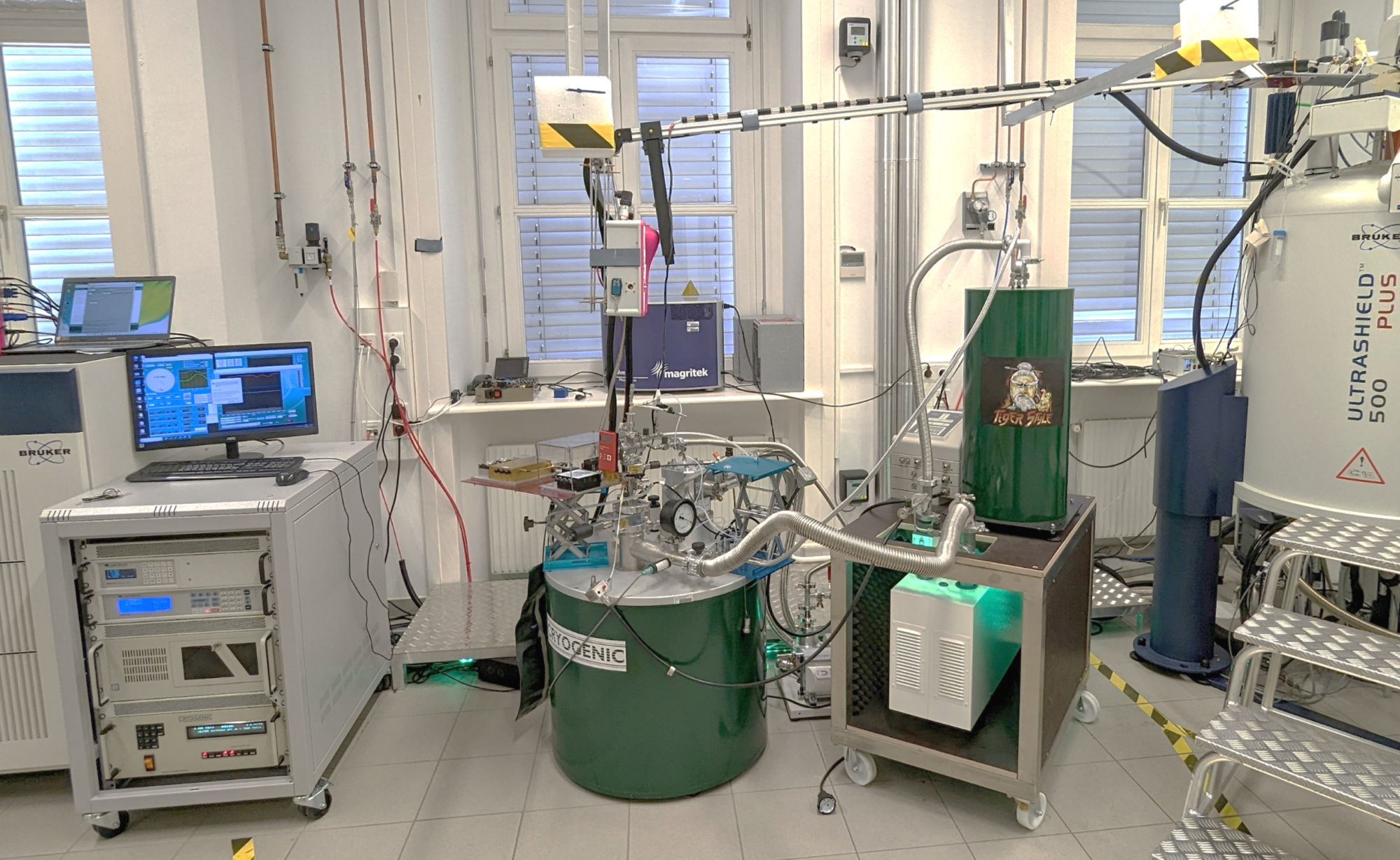
Dennis Kurzbach serves as Associate Professor and Deputy Head of the NMR Center at the University of Vienna's Faculty of Chemistry, Institute of Biological Chemistry. His research pioneers magnetic resonance methodologies including NMR, EPR, and hyperpolarization techniques to solve critical challenges in chemical and biological sciences, with significant contributions to biomimetic materials and protein dynamics. His research centers on the structural dynamics of intrinsically disordered proteins (IDPs), peptide-guided biomimetic mineralization (silica/calcium phosphate systems), and hyperpolarization-enhanced NMR for real-time monitoring of transient biological processes. The Kurzbach Spectroscopy Studio develops innovative approaches to observe short-lived intermediates in protein folding, mineral nucleation, and metabolic pathways, bridging biophysical chemistry with materials science through advanced spectroscopic techniques. Recent publications demonstrate a cohesive trajectory toward enhancing NMR sensitivity for biological applications, with hyperpolarization techniques enabling unprecedented observation of fast dynamic processes. Key thematic clusters include biomimetic material synthesis driven by peptide self-assembly, domain-specific spectroscopy for DNA-protein interactions, and machine learning integration for analyzing complex IDP behavior – all converging toward real-time molecular observation in physiological conditions. Professor Kurzbach actively supervises bachelor and master theses across organic, bioinorganic, biological, and biophysical chemistry disciplines while leading post-graduate seminars in chemical and biological chemistry. His laboratory maintains cutting-edge instrumentation through the NMR Center, focusing on methodological innovation that directly impacts structural biology and materials science research.
Vidhu Anand, MBBS, serves as a Senior Associate Consultant in the Department of Cardiovascular Diseases and Assistant Professor of Medicine at the Mayo Clinic College of Medicine in Rochester, Minnesota. She is a specialist in cardiovascular imaging with expertise in echocardiography, CT, MRI, and nuclear cardiology techniques. Dr. Anand completed her Internal Medicine residency at the University of Minnesota (2017) followed by a Cardiovascular Disease fellowship at Mayo Clinic (2021). She is board-certified in Internal Medicine, Cardiovascular Diseases, Adult Echocardiography, and Nuclear Cardiology. Her research focuses on pulmonary hypertension epidemiology and novel therapies, valvular heart disease (particularly aortic valve disease and TAVR complications), apical hypertrophic cardiomyopathy, and machine learning applications in cardiovascular diagnostics. Recent publications demonstrate her leadership in developing risk stratification tools for tricuspid regurgitation and applying artificial intelligence to echocardiographic interpretation. Dr. Anand has published extensively in high-impact cardiology journals, with recent work appearing in European Heart Journal Cardiovascular Imaging, JACC Cardiovascular Imaging, and Journal of the American Heart Association. Her research often involves large cohort studies and innovative applications of imaging technology. Young Investigator Award (runner up), European Society of Cardiology (2019) Early Career Top Investigator, American Society of Echocardiography (2019) Best Poster Award, American College of Cardiology-Minnesota Chapter (2018) Outstanding Research Award, University of Minnesota Internal Medicine Residency (2017) Dr. Anand is actively involved in professional organizations including the American Society of Echocardiography (Research Committee), American College of Cardiology (Wisconsin Chapter Board of Councilors), and the 2026 AHA/ACC Valve Guideline Writing Group. She serves as an abstract reviewer for major cardiology conferences and contributes to guideline development in her specialty areas.
Ji Chen is an Associate Professor with Tenure at the Institute of Condensed Matter Physics and Material Physics (ICMP), School of Physics, Peking University. His research focuses on developing and applying advanced computational and artificial intelligence methods to study the electronic structure, atomic structure and dynamics of condensed matter and materials. Dr. Chen received his PhD in Condensed Matter Physics from Peking University, following graduate studies at the Institute of Physics, Chinese Academy of Science, and an undergraduate degree in Physics from the University of Science and Technology of China. He completed postdoctoral training at University College London and the Max Planck Institute for Solid State Research. Dr. Chen's research interests span electronic structure theory, quantum Monte Carlo methods, deep learning applications in physics, strongly correlated systems, water and carbon structures & dynamics, and AI-assisted materials modeling. His group develops and applies advanced computational techniques to address challenging problems in condensed matter physics and materials science, with particular emphasis on neural network-based quantum Monte Carlo methods and their applications to complex physical systems. The research demonstrates significant advancements in computational methodologies that combine machine learning with quantum mechanical simulations, achieving unprecedented accuracy in modeling strongly correlated electrons and complex material properties. Dr. Chen leads the Chen Research Group at Peking University, which actively collaborates with researchers worldwide. The group maintains an active publication record in top-tier journals including Nature, Science, Physical Review Letters, and other leading physics and chemistry publications. Their work spans from fundamental method development to practical applications in quantum technologies, materials design, and energy-related research. Dr. Chen teaches undergraduate and graduate courses in computational physics at Peking University, including "Computational Physics" and "An Introduction to Computational Physics," sharing his expertise in molecular dynamics simulations, Monte Carlo calculations, and density functional theory with the next generation of physicists and materials scientists.
Professor Marc Aubreville is a Professor of Applied Computer Science with a specialization in Visual Computing at Flensburg University of Applied Sciences. Appointed in September 2024, he leads the FLAIR (Flensburg Artificial Intelligence Research Visual Computing) research group and serves as spokesperson for the STEM research team of the Schleswig-Holstein Promotional College (PKSH), representing 45 professors from six universities across the state. Aubreville's research centers on digital pathology and AI-assisted medical diagnostics, developing systems that help pathologists identify malignant tumor regions through abnormal cell division patterns. His DFG-funded "Digital Pathology" project is particularly notable as only approximately 1% of DFG funding goes to universities of applied sciences. His work began with veterinary applications using canine tumor samples before transitioning to human medicine, demonstrating the translational potential of AI in healthcare. His publication record from 2024-2025 reveals significant contributions across computational pathology, human-AI collaboration dynamics, and veterinary medical imaging. Key research themes include confirmation bias in AI-assisted diagnosis, automation bias under time pressure, and the development of comprehensive datasets for training AI in histopathology. His work bridges computer science, medicine, and veterinary science with practical applications for improving diagnostic accuracy. Key Research Contributions: AI systems to assist pathologists in identifying malignant areas in tissue samples Creation of standardized reporting guidelines for AI-based image analysis Development of datasets for mitosis detection in breast cancer Investigation of human factors in AI-assisted medical decision-making Professional Engagement: Spokesperson for PKSH STEM research team since March 2025 Collaboration with institutions in Berlin and Vienna for data collection Active participation in establishing methodological standards for AI in pathology
Hidekata Hontani is a Professor at the Graduate School of Engineering, Nagoya Institute of Technology, specializing in image processing and medical image analysis. His research focuses on computational anatomy, deep learning applications in medical imaging, and pathological image analysis, particularly for cancer diagnosis and treatment. Professor Hontani's research interests center around medical image processing , with specific focus on computational anatomy , deep learning for medical applications , pancreatic cancer analysis , and malignant lymphoma grading . His work bridges computer science and medical diagnostics, developing innovative algorithms for analyzing pathological images and creating 3D models of tumors. His recent publications (2023-2024) show a strong trend toward applying diffusion models and weakly supervised learning techniques to medical image analysis, particularly for cancer diagnosis. Key areas include pathological image analysis , tumor microenvironment modeling , anomaly detection , and multi-modal image registration , with increasing focus on lymphoma grading and pancreatic cancer analysis. Scientific Awards: Multiple Japanese Society of Medical Imaging Technology Conference Encouragement Awards (2012-2024) JAMIT Young Medical Imaging Engineering Symposium Encouragement Awards (2023) Cum Laude Poster Award, SPIE Medical Imaging (2018) Student Award, VISAPP 2017 Professor Hontani has secured multiple significant research grants, including an ongoing Grant-in-Aid for Scientific Research (B) from the Japan Society for the Promotion of Science titled Comprehensive Characterization of All Two Million Nuclei in Pathology Images for Quantitative Assessment of Atypia in Malignant Lymphoma (2025-2028). He serves on key academic committees including as Secretary of the Medical Imaging Research Group at IEICE since 2010 and as a committee member of the Pattern Recognition and Media Understanding Research Group since 2013.
Kristian Lindholm serves as a Guest Researcher within the Department of Biomedical Sciences at the University of Copenhagen, specifically affiliated with the Cluster for Molecular Imaging. His research integrates computational methods with advanced medical imaging techniques to address clinical challenges in oncology. His primary research interests include: Medical Imaging Artificial Intelligence applications in healthcare Neuroendocrine tumor diagnostics PET/CT imaging analysis Deep learning for tumor segmentation Biomedical engineering solutions Lindholm's recent work demonstrates a clear trajectory toward leveraging convolutional neural networks for precision oncology, particularly in nuclear medicine imaging. His research bridges machine learning algorithms with clinical nuclear medicine to improve tumor quantification in neuroendocrine neoplasms, reflecting growing interdisciplinary convergence between AI and medical imaging. He operates within the Cluster for Molecular Imaging, a specialized research environment focused on developing and applying advanced molecular imaging technologies for biomedical research and clinical translation.
Tadeusz Wibig is a faculty member in the Department of Experimental Physics at the Faculty of Physics, University of Lodz, Poland. His academic career focuses on particle physics and cosmic ray research, with particular emphasis on ultra-high energy cosmic rays and their interactions. Dr. Wibig's primary research interests include: Ultra-High Energy Cosmic Rays (UHECR) physics Cosmic ray ensembles and the Gerasimova-Zatsepin effect Particle interactions and shower development Cosmic Microwave Background foreground analysis Citizen science projects in physics education Instrumentation for cosmic ray detection Analysis of Dr. Wibig's publication record shows a consistent focus on cosmic ray physics spanning nearly two decades. His work demonstrates particular expertise in ultra-high energy cosmic ray phenomena, including the GZK cutoff, cosmic ray spectrum features, and cosmic ray ensembles. In recent years, he has increasingly focused on citizen science applications, developing educational projects like "Nuclear E-Cology" and CREDO-Maze that engage students in authentic research. His technical work spans theoretical modeling, simulation studies, and instrumentation development for cosmic ray detection. Dr. Wibig has been actively involved in several major collaborative projects: CREDO (Cosmic-Ray Extremely Distributed Observatory) - developing citizen science approaches to cosmic ray research JEM-EUSO (Extreme Universe Space Observatory on the Japanese Experiment Module) - space-based cosmic ray detection The Roland Maze Project - engaging high school students in cosmic ray detection Nuclear E-Cology project - integrating nuclear physics into high school curricula His educational initiatives have established networks with high schools in Łódź and international collaborations involving Polish, Thai, and Russian students. These projects combine authentic scientific research with educational goals, creating opportunities for students to participate in real physics investigations while developing scientific literacy.
Prof. Karl-Josef Langen is a Group Leader at the Institute of Neuroscience and Medicine (INM) within the Physics of Medical Imaging (INM-4) department at the Forschungszentrum Jülich . His work focuses on advancing neuroimaging techniques for brain tumor diagnostics, particularly using 18F-FET PET and multimodal imaging approaches. His research addresses challenges in distinguishing tumor recurrence from treatment-related effects, improving survival predictions, and optimizing therapeutic strategies in glioma patients. Key areas of expertise include neuro-oncological imaging , radiomics , functional connectivity analysis , and clinical trial design . He leads projects integrating machine learning for automated tumor segmentation and has contributed to international guidelines for PET/MRI applications in neuro-oncology. His team collaborates across disciplines to develop innovative imaging protocols and predictive biomarkers for personalized cancer care. Recent studies emphasize cost-effectiveness analyses of imaging modalities, validation of imaging algorithms, and preclinical model development. His work directly impacts clinical decision-making through multidisciplinary tumor boards and has implications for global neuroimaging standards.
Hanyu Wei is an Assistant Professor of Physics at Louisiana State University's Department of Physics & Astronomy, part of the College of Science. His research focuses on experimental high-energy physics, particularly neutrino experiments such as MicroBooNE, SBND, and the upcoming DUNE. He develops the Wire-Cell reconstruction paradigm for liquid argon time projection chambers (LArTPCs), enhancing neutrino event analysis and physics capabilities. His work includes cross-section measurements, sterile neutrino searches, and nucleon decay studies. Previously, he contributed to the Daya Bay reactor neutrino experiment, achieving precise θ13 mixing angle measurements and designing a supernova trigger system. He also explores supernova relic neutrino detection at the Jinping experiment. Key Research Areas : Neutrino oscillations and beyond Standard Model physics LArTPC detector technology and reconstruction algorithms Supernova neutrino detection systems MicroBooNE and DUNE collaborations Recent Projects : Wire-Cell algorithm integration with machine learning SNB and DUNE oscillation physics analyses Dark sector probes via neutrino interactions His work bridges detector innovation with fundamental physics questions, emphasizing precision measurements and novel experimental techniques.
Mariapina D'Onofrio is an Associate Professor at the Department of Biotechnology, University of Verona. Her research focuses on structural and dynamic studies of biomolecules using nuclear magnetic resonance (NMR) spectroscopy, particularly investigating protein-ligand interactions, protein-nanoparticle interactions, and the role of ubiquitination in Alzheimer's-related tau proteins. She is a member of the Gruppo Italiano di Discussione Risonanze Magnetiche and actively contributes to academic governance, including roles in the Faculty Board of the PhD in Nanoscience and Advanced Technologies. Education: PhD in Chemistry from the University of Modena. Teaching: Courses in Organic Chemistry and Elements of Chemistry for Bioinformatics, Biotechnology, and Viticultural/Oenological Science programs. Research Interests: Her work spans biological chemistry, chemical biology, and organic chemistry. Key projects include structural studies of tau protein aggregation, development of biofunctionalized nanomaterials, and exploring NMR techniques for biomacromolecules. Current grants involve projects on antiviral drug design via PROTAC technology and neuroprotective effects of nutraceuticals. Publications: Recent work includes studies on tau protein ubiquitination, nanomaterial interactions, and high-energy physics collaborations (e.g., ATLAS detector analyses). Her articles reflect interdisciplinary approaches combining biochemistry, nanotechnology, and advanced analytical techniques. Awards: No specific awards listed, but contributions to collaborative research and academic leadership are notable. Advising/Grants: Leads multiple research grants and collaborates internationally. Her lab focuses on applying NMR spectroscopy to biomedical applications. Active in spin-off companies linked to biotechnology innovations. Labs/Teams: Member of the NMR Spectroscopy research group and collaborates with the Nanomaterials Research Group on biomedical nanotechnology.
Ignacio Bravo Muñoz is a Professor at the Department of Electronics, Universidad de Alcalá (Spain), affiliated with the GEINTRA research group focusing on Electronic Engineering applications in Intelligent Spaces and Transport. He holds a PhD from Universidad de Alcalá (2007) with a thesis on FPGA-based object detection using computational vision and PCA techniques. His research spans indoor positioning systems (using LED/PSD sensors), sustainable energy frameworks for smart communities, FPGA-based hardware design , and remote laboratory platforms . Key contributions include real-time metrology for ESA's PLATO mission, cooperative demand response algorithms, and innovative pedagogical approaches integrating sustainability into digital electronics education. Recent work emphasizes edge computing for video surveillance , machine learning in human action recognition , and non-cooperative target identification using radar signatures. His interdisciplinary projects bridge electronics engineering with energy systems, biomedical applications, and educational technology. He actively collaborates with industry and academic institutions on EU-funded projects, contributing to advancements in aerospace instrumentation (PLATO FPA qualification), smart grid technologies, and STEM education innovation.
Matthijs van Berkel is an Associate Professor at Eindhoven University of Technology, affiliated with the Control Systems Technology group in Mechanical Engineering and Electrical Engineering. His research focuses on developing advanced control systems for nuclear fusion reactors, particularly tokamak devices. He specializes in real-time plasma control, machine learning applications for fusion diagnostics, and optimization of deuterium-tritium reactions. His work bridges plasma physics and engineering, with emphasis on: Feedback control systems for plasma stability Tomographic reconstruction of fusion reactions Experimental validation at major facilities (JET, ASDEX Upgrade) Machine learning-enhanced reactor monitoring He actively contributes to international collaborations like the EUROfusion consortium and has been recognized for innovations in heat loss control methods for fusion reactors.
Xicheng Wang is a Researcher (Postdoc) in the Division of Nuclear Science and Engineering, Department of Physics at KTH Royal Institute of Technology. His research focuses on computational fluid dynamics (CFD) and system-level code analysis of thermal-hydraulic phenomena in nuclear reactor safety, including thermal stratification, steam injection effects, and suppression pool dynamics. Since 2022, he has explored machine learning applications in nuclear engineering. Education: Ph.D. in Nuclear Engineering, KTH Royal Institute of Technology (2025) M.Sc. in Nuclear Energy Engineering, Tsinghua University (China) and KTH (Sweden, dual program) Research Interests: CFD modeling of thermal-hydraulic phenomena Development of effective momentum/heat transfer models for nuclear reactor safety Machine learning integration for predictive safety analysis Experimental validation of suppression pool behavior Dynamic analysis of fuel assemblies and transport casks Advising & Grants: No formal advisees listed Contributions to projects like PANDA/PPOOLEX experimental campaigns Labs/Teams: Active in the Division of Nuclear Science and Engineering’s thermal-hydraulics research group, focusing on reactor safety and CFD-experiment correlations.