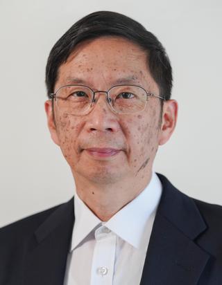
Wing-Chi E. Kwok is an Associate Professor of Imaging Sciences at the University of Rochester since 1990. His work focuses on MR safety, RF coil development, and advanced MRI techniques for clinical and research applications. Ph.D. in Physics (1990), Rensselaer Polytechnic Institute M.S. in Physics (1988), Rensselaer Polytechnic Institute B.S. in Physics (1984), Hong Kong - Non-Medical School His research spans MRI technology innovation , including high-resolution imaging, water/fat separation, and diffusion-weighted MRI. He has pioneered RF coil designs and pulse sequences for clinical systems, with patents in chemical-shift correction and quantum coherence methods. Recent publications highlight applications in RF interference correction MRI-induced heating risk assessment Chronic compression injury modeling Multimodal spine imaging Scientific Recognition : Cum Laude Award (2020) Roadie Award (2019) Magna Cum Laude Merit Award (2012) Certificate of Merit Citation (1999) Student Fellowship (1985–1989) Full Tuition Scholarship (1982–1983) Kwok holds six U.S. patents and supervises MRI quality assurance across multiple hospitals. He serves on the URMC MRI Safety Committee and is certified as an MR Safety Expert (2016). His clinical responsibilities include protocol optimization and safety evaluation for implants/foreign objects.
Daniel Herzka is an Associate Professor at the Department of Radiology , School of Medicine , Case Western Reserve University . His research focuses on high-resolution magnetic resonance imaging , quantitative cardiovascular MRI , and cardiac magnetic resonance fingerprinting . Herzka develops innovative MRI techniques for in vivo neurography and cardiovascular applications , including interventional MRI and low-field MRI systems . Doctor of Philosophy in Biomedical Engineering, Johns Hopkins University School of Medicine (2004) Master of Engineering in Biomedical Engineering, Johns Hopkins University (1999) Professional affiliations include membership in the International Society for Magnetic Resonance in Medicine (ISMRM), Society for Cardiovascular Magnetic Resonance Imaging (SCMR), Medical Image Computing and Computer Assisted Intervention Society , and American Heart Association (AHA). Herzka's research has produced significant publications in Magnetic Resonance in Medicine , Journal of Cardiovascular Magnetic Resonance , and Radiology , primarily focusing on cardiac imaging , low-field MRI , and interventional radiology . His recent work explores accelerated T2 mapping , low-rank reconstruction , and cardiac interventional devices . Herzka's scientific contributions span MRI physics , sequence optimization , and clinical translation of imaging technologies . Key collaborators include researchers from National Institutes of Health , Johns Hopkins University , and University Hospital Bonn . Developed real-time free-breathing cardiac imaging with self-calibrated radial GRAPPA Innovated sorted Golden-step phase encoding for self-gated cine MRI Created analytical polyhedral MRI phantoms for imaging validation
Seong-Eun Kim, PhD, is a research scientist with 25 years of experience specializing in advanced MRI techniques for clinical applications. Affiliated with the Department of Radiology & Imaging Sciences, his work bridges academic research and clinical validation. He holds a PhD from the University of Colorado Boulder and completed postdoctoral training at the University of Utah School of Medicine and University of Colorado School of Medicine. Research Focus: Dr. Kim develops novel MRI methodologies for vascular and neurological disorders, with emphasis on carotid plaque characterization, diffusion-weighted imaging optimization, intravascular contrast agents, and quantitative thermometry. His publications demonstrate consistent innovation in pulse sequence design (e.g., 3D Star of Star, STIR-Prep SOS) for improved diagnostic accuracy in stroke, cancer, and atherosclerosis. Key Contributions: Pioneering work includes rabbit models for intracranial atherosclerosis validation, methemoglobin as an MRI contrast agent, and motion-insensitive techniques for intraplaque hemorrhage detection. His recent research explores MRI thermometry, multi-scanner reproducibility studies, and clinical applications in head/neck cancer.
Glyn Johnson is a Professor of Clinical MRI Physics at Norwich Medical School, University of East Anglia, and an Adjunct Associate Professor of Radiology at New York University School of Medicine. He is also an Honorary Professorial Fellow and a member of Cancer Studies at UEA. His career spans leading institutions in the UK and the US, including the University of Aberdeen, Columbia University, and NYU, where he led the brain tumour imaging research group. BA in Natural Sciences (Physics and Theoretical Physics), University of Cambridge (1977) MSc in Medical Physics, University of Aberdeen (1978) PhD in Medical Physics, University of Aberdeen (1981) His research focuses on developing quantitative MRI techniques to improve diagnostic accuracy in cancer, particularly by distinguishing benign from malignant lesions. His work addresses challenges in lesion characterization using advanced MRI methods such as perfusion, diffusion, and sodium imaging. He has made seminal contributions to MRI physics, including co-developing spin warp imaging during his PhD. His recent publications show a strong trend in applying diffusion MRI and perfusion modeling to prostate and brain cancers, with increasing emphasis on computational and Monte Carlo modeling of tissue microstructure. He also contributes to interdisciplinary research linking nutrition, cognitive ageing, and neuroimaging. Notable scientific engagement includes membership in the ISMRM annual meeting program committee (2005–2007), ongoing service on NIH review panels, and extensive peer-review activity for academic journals since 1984. He has led multiple research projects funded by the National Institutes of Health and Abbott Nutrition, focusing on brain tumour imaging, cognitive ageing, and prostate cancer detection. His work is cited in policy and media, reflecting its translational impact. He is actively involved in collaborative research and continues to publish and secure grants, indicating ongoing academic leadership. Johnson is affiliated with research teams at UEA and NYU, and his projects often involve multi-institutional collaborations, particularly in neuroimaging and cancer studies.
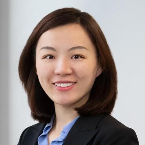
Dan Ma is an Associate Professor of Neurosurgery and Biomedical Engineering at Duke University School of Medicine, with a focus on advancing quantitative Magnetic Resonance Imaging (MRI) technology. His research bridges MR physics, signal modeling, artificial intelligence, and clinical translation to improve disease diagnosis and treatment. Education: Ph.D., Case Western Reserve University School of Medicine (2015) His lab specializes in Magnetic Resonance Fingerprinting (MRF), a non-invasive technique for quantifying tissue properties, and develops multi-parametric MR methods for clinical applications in epilepsy, brain tumors, breast cancer, and prostate cancer. Recent work includes technical innovations in SyntheticMR implementation for radiation therapy and 3D MRF for dynamic contrast-enhanced imaging. Scientific Awards: Senior Member, National Academy of Inventors (2023) Junior Fellow, ISMRM (2017) I.I. Rabi Young Investigator Award, ISMRM (2016) Dan Ma is a core member of Duke's Center for Brain Imaging and Analysis. His research integrates AI into MRI post-processing and explores motion-robust scans for neonatal imaging. Publications highlight his contributions to pulse sequence design, relaxometry standardization, and cortical dysplasia characterization.
Sydney Williams is an Honorary Lecturer at the University of Glasgow's School of Psychology & Neuroscience (SPN) and an Assistant Professor at Universidad Rey Juan Carlos in Madrid, Spain. They collaborate with the Imaging Centre of Excellence (ICE) at the Queen Elizabeth University Hospital. Williams holds a PhD in Biomedical Engineering from the University of Michigan, and MSc degrees in Electrical and Biomedical Engineering from the same institution, alongside an undergraduate degree in Biomedical Engineering from Illinois Institute of Technology. Affiliations: University of Glasgow (Honorary Lecturer), Universidad Rey Juan Carlos (Assistant Professor) Research Focus: Advanced MRI techniques, RF pulse design, parallel transmission (pTx), high-field MRI (7T), neurovascular imaging, and SAR management. Research interests center on optimizing MRI technology, particularly in parallel-transmit arrays, RF coil design, and improving diffusion-weighted imaging. Their work addresses challenges in high-field MRI such as SAR management, B1+ shimming, and motion correction. Recent projects include developing novel neurovascular coils and improving multi-shot diffusion imaging repeatability. Publications span 43 works since 2013, focusing on topics like pTx array design, RF pulse optimization, and clinical MRI applications. Key grants include funding from the Medical Research Council for 'Cortical layer-specific imaging' (2025-2029) and the Biotechnology and Biological Sciences Research Council for 'Parallel Transmission on a NextGen 7T Scanner' (2022-2023). Williams advises on grants and collaborates with teams at ICE and Glasgow's SINAPSE imaging platform. Their lab focuses on advancing ultra-high field MRI technologies for clinical and research applications.
Steven Beyea is a Professor at Dalhousie University, holding joint appointments in the Department of Physics and Atmospheric Science, Department of Diagnostic Radiology, and School of Biomedical Engineering. He leads the Biomedical Translational Imaging Centre (BIOTIC) , focusing on developing and clinically translating novel diagnostic imaging technologies. His work integrates MRI, MEG, and multimodal imaging to advance pre-surgical functional neuroimaging, abdominal/pelvic cancer diagnostics, and biomarker-driven patient stratification. Research Interests : His interdisciplinary research spans compressed sensing algorithms for parametric mapping, automated analysis of functional neuroimaging data, and machine learning applications in healthcare. Projects include high-resolution liver iron/fat quantification without a priori assumptions and enhancing reliability in pre-surgical brain mapping. Infrastructure includes clinical 3T MRI/MEG and preclinical PET/SPECT/CT systems strategically located in Halifax’s major hospitals. Key Projects : Compressed Sensing for High-Temporal-Resolution Parametric Mapping Algorithms for Functional Neuroimaging Reliability in Pre-Surgical Mapping Novel MRI Pulse Sequences for Iron/Fat Quantification Machine Learning for Patient Stratification using MRI/MEG Grants & Labs : As head of BIOTIC, he oversees translational research infrastructure. Ongoing work explores imaging biomarkers for neurological diseases and cognitive impairment in systemic lupus erythematosus.
Dr. Keigo Kawaji is an Associate Professor of Biomedical Engineering at Illinois Institute of Technology (IIT), affiliated with the Armour College of Engineering. His research focuses on novel MRI-based biomarkers, BME instrumentation, and educational pedagogy. He holds memberships in RSNA, TERMIS, ISMRM, and the American Heart Association. Notable awards include the NIH K25 Award (2019) and FSCMR Fellowship (2019). Education: Ph.D. Biomedical Engineering (Cornell, 2012), B.S.E./B.A. (Duke, 2007) Research: Specializes in cardiac and neurovascular MRI, quantitative biomarkers, and tissue engineering. Leads the MRTD Lab, emphasizing translational research between engineering and medicine. Recent work includes developing AI-driven cardiac MRI analysis, optimizing MRI protocols for implantable devices, and creating bioengineered vascular grafts. Active in grants such as the NIH K25 and NSF i-Corps programs.
Dominique Sugny is a Professor at Université de Bourgogne and a Hans Fischer Fellow at the TUM Institute for Advanced Study (2015 appointment). He leads the Optimal Control and Medical Imaging focus group, collaborating with Prof. Steffen Glaser at TUM. His primary affiliation is the Laboratoire Interdisciplinaire Carnot de Bourgogne (ICB). Education & Career: PhD in Theoretical Physics (2002), Laboratory of Spectrométrie Physique, Grenoble Habilitation à Diriger des Recherches (2009) Maître de Conférence (2003–2014) and Professor (2014–present) at Université de Bourgogne Visiting Researcher at TUM (2012) Research Focus: Dominique Sugny develops optimal control methods for quantum and classical systems, with applications to molecular dynamics, nuclear magnetic resonance (NMR), and medical imaging. His work bridges theoretical physics, applied mathematics, and collaborations with experimentalists. Key areas include: Quantum control of spin systems and molecular processes Optimal pulse design for MRI contrast enhancement Nonlinear optics and terahertz spectroscopy Time-optimization algorithms for quantum systems Publications & Impact: His recent work emphasizes MRI optimization, quantum control algorithms, and molecular dynamics. Notable contributions include time-optimal RF pulse designs, quantum-classical analogues (e.g., tennis racket effect), and terahertz-driven molecular orientation. Labs & Collaborations: Affiliated with the ICB lab, he collaborates with mathematicians, physicists, and engineers to advance control theory applications in both fundamental and applied sciences.
Dr. James Ross is a Lecturer in Medical Physics at the School of Medicine, Medical Sciences and Nutrition, University of Aberdeen. He is based in the Medical Physics Building at the Foresterhill Campus and has been a faculty member since 2020. His research focuses on advanced magnetic resonance techniques, including Zero-Field MRI and Field-Cycling Imaging (FCI), with clinical applications in cardiac energetics. MPhys in Theoretical Physics, University of St Andrews (2009) MSc in Medical Physics, University of Aberdeen (2011) PhD in Medical Physics, University of Aberdeen (2016) Dr. Ross's research lies at the intersection of physics and medicine, particularly in developing novel MRI methodologies. His work includes leading pulse sequence development for the third-generation FCI scanner commissioned in 2023/24 and applying 31P magnetic resonance spectroscopy to study heart metabolism non-invasively. He continues to advance imaging techniques for biomedical and clinical translation. Although no publications are listed in the provided text, his ongoing projects suggest active contributions to the fields of medical imaging and magnetic resonance technology. Scientific Affiliations and Memberships: Member, School Ethics Review Board (2020–present) Vice-Chair, School Ethics Review Board (2023–present) Chartered Physicist (CPhys) Member, Institute of Physics (MInstP) Member, Institute of Physics and Engineering in Medicine (MIPEM) Dr. Ross has progressed through research roles into a permanent academic position, indicating a strong research and teaching trajectory. He collaborates with Professor Dana Dawson and contributes to ethical oversight in his School. While no grants or student supervision are explicitly mentioned, his leadership in major technical developments suggests active involvement in funded research. His work is conducted within the Aberdeen Biomedical Imaging Centre (ARI), where he plays a key role in advancing next-generation MRI systems. The development and commissioning of new imaging hardware and software place him at the forefront of translational medical physics research.
Eva Alonso Ortiz is an Assistant Professor in the Department of Electrical Engineering at Polytechnique Montréal and co-director of the NeuroPoly neuroimaging research laboratory. She holds affiliations with CHU Sainte-Justine and the Institute of Biomedical Engineering, with expertise in magnetic resonance imaging (MRI) physics and biophysical modeling. Her educational background includes a Ph.D. in Physics (2015) and M.Sc. in Medical Radiation Physics (2009) from McGill University. Ph.D. in Physics (2015), McGill University M.Sc. in Medical Radiation Physics (2009), McGill University B.Sc. in Physics (2009), McGill University Her research focuses on advanced MRI techniques for brain and spinal cord microstructure characterization, including quantitative susceptibility mapping, biophysical modeling, and machine learning applications. Special emphasis is placed on field homogenization (B0 shimming) and parallel transmission optimization for ultra-high field (7T) MRI systems. Recent publications demonstrate expertise in multi-center MRI benchmarking, spinal cord imaging, and open-access data initiatives. Key collaborators include Julien Cohen-Adad and international research teams across Canada and Europe. Scientific Awards NSERC Postdoctoral Fellowship (2021-2023) TransMedTech Postdoctoral Excellence Award (2020-2022) ISMRM Magna Cum Laude Awards (2015, 2020) FRQNT Doctoral Scholarship (2011-2016) She supervises 3 Ph.D. and 6 Master's students while maintaining active collaborations with neuroimaging research groups at Montreal Neurological Institute and Hyperfine.io portable MRI developers.
Nicholas Dowell is Associate Professor in Imaging Physics (Clinical Neuroscience) at Brighton and Sussex Medical School (BSMS), based at the Clinical Imaging Sciences Centre (CISC). He is a member of the MRI Physics group and holds an active research and teaching profile in quantitative MRI and clinical neuroscience. His educational background includes a PhD in Solid-State Nuclear Magnetic Resonance spectroscopy from the University of Exeter (2004), following undergraduate studies in Chemistry at the same institution. Dr Dowell's research focuses on developing and applying advanced MRI techniques to study aging, dementia, inflammation, and neurological conditions like Multiple Sclerosis and Alzheimer’s disease. His work emphasizes non-invasive biomarkers derived from diffusion MRI, quantitative magnetization transfer, and MR pulse sequence design. Key research themes include blood-brain barrier integrity, neuroimmunometabolism, and functional hyperactivity in APOE ε4 carriers. The recent publications (2021–2025) reflect a strong trend in neuroimaging applications to neurodegenerative and psychiatric conditions, with increasing focus on the intersection of immunity, metabolism, and brain function. His work spans technical MRI development and clinical translation, particularly in aging, HIV-related depression, joint hypermobility syndromes, and early Alzheimer’s pathology. Scientific recognition includes being a Fellow of the Higher Education Academy. He has received competitive research funding from Alzheimer’s Research UK and the BBSRC. Dr Dowell is actively involved in teaching, delivering modules on Academic Skills, Quantitative MRI, Matlab programming, functional MRI, and clinical imaging physics. He supervises research but no specific students are listed. His research is conducted within the Clinical Imaging Sciences Centre, a multidisciplinary team focused on advancing MRI for clinical applications.
Ethan MacDonald is an Associate Professor in the Department of Biomedical Engineering and Department of Electrical and Software Engineering at the Schulich School of Engineering, University of Calgary. He also holds an Adjunct Associate Professor position in the Department of Radiology at the Cumming School of Medicine and is a Full Member of both the Hotchkiss Brain Institute and the Alberta Children's Hospital Research Institute. His research program focuses on three core themes: software and hardware for MRI image acquisition and reconstruction, big data science for biomedical applications, and modeling brain circuits to inspire next-generation intelligent algorithms. Dr. MacDonald completed his technical diplomas in electronics engineering at Nova Scotia Community College, followed by a BEng in Electrical Engineering at Lakehead University where he received the Dean Brawn Medal for highest ranking graduating student and the Professional Engineers of Ontario Medal. He then pursued graduate studies at the University of Calgary, earning his MSc (2010) and PhD (2014) in Biomedical Engineering, followed by a Postdoctoral Fellowship in Radiology (2020). He was appointed as an Assistant Professor in 2020 and became a founding member of the Department of Biomedical Engineering in 2021. His research encompasses diverse applications of Magnetic Resonance Imaging, including endovascular catheter tracking, quantitative cerebrovascular imaging, and imaging of brain development and aging physiology. He specializes in acquisition and reconstruction of MRI data, pulse sequence programming, and brain imaging methods such as vascular imaging, functional MRI, and diffusion tensor imaging. His work integrates big data analytics, machine learning, and computational modeling for applications in neuroinformatics, brain morphology, aging physiology, and genetics. Dr. MacDonald's publications demonstrate significant contributions to age prediction models using cerebral blood flow and cortical thickness measurements, cerebrovascular reactivity analysis, machine learning applications in neuroimaging, and novel MRI techniques for vascular and brain imaging. His research shows a consistent trajectory toward integrating advanced computational methods with sophisticated MRI techniques to address fundamental questions in neuroscience and clinical applications. Dean Brawn Medal for highest ranking graduating student Professional Engineers of Ontario Medal for Academic Achievement Dr. MacDonald actively supervises graduate students in Electrical and Computer Engineering and Biomedical Engineering programs. His lab emphasizes skill development in programming, writing, and presentation skills, while promoting a culture of healthy work-life balance, equity, diversity, and inclusion. He serves on university strategic initiatives including Brain and Mental Health (2015-2021), Child Health and Wellness (2020-2025), Engineering Solutions for Health (2015-2021), and One Health (2020-2025). His teaching includes courses on sensor systems, biomedical imaging, and advanced data analytics.
Dr. Jonathan Phillips is the Head of Magnetic Resonance Physics at Swansea Bay University Health Board (SBUHB) and an Honorary Associate Professor at Swansea University Medical School. He leads the MRI physics training program for NHS Wales Clinical Scientists in Training and serves as the Magnetic Resonance Safety Specialist (MRSE) for Swansea University and SBUHB. His research focuses on quantitative imaging, particularly non-Gaussian diffusion-weighted imaging and MRI safety. He teaches modules such as PMPM04: Medical Imaging and PMPM16: Advanced MRI Physics. Phillips is a Welsh Crucible alumnus and actively collaborates across disciplines. His work includes developing tissue-mimicking phantoms for diffusion kurtosis imaging and advancing MRI techniques for clinical applications. Education & Training: Joined Swansea University Medical School in 2012. Completed scientific training through NHS Wales programs. Research Interests: Explores fundamental MRI physics, clinical applications of diffusion-weighted imaging, and MRI safety protocols. Recent work emphasizes improving diagnostic accuracy through advanced MRI techniques and phantom development. Publications: Key contributions include studies on diffusion kurtosis imaging phantoms, prostate cancer MRI assessment, and MRI safety methodologies. His work bridges theoretical physics and clinical practice, with applications in oncology, urology, and surgical techniques. Teaching: Module leader for Medical Imaging (PMPM04) and Advanced MRI Physics (PMPM16). Trains radiologists and clinical scientists through practical MRI scanner experience and theoretical instruction.
Gary H. Glover is Professor of Radiology at Stanford University School of Medicine, with courtesy appointments as Professor of Electrical Engineering and Professor of Psychology. He also holds professorships in the Neurosciences and Biophysics programs and serves as Director of the Radiological Sciences Laboratory at the Lucas Center. His academic career spans over five decades with significant contributions to medical imaging physics. Dr. Glover earned his B.S. (1964), M.S. (1965), and Ph.D. (1969) in Electrical Engineering from the University of Minnesota, where his dissertation focused on "Millimeter-wave interaction with an InSb Magnetoplasma." His educational background laid the foundation for his pioneering work in MRI physics and engineering. Dr. Glover's research focuses on the physics and mathematics of Magnetic Resonance Imaging, particularly rapid MRI scanning methods using spiral and other non-Cartesian k-space trajectories for dynamic imaging of brain function. His work develops pulse sequences and processing methods for mapping cortical brain function by imaging metabolic responses to stimuli, with applications in basic neuroscience and clinical settings. His research has significantly advanced the understanding of hemodynamically driven increases in oxygen content in activated cortex, using pulse sequences sensitive to paramagnetic behavior of deoxyhemoglobin. Analysis of Dr. Glover's recent publications reveals a consistent focus on improving fMRI techniques, with particular emphasis on physiological noise correction, spiral imaging methods, and BOLD signal analysis. His work spans technical MRI physics, physiological monitoring, and clinical applications in neuroimaging and breast imaging. The publications demonstrate his continued leadership in addressing fundamental challenges in MRI acquisition, reconstruction, and physiological confound effects. General Electric Company Steinmetz Award (1985) Fellow, American Institute for Medical and Biological Engineering (1997) President, ISMRM (1998) Gold Medal, ISMRM (2000) RSNA Outstanding Researcher Award (2001) Member, National Academy of Engineering (2006) Outstanding Teacher Award, ISMRM (2010) ISMRM Lauterbur Lecturer (2018) Dr. Glover has advised numerous graduate students and postdoctoral researchers, with his research group including members such as Hyemin Han, Emily Ferenczi, and Haisam Islam. His laboratory has received substantial funding from NIH and other sources to advance MRI technology. The Radiological Sciences Laboratory under his direction has been a hub for innovation in MRI physics, developing techniques like spiral-in/out imaging, physiological noise correction methods (RETROICOR), and specialized software tools for fMRI analysis. The Radiological Sciences Laboratory at the Lucas Center, directed by Dr. Glover, has maintained a collaborative research environment focused on developing advanced MRI techniques. The lab has produced numerous software tools including the fmriutil package for fMRI analysis, retroicor for physiological noise correction, and specialized reconstruction algorithms. Dr. Glover's team has consistently bridged engineering physics with clinical applications, maintaining strong collaborations across Stanford and with external institutions.