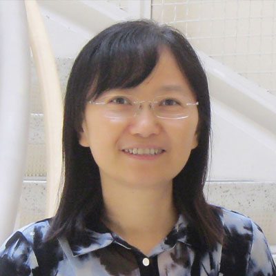
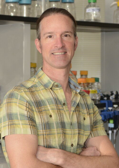
Amrinder Nain is a Professor in the Department of Mechanical Engineering at Virginia Tech. His research focuses on bio-inspired engineering, nanotechnology, and mechanobiology, with emphasis on understanding cellular interactions with fibrous environments. He leads the Spinneret-based Tunable Engineered Parameters (STEP) Lab, developing advanced materials and biomaterials for biomedical applications. Nain holds a Ph.D. in Mechanical Engineering from Carnegie Mellon University (2007) and has held faculty positions at Virginia Tech since 2009. Education: Ph.D., Mechanical Engineering, Carnegie Mellon University, 2007 M.S., Chemical Engineering, Carnegie Mellon University, 2007 B.E., Mechanical Engineering, Manipal Institute of Technology, 1990 Research Interests: Cellular dynamics and tissue engineering Nanofiber-based biomaterials Biomechanics of cancer and cellular migration Organ-on-a-chip technologies Advanced manufacturing of fibrous scaffolds Awards & Collaborations: John Jones Faculty Fellowship NIH-funded collaborations on blood-brain barrier models and cancer cell migration Global partnerships with institutions like Weizmann Institute of Science and Nara Institute of Science and Technology Labs & Teams: The STEP Lab pioneers nanofiber platforms to study cell-fiber interactions, with applications in drug testing and tissue engineering. Current projects include coiling dynamics of cellular protrusions, mitotic spindle orientation, and ultra-thin nanofiber mimics of biological membranes.
David G. Drubin is the Ernette Comby Chair in Microbiology and a Professor of Cell Biology, Development and Physiology at the University of California, Berkeley. He is also an affiliate of the Division of Genetics, Genomics and Development, with his lab focusing on molecular mechanisms of actin assembly and membrane trafficking in human stem cells, organoids, zebrafish, and budding yeast. Affiliation: Department of Molecular and Cell Biology, UC Berkeley Research Focus: Actin-mediated membrane trafficking, clathrin-mediated endocytosis, cytoskeletal dynamics, genome editing, stem cell differentiation, and yeast genetics His lab employs real-time imaging, genome editing, mathematical modeling, and biochemical reconstitution to study endocytic mechanisms. Key findings include the role of membrane curvature in endocytosis and the translation of yeast discoveries to mammalian systems. Google Scholar publications up to 2025 highlight his work on myosin-I, actin networks, and membrane biophysics. Lab members include students and researchers like Sun Hae Hong, Yansong Miao, and Nate Krefman. The lab has produced educational videos (e.g., DNA gel training) and maintains active research directions in actin force generation and organelle inheritance pathways.
Zhuhao Wu serves as Assistant Professor of Neuroscience at the Brain and Mind Research Institute, Weill Cornell Medical College since 2022. His research integrates neurovascular biology, neural circuit mapping, and neurodegenerative mechanisms to understand brain organization and disease processes. Education: Ph.D. in Neuroscience, The Johns Hopkins University School of Medicine (2011) B.S. in Biological Sciences, Tsinghua University, China (2003) Research Focus: Dr. Wu pioneers multi-scale investigations of neurovascular coupling , brain-wide circuit organization , and neurodegenerative pathways . His lab employs whole-brain imaging , single-cell transcriptomics , and genetic engineering in murine models to dissect mechanisms of stroke recovery, tau pathology, and developmental disorders. Current work emphasizes regional blood-brain barrier heterogeneity and axon degeneration pathways with therapeutic implications. Publication Trends: Recent work (2023-2025) reveals three convergent themes: (1) neurovascular dynamics in health/disease, (2) high-resolution brain atlasing techniques, and (3) molecular mechanisms of neurodegeneration. Publications in Cell , Nature , and Neuron demonstrate methodological innovation in circuit mapping and translational relevance to stroke, Alzheimer's, and autism spectrum disorders. Grant Portfolio: Principal Investigator Subaward: NINDS R01 Investigating Neurobiology of Early Cognitive Impairment (2024-2029) Principal Investigator Subaward: NINDS R01 Mechanisms of anosmia in COVID-19 (2023-2028) Principal Investigator Subaward: NINDS BRAIN CONNECTS Center for Large-scale Imaging (2023-2028) Principal Investigator Subaward: NINDS Global mapping of DDX3X mutation circuits (2023-2028) Principal Investigator Subaward: NIAID single-cell encephalitis pathogenesis (2023-2026) Dr. Wu leads a multidisciplinary team within the Brain and Mind Research Institute focused on developing HOLiS (whole-brain staining/clearing pipeline) and TrailMap for neural circuit analysis. His lab collaborates extensively on NIH BRAIN Initiative projects advancing large-scale connectome mapping.
Verena Kriechbaumer is a Senior Lecturer in Biotechnology and Plant Sciences at the School of Biological and Medical Sciences, Oxford Brookes University . She is Deputy Director of the Oxford Brookes University Centre for Bioimaging and a leading expert in plant endoplasmic reticulum (ER) structure, membrane proteins, and auxin biosynthesis, utilizing biochemical techniques, high-resolution live cell imaging, and interdisciplinary approaches. Research Focus: Plant cell biology, ER architecture, auxin metabolic pathways, protein-membrane interactions, bioinformatics, and translational projects such as engineering plants to convert methane into biofuel. Key Techniques: FRET-FLIM, light sheet microscopy, single-particle tracking, and optogenetics. Publication Trends: Recent studies emphasize ER-membrane contact sites, organelle interaction networks, and the role of reticulons in viral trafficking and methane monooxygenase expression. Collaborative work spans physics, bioenergy, and industrial biotechnology. Scientific Awards: Fellowship from Korean Federation of Science and Technology Societies (2013) Santander Travel Fellowship (2018) Oxford Brookes Research Excellence Award (2020-21) Grants: Leverhulme Trust grant for "pMMO in plants" (2015-2017), STFC Harwell facility grants (2017-2021), BBSRC funding (2021-2026), and industry collaborations with Porton Biopharma Ltd. Labs & Teams: Leads the Endomembrane Structure and Function Group , collaborates with physicists at STFC Harwell Campus, and contributes to European Commission-funded projects like "Advanced Training for Next Generation Scientists in Spatio-Temporal Imaging."
Timothy Holy, PhD is the Alan A. & Edith L. Wolff Professor of Neuroscience and Vice Chair of Research at Washington University School of Medicine. He leads the Holy Lab, which focuses on the neural mechanisms of olfactory coding and the development of innovative imaging technologies for neuroscience research. His educational background includes a BA in Mathematics and Physics (summa cum laude) from Rice University (1991), an MA in Physics from Princeton University (1992), and a PhD in Physics from Princeton University (1997) under thesis advisor Stanislas Leibler. Dr. Holy's research spans multiple domains of neuroscience with particular emphasis on the olfactory system of mice. His lab has pioneered light sheet microscopy for calcium imaging, enabling simultaneous recording from tens of thousands of neurons. More recently, they developed PhOTseq, a technique for optically tagging neurons for later sequencing. His work bridges physics, neuroscience, and computational approaches to understand how sensory systems process information. He is also among the world's foremost creators of the Julia programming language, which has gained exponential adoption in scientific computing. Analysis of his recent publications reveals a strong focus on neural coding in decision-making circuits, olfactory processing, and the development of computational tools for biological research. His work increasingly integrates machine learning approaches with traditional neuroscience techniques. Distinguished Teaching Service Award (2005, 2008, 2014) McKnight Technological Innovation in Neuroscience Award (2007) St. Louis Academy of Sciences Innovation Award (2009) NIH Director's Pioneer Award (2009) Society for Neuroscience Research Award for Innovation in Neuroscience (RAIN) (2009) Dr. Holy has mentored numerous students and postdoctoral researchers who have gone on to establish their own research programs. His lab has secured significant grant funding supporting technology development and fundamental neuroscience research. Current projects include investigating cellular mechanisms of individuality and plasticity, navigation using olfactory cues, and developing new mathematical tools for optimization and machine learning. The Holy Lab combines a focus on understanding neural circuits and behavior with a willingness to pioneer new technologies. It maintains strong collaborations across disciplines, particularly in the development and application of the Julia programming language for biological research.
Dr. Caroline Muellenbroich is a Senior Lecturer at the School of Physics & Astronomy, University of Glasgow, where she develops advanced microscopy techniques for cardiac and neuro imaging. She joined the university in 2018 and is based at the Advanced Research Centre. Her work bridges physics, engineering, and biomedical sciences to create innovative imaging solutions. Dr. Muellenbroich received her physics education at the University of Heidelberg, Germany, and earned her PhD from the Institute of Photonics, University of Strathclyde, Glasgow in 2012. Her doctoral research focused on adaptive optics in advanced microscopy techniques. She then pursued postdoctoral research at the Biophotonics group at the European Laboratory for Nonlinear Spectroscopy (LENS) in Florence, Italy, where she implemented confocal light-sheet microscopy for whole mouse brain imaging and functional calcium imaging in Zebrafish. From 2016-2018, she worked as a researcher with the Italian National Institute of Optics, part of the Italian National Research Council. Dr. Muellenbroich's research focuses on developing and applying advanced optical imaging techniques, particularly light-sheet microscopy, for biomedical applications. Her work spans neuroscience and cardiology, with a strong emphasis on whole-brain imaging in model organisms and cardiac electrophysiology studies. She has made significant contributions to improving imaging fidelity, developing artifact removal techniques, and creating open-source microscope hardware. Her research bridges fundamental physics with practical biomedical applications, enabling new discoveries in brain function and cardiac physiology. Analysis of her recent publications reveals a strong focus on light-sheet microscopy applications in neuroscience and cardiology. Her work addresses technical challenges in whole-brain imaging, artifact reduction, and the development of novel optical approaches for studying brain activity and cardiac function. She has made important contributions to the field through both technical innovations in microscopy hardware and novel applications of these techniques to biological problems. Dr. Muellenbroich is actively involved in developing open-source approaches to microscope hardware, as evidenced by her 2022 Nature Methods publication "CAD we share? Publishing reproducible microscope hardware." Her research has been supported by various grants, though specific funding sources are not detailed in the available information. She leads research efforts in developing advanced microscopy techniques for cardiac and neuro imaging, working with interdisciplinary teams that include physicists, engineers, biologists, and medical researchers. Her laboratory likely focuses on pushing the boundaries of optical imaging to address challenging biomedical questions in brain function and cardiac physiology.
Kate McDole is a Researcher at the MRC Laboratory of Molecular Biology, University of Cambridge. Her lab focuses on understanding how mechanical forces shape early mammalian embryos, combining live-imaging, genetics, and computational tools to study tissue morphogenesis at single-cell resolution. Her research investigates the transformation of simple cell populations into complex structures like the gut tube and heart, emphasizing the coordination of mechanical forces with gene expression. She pioneered an advanced light-sheet microscope to visualize post-implantation mouse development over multiple days, enabling detailed analysis of cell fate and tissue dynamics. The lab's work includes video demonstrations of their imaging systems and specific studies on cardiac and foregut development. Their publications highlight innovations in embryonic imaging and lineage analysis. Group members include Sonia Agüera Gonzalez, Katharine Goodwin, Jenny Kretzschmar, Ewa Paluch, and Henry Westmacott. Future goals involve creating a comprehensive force-map of embryogenesis to improve synthetic tissue engineering.
Brian S.Y. Kim serves as Assistant Professor of Materials Science and Engineering and Physics at the University of Arizona, holding a joint appointment since January 2024. Previously, he conducted postdoctoral research at Columbia University (2018-2023) in Mechanical Engineering and Physics. His laboratory focuses on atomic-scale engineering of quantum materials for next-generation electronic and photonic technologies. His educational background includes: PhD in Electrical Engineering, Stanford University (2018) MS in Electrical Engineering, Stanford University (2013) BS in Electrical Engineering, Northwestern University (2011) Professor Kim's research spans 2D quantum materials and heterostructures , experimental condensed matter physics , and advanced nanofabrication . His group employs robotic nano-manufacturing systems, reconfigurable device architectures, and nano-optical imaging to investigate emergent phenomena in van der Waals materials. Key methodologies include plasmonic cavity engineering, moiré superlattice fabrication, and atomic-scale device characterization. His publication record demonstrates consistent contributions to quantum materials science, with recent emphasis on plasmon-exciton interactions in layered antiferromagnets, novel FET architectures using 2D transition metal dichalcogenides, and thermal transport phenomena in nanostructured materials. The work bridges fundamental condensed matter physics with practical device applications. His scientific recognition includes: Outstanding Young Researcher Award (2024) from AKPA/KPS APL Photonics Early Career Editorial Advisory Board membership (2025-26) Research funding includes the Vertically Integrated Projects Catalyst Seed Fund Award (2024) for "twisting two-dimensional atomic sheets." The Kim Lab actively mentors undergraduate researchers like David Tashchyan and collaborates with institutions including Kyung Hee University and Sungkyunkwan University. Current projects focus on developing programmable hyperbolic polaritons and Fermi-level engineered 2D electrodes. The Brian SY Kim Lab operates within UArizona's Materials Science and Engineering department, utilizing state-of-the-art facilities for nanofabrication and quantum device characterization. Ongoing initiatives explore cavity-altered superconductivity and magnetically confined excitons, with potential applications in quantum computing and ultra-sensitive photodetection.
Jau-Nian Chen is a Professor in the Department of Molecular, Cell and Developmental Biology at the University of California Los Angeles within the College of Letters and Science. His research focuses on the molecular mechanisms underlying cardiac development, with a particular emphasis on using zebrafish as a model system to study heart formation, function, and disease. Chen's research program centers on cardiac development and gene regulatory networks, with significant contributions to understanding transcriptional regulation in heart development. His laboratory investigates how transcription factors and chromatin regulators control cardiac specification, morphogenesis, and function. A major focus of his recent work involves the Rtf1 component of the PAF1 complex and its role in transcriptional elongation during cardiogenesis. His research spans multiple areas including cardiac arrhythmia mechanisms, mitochondrial calcium handling in cardiomyocytes, and the genetic regulation of cardiac proliferation during development and regeneration. Analysis of his recent publications reveals a strong focus on transcriptional regulation in cardiac development, particularly through the PAF1 complex and Rtf1. His work increasingly integrates molecular genetics with functional physiology in zebrafish models to understand both normal cardiac development and disease mechanisms. The research spans from basic developmental mechanisms to potential therapeutic applications, with particular emphasis on calcium signaling, transcriptional control, and cardiac regeneration. Dr. Chen has been Principal Investigator on multiple NIH-funded research projects, including the current R01 grants "Impacts of transcription elongation on cardiac gene regulation during homeostasis and regeneration" (R01HL155905) and "Rtf1-dependent transcriptional regulation of heart development" (R01HL140472). His research program has been continuously funded by the National Institutes of Health since 2001, demonstrating sustained scientific impact in the field of cardiovascular development.
Malika Datta is a Research Fellow in Neuroscience at Yale School of Medicine, Yale University. Her work focuses on advanced neuroimaging techniques and molecular mechanisms in neuroscience. She holds appointments in the Neuroscience department and contributes to interdisciplinary research in biomedical engineering and pharmacology. Her research interests include light-sheet microscopy innovation for large-scale biological imaging, dopaminergic system analysis under drug exposure, and genome editing via focused ultrasound. She has published extensively on topics such as Norrin-mediated astroglia-neuron interactions and optogenetic systems development. Recent publications highlight her contributions to scalable microscopy technologies and brain mapping methodologies. Her work bridges engineering and basic neuroscience, with applications in drug impact studies and neurotechnology. No scientific awards are explicitly mentioned in the provided information.
Collin K. Kieffer is an Assistant Professor of Microbiology at the University of Illinois Urbana-Champaign, affiliated with the School of Molecular & Cellular Biology, Carl R. Woese Institute for Genomic Biology, and Grainger College of Engineering. His research focuses on HIV pathogenesis using advanced imaging techniques, including multiscale tissue imaging to study viral dissemination and latent reservoirs in humanized animal models and human patient samples. He holds a B.S. in Bacteriology and Genetics from the University of Wisconsin-Madison, a Ph.D. in Biochemistry from the University of Utah, and completed postdoctoral training in Oncology at the University of Utah and in Biology and Biological Engineering at Caltech. Research Interests : Dr. Kieffer’s work integrates cutting-edge imaging methods such as tissue clearing, electron microscopy, and light-sheet fluorescence microscopy to visualize HIV infection dynamics. His lab investigates mechanisms of HIV spread, reservoir persistence under antiretroviral therapy, and evaluates treatment efficacy. Recent projects explore viral interactions with immune cells in tissues, engineering anti-viral red blood cells, and SARS-CoV-2 spike protein biology. Labs & Affiliations : The Kieffer Lab leads interdisciplinary research at the intersection of virology and imaging, collaborating with institutions such as the Carl R. Woese Institute for Genomic Biology. His work bridges basic science and translational medicine, aiming to develop therapeutic strategies against HIV and other viral pathogens.
G. Ulrich Nienhaus is a Professor at the Institute of Applied Physics , Karlsruhe Institute of Technology (KIT) , and leads a research group focused on Biophysics and Nanoscopy . His work integrates physics, biology, chemistry, and computational methods to develop advanced light microscopy techniques with high spatial and temporal resolution. Key research areas include fluorescent protein engineering , single-molecule spectroscopy , super-resolution microscopy , and nanoparticle-biomolecule interactions . His group investigates molecular processes in living cells , protein folding , ligand dynamics , and quantitative imaging for biomedical and material science applications. Scientific Contributions span decades, with recent work highlighting innovative STED microscopy methods, DNA origami-based distance rulers , and fluorescent nanocluster applications . Publications emphasize biomolecular dynamics , nanoparticle corona formation , and live-cell imaging tools .
Alexandros Poulopoulos, PhD, serves as Associate Professor in the Department of Pharmacology & Physiology at the University of Maryland School of Medicine. His research integrates synthetic biology with developmental neuroscience to pioneer molecular therapeutics for neurogenetic disorders through advanced CRISPR-based genome editing technologies. Education: BSc in Biology, University of Athens, Greece (2003) PhD in Neuroscience, University of Göttingen, Germany (2008) Postdoctoral Fellow, Max Planck Institute for Experimental Medicine (2009) Postdoctoral Fellow (EMBO fellow), Massachusetts General Hospital (2012) Postdoctoral Fellow (HFSP fellow) and Research Associate, Harvard University (2016) Dr. Poulopoulos' research focuses on cortical development, synaptogenesis, and neurogenetic disease mechanisms. His lab develops precision CRISPR agents like Cas9-RC for in vivo somatic genome editing, targeting conditions including epilepsy, autism, schizophrenia, and neurodegeneration. Key investigations explore mTOR signaling pathways, cell adhesion molecules (particularly Neuroligin), and CRISPR delivery systems using in utero electroporation. His work bridges fundamental synaptic biology with therapeutic applications for brain disorders. Analysis of recent publications (2023-2025) reveals three dominant research trajectories: 1) Advancement of prime editing technologies for modeling rare epilepsies (particularly GRIN2A-related disorders), 2) Elucidation of synaptic organization mechanisms through phosphorylation-dependent neuroligin localization and axon guidance principles, and 3) Development of novel delivery platforms including focused ultrasound-mediated blood-brain barrier penetration and nanoparticle systems. These efforts demonstrate a clear progression from basic synaptic biology toward clinically translatable genome editing therapies. Scientific Awards: NIH TARGETED Challenge, phase II winner (2025) Society for Neuroscience Greater Baltimore Chapter President (2024) GPILS Teacher of the Year Award, University of Maryland (2020) NIH Director's New Innovator Award (2019) Harvard Distinction in Teaching Award (2015) Human Frontier Science Program Fellowship (2012) EMBO Fellowship (2010) Max Planck Society Otto Hahn Medal (2009) Dr. Poulopoulos leads the Poulopoulos Lab (poulab.org), which operates within the University of Maryland's Center for Innovative Medicine. His team comprises postdoctoral fellows, graduate students, and research technicians focused on CRISPR agent development and neurogenetic disease modeling. Current funding includes NIH New Innovator Award support for precision genome editing platforms and recent success in the NIH TARGETED Challenge for rare epilepsy therapeutics. He actively mentors PhD candidates through the Graduate Program in Life Sciences (GPILS) and serves as course director for advanced neuroscience modules. The lab employs cutting-edge approaches including single-cell transcriptomics of neuronal compartments, in utero prime editing, and light-sheet imaging of developing cerebellar circuits. Collaborations with clinical neurologists at UMMC and industry partners accelerate translation of their CRISPR-Cas9-RC system toward correcting genomic lesions in neurodevelopmental disorders, with particular emphasis on patient-specific epilepsy models.
Greg D. Field is an Adjunct Associate Professor of Neurobiology at Duke University and a Faculty Network Member of the Duke Institute for Brain Sciences. His laboratory investigates retinal processing of visual scenes, focusing on functional connectivity, light adaptation, circadian rhythms, and retinal degenerative conditions. Using multi-electrode arrays, transgenic mouse models, and chemogenetics, he examines how retinal circuits encode visual information and how degeneration impacts signaling. Recent work addresses neural adaptation mechanisms, retinal mosaics, and potential therapies for retinal diseases. Research Themes : Retinal circuit organization, neural adaptation, visual signal processing, retinal degeneration, and optogenetics Technologies : Multi-electrode recordings, light-sheet microscopy, computational modeling, and transgenic approaches The 15 most recent publications (2010-2025) demonstrate his work on retinal ganglion cell function, visual hierarchy encoding, and therapies for photoreceptor degeneration. Grants from the National Institutes of Health (2015-2025) support studies on neural population mapping, comparative biology, and retinal circuit restoration. His work bridges theoretical neuroscience with clinical applications, particularly in developing interventions for blindness.
Jan Huisken is a Humboldt Professor for Multiscale Biology at the Georg-August-Universität Göttingen, affiliated with the Johann Friedrich Blumenbach Institute of Zoology and Anthropology. His research focuses on advanced light sheet microscopy techniques for biomedical and developmental biology applications. Role: Humboldt Professor University: Georg-August-Universität Göttingen Department: Johann Friedrich Blumenbach Institute of Zoology and Anthropology Research interests include light sheet microscopy , biomedical imaging , and developmental biology with a strong emphasis on zebrafish models. He develops tools for tissue clearing , image processing , and 3D microscopy . The 15 most recent publications analyze innovations in light sheet microscopy, tissue clearing protocols, and computational methods for image restoration. These works span fields such as optical imaging , developmental cardiology , computational biology , and biomedical instrumentation . Huisken contributes to open-source microscopy systems like 'Flamingo' and 'BigFUSE,' aiming to democratize access to advanced imaging technologies. His work integrates engineering, computer science, and biology to solve complex imaging challenges.