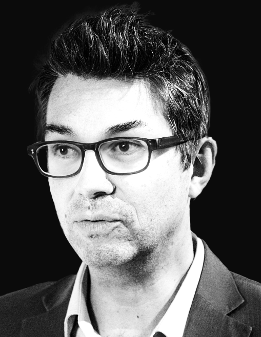
Stephane Cotin is a Research Director at Inria and leader of the MIMESIS team, specializing in real-time physics-based medical simulations. His work focuses on surgical training, planning, and image-guided therapy, with over 200 scientific articles and the development of the open-source SOFA framework. He co-founded InSimo, Twinical, and EVE, and previously held roles at Harvard Medical School and Mitsubishi Electric Research Lab. Cotin’s research bridges imaging, robotics, and medicine to improve healthcare outcomes, emphasizing patient-specific biophysical modeling and real-time computation. His awards include the Academy of Sciences Award (2018) and Dirk Bartz Medical Prize (2015). He has advised numerous PhD students and led projects like MediTwin and PREMYOM, advancing digital twin technologies for precision medicine.
Benedikt Günther is a research scientist at the Technical University of Munich (TUM) working within the Chair of Biomedical Physics led by Prof. Dr. Franz Pfeiffer. His research focuses on the Munich Compact Light Source (MuCLS), a laboratory-scale inverse Compton X-ray source that provides synchrotron-like radiation for biomedical applications. Günther plays a key role in developing, optimizing, and characterizing this innovative technology, contributing to both its fundamental physics and practical medical applications. His primary research interests center around X-ray physics and imaging techniques, particularly laser enhancement cavities for inverse Compton X-ray sources, X-ray microscopy, dynamic phase-contrast imaging, and X-ray spectroscopy. Günther's work bridges fundamental physics with practical medical applications, developing instrumentation that brings synchrotron-quality imaging to conventional laboratory settings. His research has significant implications for improving medical diagnostics while making advanced imaging techniques more accessible. Analysis of Günther's publication record reveals a consistent focus on advancing compact X-ray source technology and its applications. His work demonstrates expertise in both theoretical modeling and experimental implementation, with publications spanning instrument development, imaging techniques, and specific medical applications. The research shows progression from fundamental source characterization to increasingly sophisticated biomedical applications, particularly in breast imaging, dental diagnostics, and materials science. 2019 Best Poster Award at the combined meeting of the 68th Denver X-ray Conference (DXC) & 25th International Congress on X-ray Optics and Microanalysis (ICXOM) for 'Full-Field Structured Illumination Super-Resolution X-ray Transmission Microscopy' Günther regularly presents his work at major international conferences including the International Particle Accelerator Conference, High-Brightness Sources and Light-driven Interactions Congress, and specialized X-ray imaging meetings. His research is conducted within the Munich Compact Light Source facility, a collaborative project involving physicists, engineers, and medical researchers working to develop laboratory-scale synchrotron technology for widespread biomedical use.
Steffi Colyer is a Senior Lecturer in Biomechanics at the Department for Health, University of Bath. She is affiliated with the Centre for Health and Injury and Illness Prevention in Sport and the Bath Institute for the Augmented Human. Her research is supported by major grants from EPSRC and ESA, focusing on elite athletic performance, rehabilitation, and motion analysis technologies. Her research interests center on biomechanics of athletic performance, particularly in sports such as skeleton, badminton, and sprinting. She investigates the kinetic and kinematic determinants of elite performance, develops markerless motion capture systems for real-world analysis, and applies musculoskeletal modelling to understand internal loading and adaptation in normal and simulated gravity environments. Her work bridges sports science, engineering, and rehabilitation. The recent trend in her publications shows a strong focus on markerless motion analysis, pose estimation, musculoskeletal modelling, and the biomechanics of sprinting and racket sports. She leverages advanced computational methods, including deep learning and in silico simulations, to improve performance analysis and injury prevention. Her scientific awards include: ISBS New Investigator Award finalist (oral) (co-author), 2022 Departmental Staff Award for Innovation in Learning and Teaching, 2025 She has supervised multiple research students and projects, including PhD and postdoctoral work, and is actively involved in peer review for journals such as Journal of Sports Sciences , Scientific Reports , and Journal of Biomechanics . She leads the IAA project on markerless motion capture for skeleton push-start analysis and contributes to the CAMERA initiative, a major interdisciplinary research center focused on motion analysis and virtual reality applications. Her research is conducted within the Centre for the Analysis of Motion, Entertainment Research and Applications (CAMERA), where she collaborates with computer scientists, engineers, and sports scientists to develop and apply cutting-edge motion capture technologies in real-world settings.
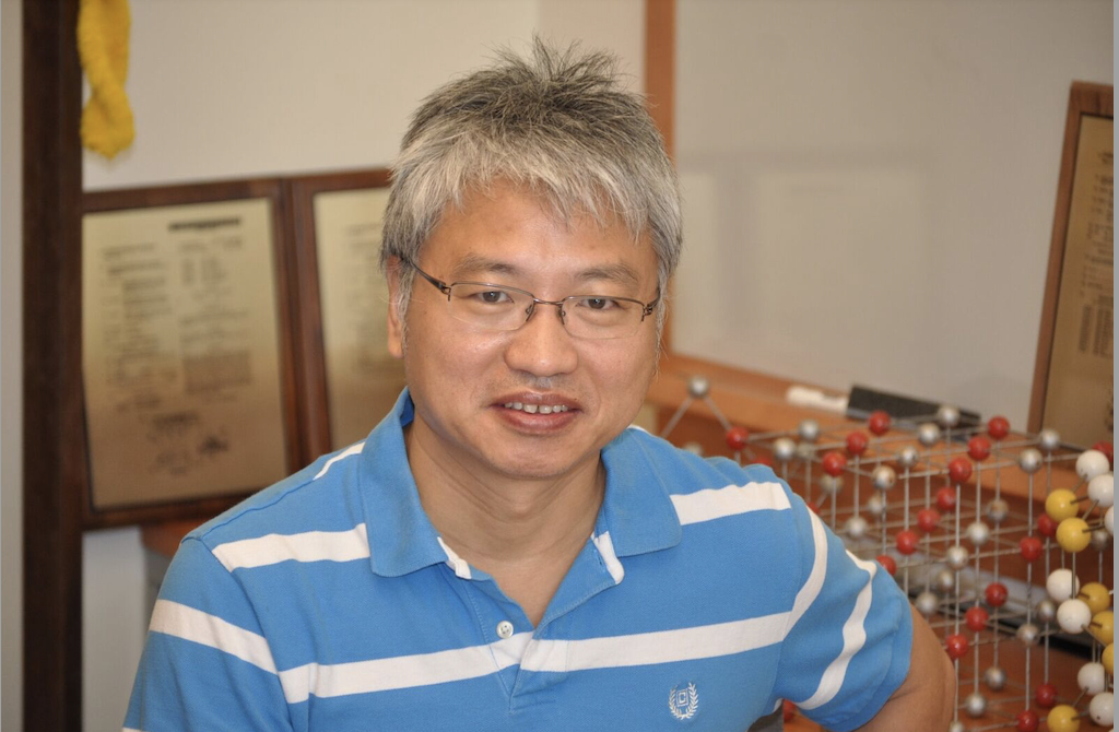
Giulio Dagnino is Associate Professor of Robotics and Mechatronics at the University of Twente and concurrently holds an appointment at the Digital Society Institute. His research integrates medical robotics, real-time perception and haptics to create MR-compatible platforms for endovascular surgery, earning an h-index of 17 and 971+ citations. Education & Career: PhD (details not specified in source) leading to faculty appointment at University of Twente. Promoted to Associate Professor with cross-appointments in Robotics & Mechatronics and Digital Society Institute. Research Interests: Prof. Dagnino’s core interest is medical robotic systems that can operate safely inside an MRI scanner. His work spans haptic guidance, real-time computer vision, soft robotic actuation, synthetic data generation and surgical simulation. By combining ferrofluid actuation, electromagnetic tracking and deep-learning-based scene understanding, he aims to reduce ionizing radiation exposure, enhance navigation accuracy and shorten procedure times for minimally invasive endovascular interventions. Publications Trend: Across 44 outputs (2010-2025) the portfolio reveals a clear evolution from early vision-based microsurgery and fracture-robot systems (2010-2016) toward holistic endovascular platforms integrating MR guidance, haptics and autonomy. Recent 2024-25 papers cluster around (i) synthetic data & scene understanding for surgical AI, (ii) MR-safe robot design and tracking, and (iii) translational studies bringing CathBot and related platforms closer to clinical use. Scientific Awards: Best Design Award – Hamlyn Symposium 2019 (with team) Best Innovation Award – ICRA 2018 Best Paper Award – CURAC 2019 IEEE ICRA Best Paper Award in Medical Robotics – 2016 Grants & Projects: Although explicit grant numbers are not listed, the continuous outputs, patents, multi-institutional collaborations (UK, Germany, Estonia, Canada) and press releases imply sustained funding from EU, Dutch and UK research councils as well as industrial partnerships. Labs & Teams: He leads activities within the Robotics and Mechatronics group at University of Twente, collaborates closely with the Digital Society Institute, and maintains international partnerships visible in co-authored papers with Imperial College London, University of Leeds, and several European hospitals.
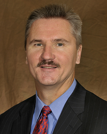
Richard D. Komistek serves as the Fred M. Roddy Professor of Biomedical Engineering and Co-Director of the Center for Musculoskeletal Research at the University of Tennessee, Knoxville, positions he has held since 2007 and 2003 respectively. His academic career spans over three decades with significant contributions to orthopaedic biomechanics and joint replacement technology. Dr. Komistek earned his PhD (1992), MSME (1989), and BSME (1988) from the University of Memphis, establishing a strong foundation in mechanical engineering principles applied to biomedical challenges. His research program focuses on advanced biomechanical modeling of the musculoskeletal system, with particular expertise in in vivo kinematic analysis of total joint replacements. Key research areas include failure mode analysis of arthroplasty implants, closed-loop control systems of human movement, and mathematical modeling of joint mechanics. His work bridges engineering principles with clinical orthopaedics to improve implant design and patient outcomes. Analysis of his publication record reveals consistent innovation in total knee arthroplasty research, with emphasis on tri-condylar designs, mobile-bearing systems, and in vivo measurement techniques. His work demonstrates evolving trends toward high-flexion implants, gender-specific considerations, and advanced fluoroscopic tracking methods to optimize joint replacement performance. Dr. Komistek's contributions have been recognized through prestigious awards including: University of Tennessee Research and Creative Achievement Award (2015) Multiple Research Fellow Awards (2005-2012) Knee Society Conventry Award (2003) ESB Clinical Biomechanics Award (1996-1998) Multiple Clinical Orthopedics Multimedia Awards Journal of Clinical Biomechanics Award (2007) As Co-Director of the Center for Musculoskeletal Research, he leads interdisciplinary teams developing next-generation diagnostic tools including mobile tracking fluoroscopy and implant diagnostic devices. His patented technologies focus on improving total hip and knee arthroplasty through innovations in load sensing, infection detection, and wear monitoring.
Kawal Rhode is a Professor in Biomedical Engineering and the Head of Education at the School of Biomedical Engineering & Imaging Sciences , King’s College London. His work bridges engineering, clinical practice, and education, with a focus on image-guided interventions, medical robotics, and 3D printing in healthcare. He leads the educational strategy for multiple taught programs, including the BEng/MEng in Biomedical Engineering and MSc programs in Healthcare Technologies and Clinical Sciences. His educational background includes a BSc in Basic Medical Sciences and Radiological Sciences from Guy's & St. Thomas' Hospitals Medical School (1992) and a PhD in arterial blood flow analysis from University College London (2006). He joined King’s in 2001 as a postdoctoral researcher and progressed through academic ranks to full Professor in 2016. Prof. Rhode’s research centers on image-guided interventions , medical robotics , and innovative pedagogy . His team develops intelligent systems for catheter-based procedures, robotic ultrasound, and simulation platforms using 3D printing. His work integrates AI, biomechanics, and translational engineering to improve clinical outcomes. His recent publications (2024–2025) reflect a strong trend in autonomous robotic systems , AI-driven image analysis , and simulation-based training , particularly in cardiac and interventional applications. These works span journals and conferences in medical imaging, robotics, and biomedical engineering, showcasing interdisciplinary innovation. Wellcome EPSRC Centre for Medical Engineering (Co-Investigator) Three-Dimensional Hybrid Guidance System for Cardiac Interventional Procedures (PI, EPSRC-funded) SIE CDT: Haemodynamics of complex aortic aneurysms (Co-I, Artivion Inc) He leads the Success for Black Engineers initiative, funded by the Royal Academy of Engineering, to improve diversity and inclusion in engineering education. This includes outreach, mentoring, industry engagement, and wellbeing support for Black students. He has supervised numerous students and collaborators, many of whom appear as co-authors on recent publications. His lab is part of a broader network within the School of Biomedical Engineering & Imaging Sciences, collaborating with clinicians at St Thomas’ Hospital and industry partners.
Sero Andonian is a Professor in the Department of Surgery at McGill University's Faculty of Medicine and Health Sciences and an Associate Investigator at the Research Institute of the McGill University Health Centre (RI-MUHC) at the Glen site. He specializes in Adult Urology within the Department of Surgery at the MUHC, with a primary focus on the Metabolic Disorders and Complications Program. Dr. Andonian's research spans multiple critical areas in urology, with particular emphasis on Endourology , kidney stone disease , radiation safety , medical education , minimally invasive surgery , and laparoscopy . His work has significantly advanced understanding of kidney stone disease management, patient quality of life impacts, and innovative surgical techniques. He has developed assessment tools like the Canadian Endourological Group Stent Symptom Score (CEGSSS) and contributed to national practice guidelines. Analysis of Dr. Andonian's publication record reveals a consistent focus on improving clinical outcomes for stone disease patients. His research encompasses epidemiological studies, surgical technique comparisons, quality of life assessments, and radiation safety protocols. Recent work includes investigations into vacuum-assisted versus traditional percutaneous nephrolithotomy, fluoroless ureteroscopy techniques, and the impact of bilateral stone disease on patient outcomes. As an active member of the Metabolic Disorders and Complications Program at RI-MUHC, Dr. Andonian collaborates with multidisciplinary teams to investigate the metabolic factors underlying urological conditions. His research bridges clinical practice with scientific investigation, ensuring that patient care is informed by the latest evidence-based approaches. He also contributes significantly to urology education through simulation-based training development and assessment of surgical competencies.
Jacques Van Dam, MD, PhD, is a Professor of Medicine (Clinical Scholar) at the Keck School of Medicine of the University of Southern California. He specializes in advanced gastrointestinal endoscopy, focusing on pancreatic, esophageal, gastric, and colorectal cancers, as well as precancerous conditions like Barrett’s esophagus and pancreatic cystic neoplasms. His clinical expertise includes diagnostic and therapeutic gastrointestinal endoscopy, ERCP, and EUS. Dr. Van Dam holds academic titles from Georgetown University (MD, PhD), Harvard Medical School (residency/fellowship), and specialized training at the Cleveland Clinic. He has been recognized with major awards including the AMA Inspirational Physician Award (2014) and the ASGE Distinguished Service Award (2015). His research spans translational medicine, endoscopic innovation, and cancer diagnostics. His work emphasizes minimally invasive procedures, including EUS-guided therapies and optical biopsy techniques. Dr. Van Dam has contributed to over 150 peer-reviewed publications, focusing on endoscopic innovations, cancer detection, and device development. He serves as a past president of the American Society for Gastrointestinal Endoscopy (ASGE) and other professional societies.
Philippe Cinquin is a Professor of Medical Informatics and Director of TIMC-IMAG (UMR5525), a CNRS-Université Joseph Fourier research unit. He leads the CAMI (Computer Assisted Medical Interventions) Labex and co-heads INSERM's CIC-IT 803 clinical investigation center. With a PhD in Applied Mathematics and Medical Doctor qualification, he pioneered surgical robotics and computer-aided interventions since 1984, benefiting over 100,000 patients through startup innovations. His recent work focuses on symbiotic implantable devices, energy scavenging for medical implants, and hydrogen therapy. He received the 2013 CNRS Innovation Award and 2014 Ambroise Paré Surgical Award. Research interests span biomimetic devices, medical robotics, and innovative therapies including transdermal hydrogen delivery. He has developed implantable systems like intestinal reactors and biosensors, with applications in diabetes treatment and pandemic response. His work integrates mathematics, signal processing, and nanotechnology to advance minimally invasive surgery and autonomous medical implants. Key contributions include virtual fluoroscopy systems for navigation, enzymatic biofuel cells, and reusable PPE sterilization methods during the pandemic. His interdisciplinary teams collaborate across engineering, medicine, and computer science to address clinical challenges through technological innovation. Education: PhD in Applied Mathematics; Medical Doctorate Grants & Startups: Founded multiple companies commercializing surgical navigation systems and diagnostic devices Labs: TIMC-IMAG, CAMI Labex, CIC-IT 803 clinical innovation center
University of North Carolina at Chapel HillUnited States
Professor Jianping Lu is a leading academic at the University of North Carolina at Chapel Hill, affiliated with the Department of Physics and Astronomy within the College of Arts and Sciences. His work focuses on advancing medical imaging technologies, particularly in X-ray and computed tomography (CT) systems, with a strong emphasis on carbon nanotube (CNT) X-ray sources. He holds a Ph.D. in Physics from the City University of New York (1988). Education: Ph.D. in Physics, City University of New York, 1988 Research Interests: Development of novel imaging systems, including stationary tomosynthesis and multisource CBCT Optimization of X-ray technology for clinical applications (e.g., oncology, cardiology, dentistry) Integration of artificial intelligence (AI) for diagnostic accuracy and automated analysis Portable and low-cost medical imaging solutions Recent Work Trends: His 2025 publications highlight advancements in AI-driven diagnostics for pancreatic cancer, improved contrast in adaptive radiation therapy, and stationary chest tomosynthesis systems. Key innovations include low-cost dual-energy CBCT and carbon nanotube-based X-ray arrays, which enhance image quality while reducing radiation exposure. His 2024 studies further explore cardiac imaging, dental tomosynthesis, and system optimizations for clinical adoption. Awards: None explicitly listed in the provided text. Advising & Grants: While student advisees are not listed, his research is likely supported by grants focusing on medical imaging innovation. Collaborations span physics, engineering, and clinical departments to bridge technical and clinical challenges. Labs/Teams: Likely affiliated with UNC’s imaging research groups, particularly those developing CNT X-ray technologies and clinical imaging systems for cancer and cardiovascular applications.
Mayo Clinic College of Medicine and ScienceUnited States
Stephen J. Riederer, Ph.D., is a Professor of Radiology at Mayo Clinic, holding dual appointments in the Department of Radiology and the Department of Physiology & Biomedical Engineering. He leads the Magnetic Resonance Laboratory, focusing on advancing MRI physics and clinical applications. His research emphasizes high-resolution prostate MRI, super-resolution T2SE imaging, and contrast-enhanced magnetic resonance angiography (CE-MRA). Dr. Riederer has developed fast-scanning techniques, real-time signal processing, and parallel acquisition methods, many of which are now industry standards. Education: B.A. in Mathematics, University of Wisconsin-Madison SM in Nuclear Engineering, MIT Ph.D. in Medical Physics, University of Wisconsin-Madison Research Interests: Dr. Riederer’s work bridges MRI physics and clinical implementation. Key areas include: Prostate cancer imaging via high-resolution T2SE and DCE-MRI Super-resolution MRI for improved anatomic detail Real-time MRI scanning and interactive triggering Parallel acquisition techniques and coil array optimization Publications & Impact: Over 300 peer-reviewed articles highlight his contributions to MRI innovation. Recent work focuses on AI-driven prostate MRI quality assessment and coil array improvements. His methods are widely adopted in commercial MRI systems. Awards & Leadership: Gold Medal (International Society for Magnetic Resonance in Medicine, 2002) President, Society of Magnetic Resonance Angiography (2008) George M. Eisenberg Professor I, Mayo Clinic (2024) Advising & Grants: Mentor to over two dozen doctoral students. Active in training via courses at the Mayo Clinic Graduate School. Leads grants on prostate MRI super-resolution and spatiotemporal imaging, funded by NIH and the U.S. Army. Labs & Affiliations: Part of the Center for Advanced Imaging Research, collaborating across radiology, biomedical engineering, and oncology. Facilities include state-of-the-art MRI scanners and imaging laboratories.
Ramez N Abdalla, MD, PhD, is an Assistant Professor of Radiology specializing in Interventional Neuroradiology at Northwestern University's Feinberg School of Medicine. His clinical focus includes endovascular management of cerebrovascular diseases, spinal interventions, and acute stroke treatment. He completed his MD, MS, and PhD at Ain Shams University, followed by postgraduate training in Egypt and fellowships at Northwestern University in Interventional Neuroradiology. Dr. Abdalla’s research emphasizes advanced imaging techniques (e.g., 4D Flow MRI) for optimizing stroke care, hemodynamic analysis in intracranial atherosclerosis, and novel treatments for cerebral vasospasm. Education: MD: Ain Shams University, Egypt (2010) MS: Ain Shams University (2015) PhD: Ain Shams University (2023) Fellowships: Ain Shams University (2018–2022) and Northwestern University (2019–2022) Research Interests: Endovascular treatment of neurovascular diseases 4D Flow MRI for cerebrovascular hemodynamics Stroke intervention outcomes Imaging biomarkers for intracranial atherosclerosis Publications reflect his focus on neurovascular imaging and clinical trials comparing endovascular vs. surgical treatments. Notable awards include the 2019 LINNC Travel Grant and Egyptian Ministry research scholarships. He is an active member of societies like the American Society of Neuroradiology and Society of Neurointerventional Surgery. Grants and Collaborations: His work is supported by institutional and registry-based research (e.g., NVQI-QOD). He has contributed to multi-institutional studies on stroke thrombectomy outcomes and ARUBA-eligible AVM treatments. No external industry relationships were disclosed for 2024. Labs/Teams: Collaborates with neurovascular imaging and interventional research groups at Northwestern, focusing on translational applications of advanced MRI and AI-driven diagnostic tools.
Aymeric Becq is an Associate Professor at Sorbonne University and a clinical practitioner in gastroenterology and endoscopy. He works at Henri Mondor Hospital (APHP, University of Paris Est Creteil) and Cochin Hospital (APHP, University of Paris). Since January 2020, he is a PhD student affiliated with the AGATHE Team, INSERM Unit U1150, and the Institute of Intelligent Systems and Robotics (ISIR). University Hospital Practitioner (Gastroenterology, Henri Mondor Hospital) PhD Student (Doctoral School SMAER, ISIR Laboratory) Research Focus: Medical Imaging, Endoscopy, and Gastroenterology His research bridges clinical practice and robotics, focusing on computer-aided detection in endoscopic imaging, 3D biliary reconstruction, and microbiota modulation in hepatocellular carcinoma. Recent work includes optimizing fluoroscopic image analysis and developing AI-driven tools for ERCP procedures. His publications since 2022 highlight trends in AI applications for medical imaging, particularly in guidewire detection, instrument segmentation, and biliary system modeling. Collaborations span INSERM, APHP hospitals, and institutions like Harvard Medical School.
Leendert-Jan W. Ligtenberg is a Researcher in the Robotics and Mechatronics department at the University of Twente , affiliated with the TechMed Centre . His work focuses on medical robotics, particularly magnetic actuation and wireless control systems for endovascular and gastrointestinal applications. Research Areas : Robotics, Mechatronics, Magnetic Actuation, Biomedical Engineering. Collaborations : Radboud University Medical Center (2024-2025). Scientific Activity Trends : Recent publications highlight advancements in untethered magnetic robots for clinical settings, including X-ray-guided control, dynamic modeling of capsules, and hybrid thrombus fragmentation techniques. Subfields include fluoroscopy integration , gastrointestinal navigation , and medical device simulation .
Dr. Martin Dierolf is a researcher at the Technical University of Munich (TUM), working within the Department of Physics and the Chair of Biomedical Physics led by Prof. Dr. Franz Pfeiffer. He is actively involved in research related to X-ray imaging, particularly focusing on the Munich Compact Light Source (MuCLS) and its applications in biomedical research. His work spans both the optimization of the MuCLS machine performance and the development of experimental methods for biomedical applications. Dr. Dierolf's primary research interests include: Optimization of the Munich Compact Light Source (MuCLS) for biomedical research Development of experimental and algorithmic methods for ptychography Biomedical applications of ptychographic coherent diffractive imaging (PCDI) Wave-field characterization of focusing optics Scanning transmission X-ray microscopy Grating-based phase-contrast imaging techniques His recent publications demonstrate a strong focus on advancing X-ray imaging techniques, particularly using compact light sources. His work spans from fundamental physics of X-ray optics to practical medical applications, with particular emphasis on breast imaging, renal tissue analysis, and cardiovascular applications. A significant portion of his recent work focuses on the Munich Compact Light Source and how to optimize its use for various biomedical applications. Dr. Dierolf has received recognition for his academic supervision, having been awarded the Supervisory Award of the Graduate Center of the TUM Department of Physics in both 2019 and 2021. He has also received Best Poster Awards at international conferences in 2008 and 2009. As an educator, Dr. Dierolf serves as a lecturer and teaching assistant for courses in Modern X-Ray Physics at TUM. He is scheduled to teach in the Winter term 2025/26, indicating his ongoing active role at the university. His research is conducted within the framework of the Munich Compact Light Source facility, which represents a significant advancement in making synchrotron-like X-ray sources accessible in laboratory settings. This work has potential applications across multiple biomedical fields, from cancer research to cardiovascular imaging.