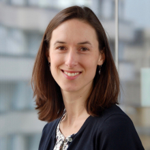
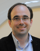
Professor Maja Pantic is a Professor of Affective & Behavioural Computing at the Department of Computing, Faculty of Engineering, Imperial College London. Her research focuses on artificial intelligence, image processing, and audio-visual speech recognition. She leads projects in multimodal systems, including facial analysis, emotion recognition, and speech-driven animation. Affiliations include the AI for Healthcare initiative, the Artificial Intelligence Network, and the Machine Learning Network. Her work addresses challenges in real-time speech enhancement, cross-modal learning, and synthetic data generation. Recent publications emphasize advancements in audiovisual speech synthesis, lip-reading, and emotion-aware systems. She has contributed to datasets like KAN-AV and SEWA DB, advancing research in face analysis and affective computing.
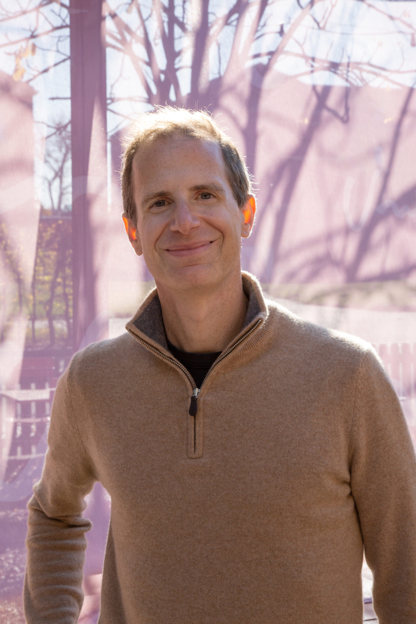
Sergey Tulyakov is the Director of Research at Snap Inc. , leading the Creative Vision team. His work focuses on enhancing creator capabilities through computer vision , machine learning , and generative AI , with applications in 2D/3D/4D video generation, editing, and personalization. He pioneered video generation frameworks like MoCoGAN and First Order Motion Model , and has been recognized for BEST IN SHOW AWARD at SIGGRAPH Real-Time Live! 2020. PhD (2012-2017): University of Trento, Italy MSc (2010): Belorusian State University of Informatics and Radioelectronics B.Eng (2009): Belorusian State University of Informatics and Radioelectronics His research interests span computer vision , generative models , 3D reconstruction , and personalization , with a focus on making large models efficient and mobile-compatible . Recent publications highlight advancements in 4D video generation , text-guided 3D composition , and lightweight architectures . Key scientific awards include the SIGGRAPH Real-Time Live! 2020 Best in Show for Interactive Video Stylization. He has also served on technical program committees for top-tier conferences like CVPR, ICCV, SIGGRAPH, and NeurIPS since 2022. His team organizes tutorials and keynotes, including courses on Deep Generative Models and Efficient Neural Networks . While no direct student names are listed, his collaborative work spans 60+ top-tier publications.
Serge Belongie is a Professor at the Department of Computer Science (DIKU) at the University of Copenhagen, where he holds dual affiliations with the Pioneer AI research section and the Image Analysis, Computational Modelling, and Geometry section. His academic position places him at the forefront of interdisciplinary research connecting computer vision with language models, geospatial analysis, and cultural understanding. Professor Belongie's research program encompasses several critical domains in modern artificial intelligence: Advanced computer vision and image analysis techniques Vision-language model integration and multimodal systems 3D point cloud processing and semantic segmentation Geospatial representation learning for environmental applications Fine-grained object recognition and detection Cultural context understanding in AI systems His recent publication record reveals a sophisticated trajectory toward developing precise control mechanisms for vision-language models, with applications spanning forensic analysis, cultural heritage preservation, and social media understanding. The research demonstrates increasing sophistication in handling cultural context and enabling fine-grained manipulation of visual content through natural language interfaces. Professor Belongie maintains an active research group producing significant scholarly output, with over 280 research publications documented in his academic profile. His work is supported by research funding that enables cutting-edge exploration in multimodal AI systems with practical societal impact. He plays a key role in the Pioneer AI center at the University of Copenhagen, which focuses on advancing artificial intelligence through interdisciplinary collaboration and innovative research approaches that bridge theoretical computer science with real-world applications.
Jingjing Zou is an Assistant Professor in Residence at the Herbert Wertheim School of Public Health & Human Longevity Science, University of California San Diego. Her research focuses on integrating statistical methodologies with medical imaging and public health challenges, particularly in cancer prevention, physical activity analysis, and radiomics. Education: PhD in Statistics, Columbia University MA in Statistics, Columbia University BS in Statistics, Peking University Research Interests: Dr. Zou's work emphasizes functional data analysis, accelerometer-based physical activity monitoring, and MRI techniques for cancer treatment evaluation. She develops statistical models to analyze longitudinal health data, with applications in cardiovascular disease prevention and oncology. Her studies often bridge clinical outcomes with advanced imaging biomarkers. Grants: NIH/NHLBI (Principal Investigator): Examining Longitudinal Changes in Accelerometer-Measured Physical Activity in Preventing Cardiovascular Disease (2024-2028) NIH R37CA249659 (Co-Investigator): Advanced diffusion MRI for cervical cancer treatment evaluation (2021-2026) NIH R01CA255780 (Co-Investigator): Whole-body radiomics for gynecologic cancers (2020-2024) Labs/Teams: Collaborates with interdisciplinary teams in radiology, oncology, and biostatistics, including co-authors like Loki Natarajan (UCSD) and Loren Mell (UCSD).
Knut Håkon Hole is an Associate Professor at the University of Oslo's Department of Radiology and Nuclear Medicine. His research focuses on diagnostic imaging applications in oncology, particularly in prostate and rectal cancers. He specializes in MRI, PET, and radiogenomics techniques to assess tumor biology, treatment response, and recurrence. Expertise: Prostate cancer imaging, neoadjuvant therapy response, tumor hypoxia, and imaging biomarkers Key affiliations: Oslo University Hospital (Rikshospitalet), Radium Hospital Research interests include: Developing MRI and PET protocols for cancer staging and recurrence detection Integrating imaging with genomic data (radiogenomics) Optimizing therapeutic approaches using imaging biomarkers Recent work highlights: Prostate cancer radiogenomics and hypoxia biomarkers (2024) MRI/PET comparisons for tumor localization (2021-2023) Neoadjuvant therapy response assessment in rectal and breast cancers (2020-2023) Publications span over 50 peer-reviewed articles with a focus on translational imaging research. Collaborates extensively with oncology and urology teams.
Christine Tardif is an Assistant Professor in the Department of Biomedical Engineering and the Department of Neurology and Neurosurgery at McGill University. As head of the McConnell Brain Imaging Centre lab at the Montreal Neurological Institute, she develops advanced MRI techniques for in-vivo brain imaging, focusing on quantitative mapping of myelin and cortical microstructure. Her work spans methodological innovation (e.g., multi-modal biophysical modeling) and translational applications across preclinical (7 Tesla) and clinical (3 and 7 Tesla) systems. Undergraduate: B.Eng. in Computer Engineering, McGill University (2004) Master's: M.Sc. in Bioengineering, Imperial College London (2006) PhD: Biomedical Engineering, McGill University (2011) Her research explores myelin dynamics in health and disease, emphasizing its role in neural conduction, brain plasticity, and cognitive functions. The lab investigates dysmyelination in psychiatric disorders (e.g., bipolar disorder) and neurodegenerative conditions (e.g., multiple sclerosis) using relaxometry , magnetization transfer , and diffusion-weighted imaging . Recent methodological work includes 3D MERMAID sequences for motion-insensitive diffusion imaging and optimization of magnetization transfer saturation maps. Current projects integrate ultra-high field MRI with histological validation in preclinical models (e.g., marmoset brain sections), aiming to bridge microstructural metrics with macro-scale brain function. Applications span Alzheimer's disease risk assessment via white matter alterations, synaptic density mapping in psychosis, and cortical laminar differentiation studies.
Robert Fulbright, MD, is Professor of Radiology and Biomedical Imaging at Yale School of Medicine with a secondary appointment in Neurology. He serves as Medical Director of the Magnetic Resonance Research Center and specializes in neuroradiology, focusing on diagnostic examinations of the brain, head and neck, spine, and peripheral nervous system using CT, MRI, and MR spectroscopy. Dr. Fulbright completed his medical degree at Baylor College of Medicine (1984), followed by residency training at Baylor College of Medicine and Columbia University, and a fellowship at Yale University School of Medicine. He is board certified in both Diagnostic Radiology (1991) and Internal Medicine (1988). His research centers on advanced magnetic resonance techniques to better understand brain function and disease mechanisms, with particular emphasis on Deuterium Metabolic Imaging (DMI) for mapping brain tumor metabolism. His work bridges radiology, neurology, and oncology to improve diagnostic capabilities and patient outcomes. Analysis of his recent publications (2023-2025) reveals three major research thrusts: metabolic imaging of brain tumors using DMI, genomic correlations with imaging findings in meningiomas, and technical innovations in MRI acquisition and processing. His work demonstrates increasing integration of AI techniques with traditional imaging modalities. As Medical Director of the Magnetic Resonance Research Center, Dr. Fulbright oversees a multidisciplinary team working at the forefront of neuroimaging technology. His clinical work focuses on neuroradiology with particular expertise in brain tumor imaging and neurological disorders.
Anna Vilanova is a Full Professor in Visual Analytics at the Department of Mathematics and Computer Science, Eindhoven University of Technology (TU/e), and is associated with the Electrical Engineering department's Signal Processing Systems. Previously, she served as Associate Professor at TU Delft (2013-2019) and Assistant Professor at TU/e (2002-2013). Her research focuses on Visual Analytics for high-dimensional data , explainable AI , and biomedical applications including Diffusion Weighted Imaging, 4D Flow, and Pangenomics. Education: Doctorate in Computer Graphics & Visualization (2001) Master in Computer Science (1997), Universitat Politècnica de Catalunya Research Highlights: Vilanova leads work on Visual Analytics systems for biomedical data, with recent publications in Diffusion MRI modeling , Tractography visualization , Explainable AI frameworks , and Pangenomic variant analysis . Her work bridges dimensionality reduction , uncertainty visualization , and medical imaging applications. Scientific Contributions: NWO-Veni grant (2005): "Visualization of global tensor information for diffusion tensor imaging" NWO-Aspasia grant (2013) Best Poster Award EuroVis (2025) Best Demo/Poster Awards (2022) Leadership & Service: Vilanova serves on the IEEE VIS Steering Committee , was EUROGRAPHICS President (2019-2022), and contributes to conferences like IEEE Visualization and EG-EuroVis . She co-founded the EAISI Health research initiative at TU/e.
Prof. Dr. Florian Knoll is a full professor in Computational Imaging at the Department of Artificial Intelligence in Biomedical Engineering (AIBE) at Friedrich-Alexander-Universität Erlangen-Nürnberg. He leads the Computational Imaging Lab, focusing on machine learning applications in medical imaging, particularly accelerating MRI through innovative reconstruction algorithms and translating them into clinical practice. His research emphasizes improving MRI speed, artifact robustness, and accessibility, alongside developing quantitative biomarkers for disease processes. Knoll's work is funded by NIH grants, including projects on machine learning for musculoskeletal imaging, MR fingerprinting, and deep learning frameworks for MRI reconstruction. He is a key figure in open science initiatives, co-creating the fastMRI dataset with Facebook AI, providing public access to over 1300 knee and 7000 brain MRI scans. He currently serves as deputy editor of Magnetic Resonance in Medicine and chairs the ISMRM Reproducible Research Study Group. His contributions extend to reproducible research, maintaining GitHub repositories with code for image reconstruction techniques (e.g., AGILE, gpuNUFFT) and educational materials. He teaches medical imaging fundamentals at FAU, integrating theoretical and practical insights for students and researchers. Grants: NIH R01EB024532, R21EB027241, P41EB017183, R01EB029957 Labs/Teams: Computational Imaging Lab, fastMRI initiative Software: GitHub repositories for MRI reconstruction (e.g., github.com/FlorianKnoll )
Mariano Cabezas is a researcher in medical imaging and computer vision, currently affiliated with Macquarie University and as an affiliate at the University of Sydney . His work focuses on automating brain MRI analysis for pathologies like multiple sclerosis, Alzheimer's disease, and tumors, with additional contributions to UAV image analysis. PhD in Computer Science (2013), University of Girona MSc in Automation, Computation, and Systems (2010), University of Girona BSc in Computer Science (2009), University of Girona Research Interests : Specializes in magnetic resonance imaging , lesion detection , deep learning , and image processing , with applications in multiple sclerosis , hearing loss , and UAV-derived ecological data . His recent work includes federated learning frameworks for cross-site MS lesion segmentation and pseudo-labeling techniques for longitudinal brain volume estimation. Publication Trends : Over the past five years, his research has emphasized federated learning (4 articles), lesion segmentation (9 articles), and UAV image analysis (3 articles), with a strong focus on clinical validation and cross-institutional collaboration. Labs & Collaborations : Contributed to the NIC-VICOROB group at the University of Girona and maintains affiliations with the Research Institute of the Hospital Vall d'Hebron (VHIR) in Barcelona and Macquarie University in Sydney. Actively develops open-source tools hosted on GitHub.
Xiaoqiang Wang is a Professor in the Department of Scientific Computing at Florida State University (FSU). His research focuses on numerical analysis, applied partial differential equations, mathematical biology, image processing, and scientific computing. He holds a Ph.D. from Pennsylvania State University (2005). His work emphasizes phase-field modeling for elastic bending energy, biological microstructures, and computational methods for complex systems. Notable contributions include advancements in centroidal Voronoi tessellation algorithms for image segmentation and high-performance computing techniques for scientific visualization. Recent publications highlight innovations in topology-preserving phase-field models, neural network-based energy minimization, and stochastic resource competition models. His research bridges theoretical mathematics with practical applications in biophysics, materials science, and biomedical engineering. Wang collaborates actively with interdisciplinary teams, contributing to FSU's computational science initiatives. His lab focuses on developing novel numerical methods and simulations for biological and physical systems, reflecting a commitment to both foundational and applied research.
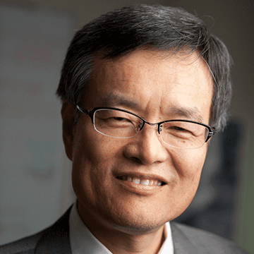
Hsiao-Dong Chiang is a Professor in the School of Electrical and Computer Engineering at Cornell University. He holds a Ph.D. in Electrical Engineering from the University of California, Berkeley, and has made significant contributions to nonlinear system theory and power system stability. His research spans theoretical development and practical applications in electric power systems, nonlinear optimization, and machine learning. B.S., Electrical Engineering, National Taiwan University, 1979 M.S., Electrical Engineering, National Taiwan University, 1981 Ph.D., Electrical Engineering, University of California, Berkeley, 1986 Chiang's research interests focus on nonlinear system theory , power system stability and control , nonlinear optimization , and their applications to modern power grids with high penetration of inverter-based resources. He is renowned for developing the BCU method and TRUST-TECH methodology , which have enabled fast direct stability assessment and global optimization in complex systems. His work bridges fundamental theory with industrial deployment through his companies, Bigwood Systems, Inc. and Global Optimal Technology, Inc. His recent publications (2024–2025) reflect a strong trend toward integrating machine learning and deep neural networks with power system analysis , particularly in state estimation, optimal power flow, and voltage control. There is a clear emphasis on handling uncertainty, non-convexity, and multi-scale dynamics in active distribution networks and integrated energy systems . His work increasingly focuses on resilience , real-time control , and user-centered methodologies for modern grid operations. Chiang has received numerous scientific honors, including: IEEE Fellow (1997) United States Presidential Young Investigator Award (1989) Multiple DOE Grid Optimization Challenge Awards (2020–2023) Best Paper Awards from IEEE Transactions and Conferences Outstanding Education Award, Cornell University (1990) He has successfully managed over 100 research projects and holds 28 U.S. and international patents. As the founder of Bigwood Systems, Inc., he has commercialized advanced software for utility companies across the U.S. and Japan. His team has published over 480 refereed papers and received more than 17,500 citations. He advises a large research group and leads innovations in computational methods for energy systems. His lab is actively involved in developing next-generation tools for grid security, optimization, and machine learning integration.
Dr. William M Holmes is a Senior Research Fellow and Senior MRI Physicist at the University of Glasgow's School of Psychology & Neuroscience, affiliated with the Glasgow Experimental MRI Centre. He holds a PhD in Physical Chemistry from the University of Nottingham and specializes in advancing MRI techniques for biomedical and physical sciences. His work bridges physics, neuroscience, and clinical applications, with a focus on cerebral blood flow imaging, glymphatic system dynamics, and disease modeling in rodents. Key roles: MRI method development, neuroimaging biomarkers, porous media analysis Leadership: Glasgow Experimental MRI Centre Research interests emphasize novel MRI applications in stroke, neurological disorders, and material science. Over 80 peer-reviewed publications demonstrate contributions to perfusion imaging, biofilm dynamics, and translational MRI techniques. Recent projects include: Quantitative arterial spin labeling methods Glymphatic system's role in multiple sclerosis Non-invasive rodent disease modeling
Jie Deng, Ph.D., is a Professor in the Department of Radiation Oncology at UT Southwestern Medical Center, where she serves as faculty in the Division of Medical Physics & Engineering. She is a certified MRI and MRI for radiation therapy medical physicist by the American Board of Medical Physics and holds a leadership role as a magnetic resonance safety officer. Dr. Deng is actively involved in both clinical and research aspects of medical imaging and radiotherapy, with a strong emphasis on integrating advanced imaging technologies into therapeutic workflows. Dr. Deng earned her academic degrees from prestigious institutions: a Bachelor of Science in Biomedical Engineering from Southeast University in China, a Master’s in Bioengineering from the University of Illinois at Chicago, and a Ph.D. in Biomedical Engineering from Northwestern University. She further enhanced her expertise by obtaining a Master of Science in Law from the Northwestern Pritzker School of Law, reflecting a multidisciplinary approach to her scientific work. Her research interests center on MRI physics , quantitative imaging , oncological imaging , and the application of artificial intelligence in medical imaging. She has pioneered work in MRI-guided radiation therapy, imaging biomarkers for therapeutic response, and AI-driven image reconstruction and artifact reduction. Her recent publications demonstrate a consistent focus on improving imaging accuracy, speed, and clinical utility, particularly in liver, pediatric, and oncological applications. The analysis of her 15 most recent articles reveals a strong trend toward deep learning-based image reconstruction , quantitative MRI biomarkers , and synthetic image generation for radiotherapy planning. Topics such as 4D-MRI, synthetic CT, motion artifact reduction, and AI fusion models dominate her scholarly output, indicating a forward-looking research trajectory centered on intelligent, fast, and precise imaging for personalized cancer therapy. Dr. Deng actively contributes to the scientific community through presentations at major conferences including the International Society for Magnetic Resonance in Medicine (ISMRM) and the American Association of Physics in Medicine (AAPM), where she shares innovations in MRI, adaptive radiotherapy, and AI integration. As an educator, Dr. Deng mentors medical physics residents and graduate students, delivering lectures on MR-only simulation, MR-linear accelerator practices, and medical imaging fundamentals. While no specific grants are mentioned in the text, her extensive publication record in high-impact journals suggests active research funding and collaborative projects. She is affiliated with key professional organizations and serves on UT Southwestern’s MRI Safety Committee, ensuring safe and effective use of MRI in clinical and research settings. Her work bridges the gap between engineering innovation and clinical application, making significant contributions to the field of radiation oncology and medical physics.
Prof. Dimitrios Karampinos is a Professor at the Technical University of Munich (TUM), leading the Experimental Magnetic Resonance Imaging group within the TUM School of Medicine and Health. He specializes in developing novel MRI techniques for quantitative biomarker discovery, focusing on musculoskeletal, metabolic, and oncological applications. His career includes a PhD from the University of Illinois (2008), postdoctoral research at UCSF (2009–2012), and leadership roles at TUM since 2012. Prof. Karampinos has pioneered advancements in MRI reconstruction, signal modulation, and biomarker validation for clinical translation. Educations: BSc in Mechanical Engineering (National Technical University of Athens, Greece), PhD in Biomedical Engineering (University of Illinois, Urbana-Champaign, 2008). Research Interests: Development of MRI measurement techniques, quantitative biomarkers for disease diagnosis, and improving therapy monitoring. Key areas include musculoskeletal disease imaging, metabolic disorder assessment, and oncology applications. His work emphasizes translating research into clinical practice through innovations like accelerated imaging, artifact correction, and AI-driven analysis. Awards: ERC Starting and Proof of Concept Grants (2015, 2019), TUM Supervisory Award (2020), ISMRM Junior Fellow (2011). Grants: Multiple ERC grants for MRI method development. Labs/Teams: Leads the Experimental Magnetic Resonance Imaging group at TUM, collaborating on clinical and technical MRI advancements.