Evelyn Lake is an Associate Professor in the Department of Radiology and Biomedical Imaging at Yale University, with affiliations to the Wu Tsai Institute and Yale Biomedical Imaging Institute. Her research focuses on functional neuroimaging, particularly using rodent models to study brain connectivity, neurovascular coupling, and disorders like Alzheimer’s disease and autism spectrum disorder. PhD in Medical Biophysics, University of Toronto (2016) Postdoctoral Associate, Yale University (2019) BSc (Honors) in Biophysics, University of Guelph (2010) Her work spans multimodal imaging techniques (e.g., simultaneous Ca2+ and fMRI), examining how genetic mutations (e.g., PTEN, KATNAL2) affect cerebrospinal fluid dynamics and neuronal connectivity. She investigates longitudinal changes in Alzheimer’s models and methodological aspects of animal neuroimaging, such as sample size optimization and awake imaging protocols. Recent publications highlight her contributions to understanding autism-related white matter abnormalities, transdiagnostic connectome predictive modeling, and the impact of neurovascular uncoupling in awake mice. Collaborations include Todd Constable, Francesca Mandino, Xilin Shen, and Xenophon Papademetris.
Leslie Valiant is the T. Jefferson Coolidge Professor of Computer Science and Applied Mathematics in Harvard University's School of Engineering and Applied Sciences, where he has held a faculty position since 1982. A foundational figure in theoretical computer science, his work bridges artificial and natural computational phenomena across multiple disciplines. His academic background includes education at: King's College, Cambridge Imperial College, London Ph.D. in Computer Science from Warwick University (1974) Valiant's research spans computational complexity , machine learning theory , parallel systems , and computational neuroscience . He pioneered the PAC (Probably Approximately Correct) learning framework that established computational learning theory as a rigorous field. His holographic algorithms work revealed deep connections between computational complexity and statistical physics, while his neuroidal model and evolvability theory provide computational explanations for cognitive processes and biological evolution. Current investigations focus on cortical computation primitives and knowledge infusion architectures. His publication trends show increasing integration of neuroscience with computational theory since 2010, with dominant themes in holographic computation (2006-2018), cortical modeling (2012-2018), and evolvability (2009-2017). The work consistently applies computational complexity analysis to biological and cognitive systems. Major recognitions include: Nevanlinna Prize (1986) for mathematical aspects of computer science Knuth Award (1997) for foundational algorithms contributions EATCS Award (2008) for theoretical computer science impact Turing Award (2010) for computational learning theory and complexity Fellowship in the Royal Society and National Academy of Sciences Valiant's research has been supported by NSF and international grants enabling cross-disciplinary work in computational neuroscience and evolutionary algorithms. While specific advisees aren't documented in source materials, his theoretical frameworks have shaped generations of researchers in machine learning and complexity theory. His current research group explores neuroidal architectures for cognitive computation, investigating how cortical circuits achieve robust information processing through in-circuit testing methodologies. Ongoing projects aim to identify fundamental computational primitives in neural systems and develop biologically inspired AI frameworks.
Dr. Hillel Adesnik is a Professor in the Department of Neuroscience at the University of California, Berkeley, and a leading researcher in the neural basis of sensory perception. His lab focuses on cortical microcircuits, optogenetics, and neural coding, with emphasis on visual processing and memory formation. Key Research Areas: Cortical Microcircuits Optogenetic Tools Gamma Band Rhythms Neural Coding Mechanisms Dr. Adesnik has pioneered high-speed optical methods like 3D-MAP and 3D-SHOT to manipulate neural activity. His work spans cortical dynamics, synaptic plasticity, and cortical layer interactions, with applications in understanding learning algorithms and sensory inference. Selected Trends from Publications: Recent preprints and papers highlight advancements in cortical VIP neuron function, channelrhodopsin structures, and inter-areal computations. His team utilizes two-photon holography, cryo-EM, and computational modeling to decode perception-related neural codes. Scientific Awards: NIH Director's New Innovator Award (2013) Dr. Adesnik's lab collaborates with institutions like NIH and develops tools for awake animal studies. Funding includes grants from the Beckman Young Investigator Program and NIH.
Istvan Mody is a Professor at the University of California, Los Angeles (UCLA) with appointments in the Department of Neurology and Department of Physiology . His research focuses on synaptic signaling in health and disease, including mechanisms of GABAergic transmission, calcium homeostasis, and their roles in neurological disorders such as epilepsy, Alzheimer's disease, Huntington's disease, stress, alcoholism, and postpartum depression. He utilizes advanced techniques like patch-clamp electrophysiology, neuroanatomical and immunohistochemical methods, and molecular biology in animal models and human brain tissue . Research Interests: Dr. Mody investigates the physiology, pharmacology, and pathology of synaptic transmission and extrasynaptic receptor activation , with a particular emphasis on GABA(A) receptors and their subunit-specific modulation. His work explores how disruptions in excitation-inhibition balance contribute to neurological diseases, including mechanisms of tonic inhibition , calcium signaling , and neurosteroid interactions . He also studies the effects of chronic stress and hormonal fluctuations on neural excitability and behavior. Publications Trends: Recent studies highlight his work on gamma oscillations in Alzheimer's models, microglial dynamics , and rehabilitation strategies for stroke. His lab develops optical tools like dqGEVI for neuronal activity monitoring and investigates human brain organoids to model network dysfunction in epilepsy and intellectual disability. Laboratory Location: 635 Charles Young Dr S, Los Angeles, CA 90095, United States.
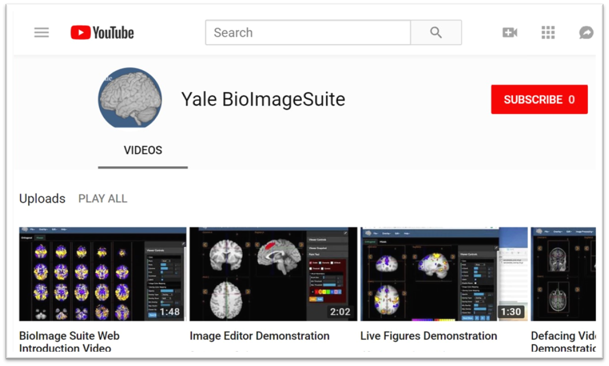
Xenophon Papademetris is a Professor of Biomedical Informatics & Data Science and Radiology & Biomedical Imaging at Yale School of Medicine. He serves as Associate Director of Biomedical Imaging Data Sciences at Yale Biomedical Imaging Institute and directs the Medical Software and Medical Artificial Intelligence Certificate Program. PhD in Electrical and Information Sciences from Yale University (2000) BA from Cambridge University (1994) Postdoctoral Fellowship at Yale University (2002) His research focuses on medical image analysis, machine learning, and biomedical software development. He has developed tools like BioImage Suite Web and contributed to standards committees at the Association for the Advancement of Medical Instrumentation (AAMI). His work spans modalities including MRI, CT, PET, and optical imaging. Recent publications emphasize neuroimaging analysis, explainable AI in healthcare, and multimodal data integration across species. He leads NIH-funded research under the BRAIN Initiative (R24 MH114805) and has authored a textbook on Medical Software published by Cambridge University Press. IEEE Senior Member Yale Brown-Coxe Postdoctoral Fellowship Harding Bliss Prize for Excellence in Engineering He directs the BioImage Suite Project, creating web-based image analysis tools using JavaScript and WebAssembly. His teaching includes both academic courses and a Coursera program on Medical Software with over 14,000 enrollments.
Dr. Rodolfo J. Flores serves as an Assistant Professor in the Department of Psychology within the College of Liberal Arts at the University of Texas at El Paso (UTEP). His primary research investigates neuromodulator function in the prefrontal cortex, with emphasis on acetylcholine and dynorphin systems during motivational conflict, stress responses, and addiction pathways. He employs advanced optogenetic and biosensor technologies to dissect neural circuitry underlying psychiatric conditions. Dr. Flores earned his Ph.D. in Social Cognitive and Neuroscience from UTEP in 2019, followed by postdoctoral training at the National Institute of Mental Health (NIMH). His work bridges molecular neuroscience with behavioral outcomes, focusing on how stress and substance exposure alter prefrontal cortical function. His research portfolio demonstrates consistent high-impact publication in top neuroscience journals including Nature Neuroscience , Neuron , and Psychoneuroendocrinology . Current work examines opioid neuropeptide dynamics, nicotine-alcohol interactions, and sex-specific hormonal influences on addiction behaviors. Methodologically, he specializes in genetically encoded biosensors and circuit-level manipulations to study threat processing and reward pathways. Dr. Flores teaches graduate and undergraduate courses including PSYC 4341 (Motivation & Emotion), thesis/dissertation supervision (PSYC 5395/6395), and research applications courses. His lab actively trains students through undergraduate research (RSRC 4033) and independent study opportunities (PSYC 4352).
Dr. Kanwaljeet S. Anand is a dual-appointed Professor of Pediatrics (Pediatric Critical Care) and Anesthesiology, Perioperative & Pain Medicine at Stanford University School of Medicine. As director of the Pain/Stress Neurobiology Lab and Jackson Vaughan Critical Care Research Fund, he serves as Editor-in-Chief of Pediatric Research and maintains active membership in Bio-X, MCHRI, and Wu Tsai Neurosciences Institute. Rhodes Scholar with D.Phil from University of Oxford Harvard postdoctoral fellowship and Boston Children's Hospital residency Founded Harmony Health Clinic - Arkansas' largest charitable medical-dental facility A translational researcher with 30+ years of impact, Dr. Anand established the first scientific framework for infant pain perception and developed novel pain assessment methodologies. Current research focuses on: Hair biomarker analysis for stress and social affiliation (cortisol/oxytocin) Machine learning systems for objective pain detection in non-verbal infants Biopsychosocial interventions for stress reduction in disadvantaged youth Neurotoxicity mechanisms of sedatives in developing brains Global NICU opioid usage patterns through the NeoOpioid Consortium His work has yielded over 260 publications and significant advances in: Pediatric pain management protocols Neonatal stress biomarker development Critical care neurobiology insights Community health initiatives AI-driven clinical decision support systems Scientific Recognition 9th Annual 'In Praise of Medicine' Public Address, Erasmus University (2014) Nightingale Excellence Award (2016) Honorary Doctorate from University of Örebro (2019) NIH SBIB-H82 Study Section Chair (2018) Multiple IASP and American Pain Society awards Swedish Academy of Medicine's Nils Rosén von Rosenstein Award (2009) St. Jude Endowed Chairholder (2010) As mentor to Med Scholar Anjali Gupta and advisor to numerous professional bodies, Dr. Anand maintains active clinical leadership in Pediatric Intensive Care while advancing computational approaches to pain detection through collaborations with Stanford's AI researchers.
Chandan J Vaidya is a Professor in the Department of Psychology at Georgetown University, directing the Developmental Cognitive Neuroscience Laboratory (DCNL). His research focuses on cognitive neuroscience mechanisms underlying adaptive behaviors, particularly implicit learning, executive control, and their dysfunction in ADHD, ASD, and other developmental disorders. Using multidisciplinary methods including fMRI, behavioral testing, and genetic analysis, he investigates how dopamine systems, brain connectivity, and environmental factors influence cognitive processes. Primary appointment: Professor, College of Arts and Sciences - Department of Psychology Education: Ph.D. from Syracuse University Research interests include neurodevelopmental disorders, neuroimaging of cognitive control, and translational neuroscience. Recent work examines striatal connectivity changes in ADHD due to stimulant use, executive dysfunction subtypes in autism, and brain correlates of reward processing in obesity. Key findings highlight hyperconnectivity in ASD, dopamine genotype influences on executive function, and age-related changes in default mode networks. Ongoing studies explore transdiagnostic models of psychopathology and precision medicine approaches in neurodevelopmental disorders. Lab activities focus on translational research bridging basic neuroscience with clinical applications. Collaborations involve pediatric neurology, psychiatry, and computational modeling.
Kamal Sen is an Associate Professor in the Department of Biomedical Engineering at Boston University, serving as Director of the Natural Sounds and Neural Coding Laboratory and Director of Admissions and Recruitment for Master’s Programs. He holds a PhD and MA in Physics from Brandeis University and a BA in Physics from Bates College. His research focuses on understanding how neurons encode natural sounds, particularly in the auditory cortex. Key areas include neural coding efficiency, hierarchical auditory processing, and the role of learning in shaping receptive fields. He developed the BOSSA algorithm to address sound segregation challenges in noisy environments, with applications for hearing aid technology. Sen’s work integrates electrophysiological techniques with theoretical approaches from signal processing, information theory, and systems theory. His lab explores neural discrimination of behaviorally relevant sounds and models cortical processing dynamics using computational frameworks. Recent studies investigate parvalbumin neuron contributions to temporal coding and cortical noise reduction in complex auditory scenes. His publications span neural circuit modeling, fNIRS applications in BCI, and biomimetic algorithms for auditory scene analysis. Research highlights include exploring schizophrenia-related gene effects on neural circuits and developing 3D neurosphere models for Parkinson’s disease.
John P. Spencer is a Professor of Psychology at the University of East Anglia , UK, and a member of its School of Psychology . He previously served as a Professor of Psychology and founding Director of the Delta Center at the University of Iowa. His research focuses on the development of executive function, working memory, attention, and word learning, with a pioneering emphasis on dynamical systems theory and dynamic neural field models of cognition and action. Sc.B. with Honors, Brown University (1991) Ph.D. in Experimental Psychology, Indiana University (1998) His work utilizes fMRI , fNIRS , and eye-tracking to explore neural-cognitive dynamics in infancy and childhood, particularly in low-resource settings. Recent articles highlight cross-situational word learning, air quality impacts on cognition, and brain network myelination studies. Scientific awards include: 2003 Early Research Contributions Award (Society for Research in Child Development) 2006 Robert L. Fantz Memorial Award (American Psychological Foundation) APA Fellow since 2007 Current projects include brain imaging centers, infant cognition in India, and theoretical advancements in dynamic systems. He leads the Developmental Dynamics Lab and collaborates with institutions worldwide.
Ruben Portugues is a Professor of Brain Circuit Function and Dysfunction at the Institute of Neuroscience, Technical University of Munich (TUM). He is a full member of the Graduate School of Systemic Neurosciences (GSN), an associate and advisory board member of the Munich Center for Neurosciences (MCN), and leads a research group focused on understanding the neural basis of behavior. His lab uses larval zebrafish as a model organism to investigate sensorimotor control, decision-making, and motor learning through whole-brain imaging and circuit analysis. His research interests lie at the intersection of systems neuroscience and behavior. He investigates how brain circuits process sensory information, integrate it with motor output, and enable adaptive and flexible behavior. Key areas include the function of the cerebellum, heading direction networks, sensorimotor transformations, and the neural mechanisms of decision-making. His lab employs cutting-edge techniques including custom-built microscopes, behavioral assays, and computational analysis. The recent publications and preprints from his lab demonstrate a strong trend in decoding distributed neural circuits underlying navigation and decision-making in zebrafish. There is a clear focus on identifying specific brain regions (e.g., interpeduncular nucleus, cerebellum) and cell types involved in processing visual, motor, and spatial information. The work increasingly emphasizes whole-brain functional imaging and the emergence of cognitive-like representations such as allocentric heading direction. FENS-Kavli Network of Excellence (FKNE) PhD Thesis Prize (awarded to student Luigi Petrucco) Ruben Portugues actively mentors PhD students, including current advisees Luigi Petrucco, Ot Prat, and Shuhong Huang, and has successfully graduated Dr. Elena Dragomir and Dr. Vilim Štih. His lab engages in extensive collaborations, hosts visiting researchers, participates in teaching (e.g., CSHL Imaging Course, Cajal Course), and secures resources for advanced research. The lab is known for building its own microscopes and software, fostering technical innovation. The Portugues Lab operates as a dynamic, interdisciplinary team that combines experimental neuroscience with computational and engineering approaches. They regularly hold retreats, participate in scientific events, and contribute to community initiatives like the Munich Brain Day. The lab is preparing to relocate to the Department of Neurobiology and Behavior at Cornell University, marking a new phase in its research trajectory.
Bryan Tripp is an Associate Professor at the University of Waterloo, specializing in computational neuroscience, deep learning, robotics, and medical AI. He leads the BRAIN Lab, which focuses on developing neural system models that interact with the physical world through robots. His research integrates neurobiological models with advanced machine learning techniques to study visuomotor processes and robotic applications. Tripp teaches courses such as Computational Neuroscience (SYDE 552), Deep Learning (SYDE 577), and Biomedical Engineering Design Workshops (BME 461/462). His lab has achieved milestones including the OREO robotic head, the first spiking neural network model for complex action planning, and comprehensive datasets for robotic grasping. His recent work emphasizes Medical AI applications, with graduate positions available. The BRAIN Lab is affiliated with the Centre for Theoretical Neuroscience and Waterloo.AI, contributing to interdisciplinary AI research initiatives.
Professor Fernando Calamante is a Professor of Biomedical Engineering at The University of Sydney and Director of Sydney Imaging Core Research Facility. He leads the National Imaging Facility node and focuses on advanced MRI methodologies, particularly Diffusion and Perfusion MRI, to study brain connectivity and neurological disorders. His work includes developing the MRtrix software, widely used in diffusion MRI analysis. He holds extensive funding (~$50M) and has been recognized with awards like ISMRM Fellowship and NHMRC grants. His research spans super-resolution imaging, brain connectomics, and clinical applications in stroke and tumors. Education: BSc (Physics, Argentina), PhD (Magnetic Resonance Imaging, University College London). Career highlights include leadership roles at The Florey Institute and ISMRM presidency (2021-2022). Research interests include: Novel MRI methods for brain connectivity and super-resolution imaging Applications of Diffusion and Perfusion MRI in neurology Integration of structural and functional connectomics Key achievements: Over 200 publications, software innovations, and leadership in global MRI societies.
Dr. Timur Alexander Yorgan is a prominent researcher at the Department of Osteology and Biomechanics at University Medical Center Hamburg-Eppendorf (UKE). Holding a Dr. rer. nat. degree, he has established himself as a leading figure in bone biology with over 60 publications spanning from 2013 to 2025. His research is deeply integrated with key institutional research areas including the Hamburg Center of Neuroscience and immunology research networks at UKE. Dr. Yorgan's research focuses on skeletal development, bone remodeling, and genetic bone disorders, with particular emphasis on Wnt signaling pathways and their role in bone homeostasis. His work spans multiple approaches from molecular genetics to translational studies, investigating osteoblast and osteocyte biology, genetic mechanisms underlying disorders like osteogenesis imperfecta, and the effects of various mutations on skeletal integrity. Recent research has expanded into the gut-bone axis, mechanobiology of bone cells, and neuro-immune interactions affecting bone metabolism. Analysis of Dr. Yorgan's publication record reveals a consistent trajectory of high-impact research in bone biology. His work increasingly explores interdisciplinary connections between bone metabolism and other physiological systems, with growing emphasis on translational approaches for therapeutic interventions. The research demonstrates sophisticated use of mouse models and molecular techniques to unravel complex bone disorders and identify potential treatment targets. Dr. Yorgan appears to be actively involved in collaborative research across multiple institutions, as evidenced by his extensive publication record with numerous co-authors from various departments and institutions. His work is frequently published in high-impact journals including Journal of Bone and Mineral Research, Nature Communications, and Bone Research. The Department of Osteology and Biomechanics at UKE, where Dr. Yorgan is based, is part of a vibrant research ecosystem that includes the Hamburg Center of Neuroscience and other key research networks focusing on immunology, oncology, and cardiovascular research. This collaborative environment enables interdisciplinary approaches to understanding skeletal disorders and developing novel therapeutic strategies.
Mark Jenkinson is a Professor of NeuroImaging at the University of Oxford's Nuffield Department of Clinical Neurosciences and also holds positions at the University of Adelaide's Australian Institute for Machine Learning and the South Australian Health and Medical Research Institute (SAHMRI). He heads the Structural Modelling and Analysis Group at the FMRIB Centre, where his research focuses on multimodal population modeling and structural brain segmentation. Education: DPhil in Robotics Research (University of Oxford, 1999) BSc (Hons I) in Mathematical Physics (University of Adelaide, 1994) BE (Hons I) in Electrical and Electronic Engineering (University of Adelaide, 1993) Professor Jenkinson's research spans two major themes: multimodal modeling of populations to describe disease processes and apply to individual patient diagnoses, and structural segmentation and analysis of brain anatomy and pathology, particularly focusing on sub-cortical structures and lesions. His work integrates advanced computational methods with neuroimaging to develop tools for understanding neurological disorders. As the developer of key components of the FMRIB Software Library (FSL), he has significantly contributed to standard neuroimaging analysis pipelines used worldwide. His recent publications demonstrate a strong focus on deep learning applications in neuroimaging, uncertainty quantification in medical AI, and advanced segmentation techniques. There's a clear trend toward developing more robust, anatomically plausible models that preserve topological structures while improving diagnostic capabilities for conditions like multiple sclerosis, Huntington's, and Parkinson's diseases. Scientific Awards: Highly Cited Researcher (Clarivate Analytics 2018-2021, Thomson Reuters 2014-2016) ISMRM Outstanding Teacher Award (2009, 2014) Teaching Excellence Award, University of Oxford (2012) David Phillips Fellowship from BBSRC (2005-2010) Professor Jenkinson has supervised over 25 doctoral students whose work spans brain segmentation, connectivity analysis, and clinical applications of neuroimaging. His research is supported by significant grants including the Medical Research Future Fund (AU$2m), Wellcome Trust Centre for Integrative Neuroimaging (£11m), and NIH Human Connectome Project (US$30m), reflecting the high impact and translational potential of his work. As head of the Structural Modelling and Analysis Group at FMRIB, Jenkinson leads a team developing the FSL (FMRIB Software Library), one of the most widely used neuroimaging analysis packages globally. His group collaborates extensively with clinical researchers on applications ranging from multiple sclerosis to traumatic brain injury, translating computational advances into clinical practice.