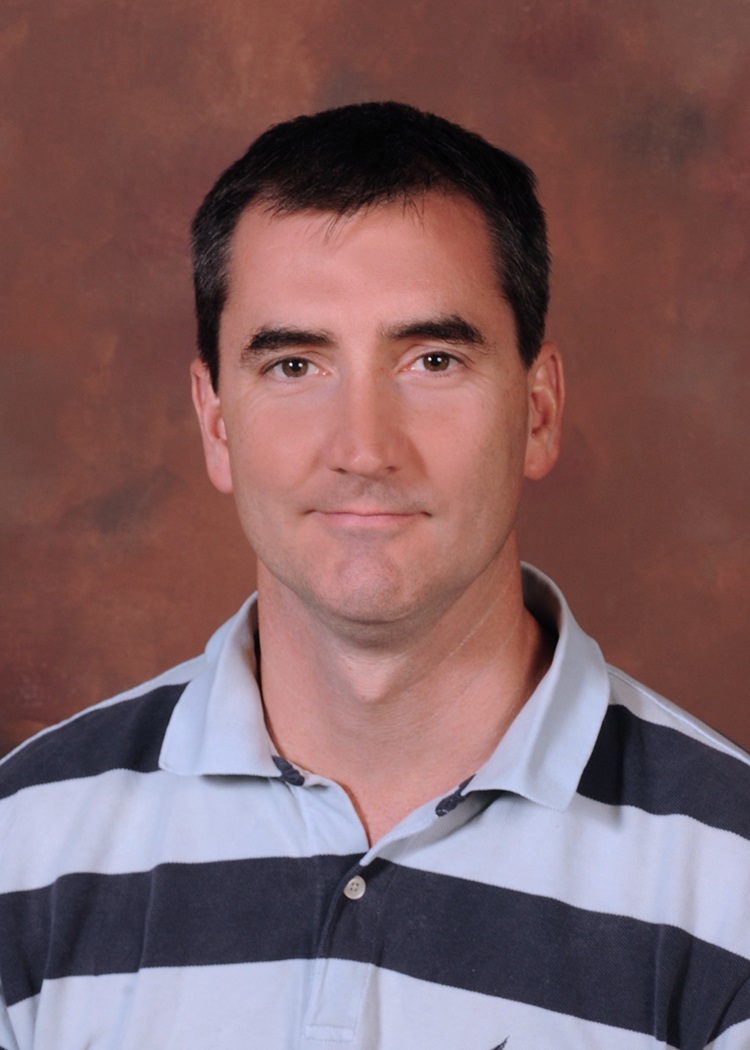
Philip O'Herron, PhD, is an Assistant Professor at Augusta University's Medical College of Georgia, Department of Physiology. His work bridges neurovascular coupling and visual processing, focusing on the interdependence between cortical neural activity and hemodynamic responses. Education: PhD in Neuroscience (2009, Johns Hopkins University), BA in Chemistry (2002, George Mason University), BA in Philosophy (2000, Christendom College) Research Themes: Dr. O'Herron's lab investigates functional hyperemia's physiological role using optogenetic vascular control. Current projects examine Alzheimer's disease vascular interactions , 3D optogenetic vessel manipulation , and vasculo-glia-neuronal coupling through collaborations with Dr. Jessica Filosa and Dr. Felipe Barros. Article Trends: Recent publications span neurovascular modeling , metabolic adaptation studies , and in vivo optical manipulation techniques , reflecting his work on vascular cognitive impairment and cortical depth-dependent processing. Scientific Awards: First prize MUSC research day (2012) Johns Hopkins neuroscience poster award (2008) Advising & Grants: Leads NIH-funded projects on Alzheimer's blood flow mechanisms (NIA R01) and neurovascular coding (NINDS R01). Mentors postdoctoral fellows in vascular neuroscience and develops models for cortical blood flow regulation.
Dr. Harvey Swadlow is a Professor in the Department of Psychological Sciences at the University of Connecticut. His primary research investigates neurophysiological mechanisms underlying sensory processing in the neocortex, with special focus on thalamocortical interactions and cortical network dynamics. Professor Swadlow's research examines thalamocortical communication pathways, cortical layer-specific processing, and state-dependent modulation of sensory systems. His work utilizes advanced in vivo electrophysiological techniques to study awake behaving models, particularly focusing on visual thalamocortical circuits and cortical inhibitory networks. Publications consistently explore thalamocortical interactions across behavioral states, cortical microcircuit organization, and sensory processing mechanisms. His research employs techniques including multi-site extracellular recording, cortical laminar analysis, and in vivo characterization of synaptic transmission dynamics.
Quentin Perrenoud is an Associate Research Scientist in the Department of Neuroscience at Yale School of Medicine. His research focuses on cortical interneurons, neural rhythms, and seizure mechanisms, with significant work on VIP interneurons, cholinergic arousal, and neurovascular coupling. Research Interests include: Cortical Rhythmic Activity Interneuron Functional Diversity Seizure-Induced Consciousness Impairment Neurovascular Response Mechanisms Frequency Spectrum Analysis of Neural Dynamics Laminar Organization of Cortical Circuits Recent Publications demonstrate expertise in: Mouse models of focal seizures Gamma oscillations in visual processing Molecular profiling of parvalbumin and nNOS interneurons Perineuronal net composition 5-HT3 receptor-mediated vascular responses Comparative analysis of cortical and hippocampal GABAergic neurons
Dr. Alicia Y Che is an Assistant Professor of Psychiatry at Yale University's School of Medicine, affiliated with the Department of Psychiatry and the Center for Brain & Mind Health. She joined Yale in 2021 after postdoctoral training at Weill Cornell Medical College and NYU. Her research focuses on neural circuits underlying social behavior and psychiatric disorders like PTSD. She holds a PhD in Physiology and Neurobiology from the University of Connecticut (2014) and a BS in Biology, Physics, and Physical Chemistry from Pacific Lutheran University (2009). Her key research projects explore oxytocin's role in social touch development and circuit dysfunction in PTSD. She investigates how sensory experiences shape cortical circuits using advanced techniques like in vivo calcium imaging and genetic manipulation. Dr. Che leads the Che Lab, which integrates molecular, electrophysiological, and behavioral approaches to study neural circuitry. Awards: NARSAD Young Investigator Award (2020), K99/R00 Pathway to Independence Award (2019). Labs/Teams: Director of Graduate Admissions for the Interdepartmental Neuroscience Program, member of the Janeway Society and Wu Tsai Institute. Her work bridges developmental neurobiology and translational psychiatry, emphasizing circuit-level mechanisms in mental health disorders.
Federico de Martino is a researcher at the Max Planck Institute for Empirical Aesthetics specializing in ultra-high field MRI techniques for mapping the human auditory pathway. His work combines computational modeling with advanced imaging to investigate sound processing from the cochlea to auditory cortical subfields at unprecedented spatial resolution. His research interests include: Auditory Neuroscience Ultra-High Field MRI (7T and 9.4T) Computational Modeling of Sound Processing Cortical Layer-Specific Processing Functional Brain Mapping of Auditory Pathways Dr. de Martino's research program investigates sound processing throughout cortical and sub-cortical areas of the human brain. He has reliably measured fMRI responses to natural sounds from multiple sub-cortical auditory nuclei including the cochlear nucleus, superior olive, inferior colliculus and medial geniculate body. Using computational models, he maps functional characteristics like tonotopic organization and spectrotemporal modulation preferences throughout these nuclei. Through high spatial resolution diffusion weighted MRI, he characterizes anatomical connections between auditory nuclei and examines their relationship to resting state functional connectivity. His work combines functional and anatomical information to parcellate the cortex into auditory fields and explore layer-specific processing in the auditory cortex. His findings demonstrate relatively stable 'columnar' tuning for frequency and temporal modulations in primary auditory cortex, with frequency tuning in superficial layers sharpening depending on task demands. His ongoing research extends these investigations beyond primary auditory cortex, with preliminary data showing that while acoustic content in natural stimuli may be processed similarly in middle and deep cortical layers, semantic information emerges in superficial cortical layers of higher order auditory regions.
Alexander Opitz serves as an Associate Professor in the Department of Biomedical Engineering at the University of Minnesota, where he leads innovative research in non-invasive brain stimulation technologies. His laboratory focuses on developing computational models to estimate electric field distributions during transcranial magnetic stimulation (TMS) and transcranial electric stimulation (TES), integrating these with neuronavigation systems to improve targeting of specific brain circuits. Opitz's research bridges engineering principles with neuroscience to address neurological and psychiatric disorders through personalized neuromodulation approaches. Opitz's primary research interests center on the biophysical and physiological foundations of non-invasive brain stimulation (NIBS). His lab develops advanced computational tools like SimNIBS for electric field simulation and NeMo-TMS for multi-scale neuron modeling, enabling precise prediction of stimulation effects from whole-brain to single-neuron levels. Current projects include closed-loop real-time TMS-EEG systems that align stimulation pulses with ongoing brain activity phases, deep-learning-based modeling for rapid TMS field estimation, and personalized rehabilitation protocols for stroke recovery in children. His work emphasizes translating improved understanding of brain physiology into clinical applications for conditions like depression and stroke. The research trends in Opitz's 15 most recent publications reveal a strong focus on personalization and precision in brain stimulation. Key themes include individual anatomical and functional predictors for NIBS response, real-time brain state-dependent stimulation, cross-species modeling frameworks, and meta-analyses of electric field effects in clinical populations. His work increasingly integrates machine learning with traditional computational methods while expanding applications to psychiatric disorders and pediatric populations, demonstrating a clear trajectory toward clinically viable personalized neuromodulation therapies. Opitz actively contributes to the scientific community through his lab's extensive resource sharing. He maintains the SimNIBS software platform for electric field simulation, develops the NeMo-TMS toolbox for neuron modeling, and hosts annual workshops on non-invasive brain stimulation methods. His lab's GitHub repository provides open access to published code, while their YouTube channel 'Brain Stimulation Science' disseminates educational content and seminar recordings. These resources support global researchers in advancing NIBS technologies and methodologies.
Scott Pluta is an Assistant Professor in the Department of Biological Sciences at Purdue University, affiliated with the College of Science. His research focuses on understanding the neural mechanisms underlying sensory perception, sensory-guided behavior, and sensorimotor loops. He employs advanced techniques such as optogenetics, viral tracing, and behavioral analysis to study neural circuits in rodents, particularly focusing on the midbrain superior colliculus (SC) and cortical networks. Education and Experience: Pluta earned his Ph.D. from the University of Virginia in 2009. He completed postdoctoral research at the University of California, Berkeley (2012–2017) and Wake Forest Medical Center (2009–2012). His lab, the Laboratory for Sensorimotor Integration, investigates how descending cortical inputs influence the SC's activity and behavior, aiming to reveal principles of neural circuit function with implications for neurological disorders like ADHD and Parkinson's disease. Research Interests: Central to his work is the study of how the SC integrates sensory, motor, and motivational signals to form a 'priority map' for spatial attention. Projects include dissecting cortico-collicular circuits using genetic labeling and optogenetic manipulation, and analyzing ascending SC-thalamic-cortical pathways to understand sensorimotor loop dynamics. His lab uses rodent whisker systems as a model for active touch and spatial navigation. Lab and Collaborations: The lab combines optogenetics, neurophysiology, and behavioral tracking to study dynamic interactions between cortical and midbrain networks. Research outcomes aim to provide foundational insights for developing treatments for neurological conditions.
Gord Fishell, Ph.D., is a Professor of Neurobiology at Harvard Medical School. His research focuses on the developmental mechanisms underlying inhibitory interneuron diversity and their integration into neural circuits, with implications for autism spectrum disorder, schizophrenia, and intellectual disability. His laboratory has pioneered tools like enhancer-AAV viral vectors for targeting interneurons across species, enabling precise manipulation of cortical circuits. Research Themes: Interneuron Specification: Genetic programs governing interneuron subtype generation during embryonic development. Circuit Integration: Local cues guiding interneurons into cortical and subcortical circuits. Pathophysiological Links: Investigating how interneuron dysfunction contributes to neurological disorders. Technological Innovation: Developing viral tools for cell-type-specific manipulation in mammals and humans. Key Contributions: The lab's work on somatostatin (SST) and parvalbumin (PV) interneurons has revealed critical roles in circuit maturation and sensory processing. Their viral toolkit, developed with Paola Arlotta, enables targeted studies of diverse cortical cell types. Future Directions: Translating developmental insights into therapies for neurodevelopmental disorders, particularly through interneuron-based interventions.
Lucy Petro is a Research Fellow at the University of Glasgow’s School of Psychology & Neuroscience. Her work focuses on cognitive neuroscience, particularly how context and prediction guide neural and cognitive processes. She uses advanced neuroimaging techniques like fMRI and electrophysiology to study visual processing, emphasizing contextual feedback signals in primary visual cortex (V1). Additionally, Petro is an accredited cognitive behavioral psychotherapist, integrating therapeutic insights with her neuroscience research to explore how internal models affect behavior and emotion. Research Interests Petro investigates predictive processing mechanisms in the brain, including how top-down signals influence perception and cognition. Her studies often involve high-field functional brain imaging to decode activity patterns related to visual scenes, occluded objects, and aphantasia. She collaborates with experts like Prof. Lars Muckli and Prof. Winrich Freiwald, examining cross-modal interactions (e.g., auditory influences on vision) and the neural basis of cognitive-emotional interactions. Grants & Collaborations Her current grant (2025–2029) from the Medical Research Council supports layer-specific imaging of context-dependent cognitive processing. She leads collaborations on topics such as cortical layer profiles for illusory experiences and the role of pyramidal cells in cognitive models. Supervision Belén Montabes de la Cruz (Postgraduate) Zirui Zhang (Postgraduate) Key Contributions Her work bridges clinical and experimental neuroscience, with publications in top journals like Current Biology , Trends in Cognitive Sciences , and Nature Communications . She explores how predictive coding underpins perception and how maladaptive models can be therapeutically altered.
Jude Mitchell is a researcher specializing in visual neuroscience and cognitive processes, focusing on the role of internal brain states like selective attention in modulating sensory processing. His work emphasizes the marmoset (Callithrix jacchus) as a model organism for studying visual perception, attention mechanisms, and neural circuitry. Collaborations include researchers at UC San Diego and The Salk Institute, exploring topics such as saccadic eye movements, cortical activity, and optogenetic techniques. Key research areas include neuronal selectivity, laminar cortical activity, and the integration of motion signals during visual tasks. His research spans studies on dopamine receptor effects on distractibility, neural correlates of reach-to-grasp movements, and the interplay between sleep-wake cycles and circadian rhythms. Mitchell’s methodologies include head-mounted eye tracking, electrophysiological recordings, and computational models of neural networks. He has pioneered techniques for studying free-moving marmosets, enabling deeper insights into visual processing dynamics and attentional modulation across cortical layers and neuron types. Notable contributions include defining attention’s impact on neuronal burstiness and correlation in macaque area V4, and establishing marmoset models for understanding human mental disorders. His work bridges primate neurobiology with translational research, addressing both fundamental questions about brain function and practical applications in neuroscience.
Hillel Adesnik serves as Associate Professor in the Department of Molecular and Cell Biology at the University of California, Berkeley, with additional affiliation in Neuroscience. His research program centers on deciphering how cortical microcircuits transform sensory input into perceptions and behaviors, utilizing cutting-edge approaches in awake behaving mice to bridge cellular mechanisms with cognitive functions. Adesnik's research investigates the neural basis of perception through three integrated pillars: (1) dissecting horizontal and vertical connections in cortical layers for sensory feature extraction, (2) developing high-resolution optical tools like 3D-SHOT for single-neuron manipulation in intact brains, and (3) analyzing cross-cortical communication for percept synthesis. His lab combines two-photon imaging, optogenetics, electrophysiology, and computational modeling to study tactile processing in barrel cortex and visual perception, revealing how specific neuron types and synaptic mechanisms generate perceptual codes. Key discoveries include layer-specific inhibitory control, supra-linear feature summation, and gamma-band synchronization mechanisms. Analysis of Adesnik's publication record shows consistent focus on cortical microcircuit dynamics across sensory modalities, with increasing emphasis on tool development since 2017. His work demonstrates how precise neural manipulations can establish causal links between circuit activity and perception, particularly through innovations in holographic optogenetics. Recurring themes include the role of somatostatin interneurons in layer-specific processing, cross-laminar interactions in feature coding, and the development of quantitative frameworks for neural population decoding. Scientific recognition includes: Chan Zuckerberg Biohub Investigator (2022 cohort) Adesnik mentors a robust research team comprising postdoctoral fellows (Lamiae Abdeladim, Janine Beyer, Conor Dorian, Will Hendricks, Uday Jagadisan, Mora Ogando, Masato Sadahiro, Kevin Sit, Savitha Sridharan, Andrea Zazzi) and graduate students (Genesis Ferrer Imbert, Courtney Kim, Madi McCloud, Ravi Srinivasan). His lab operates through structured collaboration with engineering groups for optical tool development and maintains active partnerships for disease-model applications. Funding sources include the Chan Zuckerberg Biohub and NIH grants supporting neurotechnology innovation. The Adesnik Lab maintains three core research thrusts through an integrated experimental pipeline: in vivo circuit interrogation in behaving animals, in vitro synaptic analysis, and novel optical instrument development. Current work emphasizes translating high-resolution manipulation techniques to disease models including autism and epilepsy, while expanding into multi-area cortical dynamics during complex behavioral tasks.
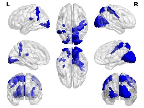
Dr. Vesna Vuksanovic is a Senior Lecturer in Health Data Science at Swansea University's School of Medicine. She also holds an Honorary Senior Lecturer position at the University of Aberdeen. Her academic career spans institutions including the University of Aberdeen and the Technical University of Berlin, Germany, where she was based prior to joining Swansea University in 2021. Research Interests: Multimodal imaging and computational modeling of healthy aging and neurodegeneration Brain connectome analysis and morphometric similarity Neurodegenerative diseases, particularly Alzheimer's disease and frontotemporal dementia Dynamic functional network analysis for dementia classification Development of computational models for understanding brain changes in neurodegenerative disorders Dr. Vuksanovic's research focuses on mapping heterogeneous changes across brain regions in healthy aging and neurodegenerative disorders, and studying disease progression in dementia patients participating in clinical trials. Her work bridges computational neuroscience, clinical neurology, and data science to develop better diagnostic tools and understanding of neurodegenerative processes. She has made significant contributions to understanding the relationship between structural and functional brain networks in conditions like Alzheimer's disease and frontotemporal dementia. Recent Publication Trends: Her publications from 2014-2024 demonstrate a consistent focus on applying advanced network analysis techniques to neuroimaging data in dementia research. Key areas include genetic factors in Alzheimer's disease, dynamic functional networks for improved diagnosis, and the degeneration patterns of specific brain networks in frontotemporal dementia. Her most recent work (2023-2024) explores the genetic basis of anatomical asymmetry in Alzheimer's disease and uses dynamic functional network analysis to improve classification of dementia subtypes. Her research shows an evolution from fundamental network science approaches to increasingly clinically relevant applications. Awards and Recognition: Co-inventor on three international patents, including 'Network methods for neurodegenerative diseases' (US17/272885) Research Funding and Supervision: Dr. Vuksanovic serves as Principal Investigator for multiple research projects including 'Brain Aging Model: Identifying neuroimaging patterns relevant to neurodegeneration' (£67,939, 2023-2024) and 'Brain Flexibility: A possible non-invasive biomarker for dementia' (£111,918, 2020-2024). She is available for postgraduate supervision and has led PhD scholarship projects focused on using brain network modules to improve dementia diagnosis. Her research portfolio demonstrates strong funding success across multiple funding bodies and international collaborations. Teaching Activities: Dr. Vuksanovic teaches several modules in health data science including Capstone Project (PM-344), Computational Science and Health Care (PMIM102/PMIM102J), Health Data Modeling (PMIM202/PMIM202J), and Advanced Machine Learning in Health Care (PMIM402J). Her teaching focuses on equipping students with practical computational skills applicable to healthcare data analysis, integrating theoretical foundations with hands-on computational approaches using tools like SPSS and R.
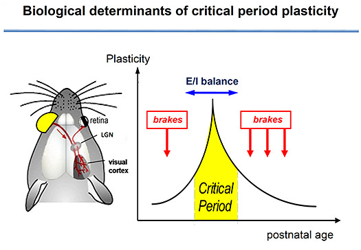
Takao Hensch is a distinguished Professor of Neurology at Harvard Medical School/Boston Children's Hospital and Professor of Molecular and Cellular Biology at Harvard University's Center for Brain Science. He serves as Director of the International Research Center for Neurointelligence (IRCN) at the University of Tokyo and leads the NIMH Silvio Conte Center for Mental Health Research at Harvard. His groundbreaking work focuses on critical periods in brain development—windows of heightened plasticity when neural circuits are most responsive to environmental input. Hensch's research has revealed how specific inhibitory (GABA) circuits trigger the onset of critical periods and how 'brake'-like factors actively prevent circuit rewiring when these periods close. His laboratory integrates molecular, cellular, and systems neuroscience to understand how early life experiences shape brain function from motor skills to language and emotions. This work has profound implications for understanding and treating neurodevelopmental disorders including autism spectrum disorders, epilepsy, and amblyopia. Analysis of Hensch's recent publications shows a consistent focus on critical period mechanisms across multiple brain systems. His work demonstrates how molecular interventions can reopen plasticity windows in adulthood, with particular emphasis on GABAergic circuits, perineuronal nets, and oxidative stress mechanisms. Recent studies examine anesthesia effects on infant brain development, sex-specific responses to early adversity, and novel pharmacological approaches for restoring neural plasticity. Order of the Rising Sun, Gold Rays with Neck Ribbon (2024) NIH Director's Pioneer Award (2007) Mortimer D. Sackler, M.D. Prize for Distinguished Achievement in Developmental Psychobiology (2016) Society for Neuroscience Young Investigator Award - Japan (Tsukahara Prize, 2001) Society for Neuroscience Young Investigator Award - US (2005) Hensch has trained numerous PhD students and postdoctoral fellows who have gone on to successful careers in neuroscience. His laboratory receives substantial funding from the National Institute of Mental Health and other sources to investigate the biological basis of critical periods and their clinical applications. Current research focuses on translating basic findings into therapeutic approaches for neurodevelopmental disorders through collaborations with clinicians at Boston Children's Hospital and computational modelers. The Hensch Lab, housed in Harvard's Northwest Building and the F.M. Kirby Neurobiology Center at Boston Children's Hospital, maintains active collaborations with researchers at the University of Tokyo, RIKEN Brain Science Institute, and other international institutions. The lab employs state-of-the-art techniques in mice to explore neural circuit development from sensory systems to prefrontal cortex, with particular emphasis on translating findings into real-world applications for pediatric care and mental health treatment.
Professor Michael Brecht holds a faculty position at the Institute of Biology, Humboldt University Berlin , where he leads research on cellular mechanisms in sensorimotor integration using rodents and elephants as model systems. His work combines in vivo whole-cell recordings , neuroanatomical reconstructions , and behavioral analyses to study active touch , social behavior , and structure-function relationships in the brain. Education & Career : Diploma in Biology, University of Tuebingen PhD with Wolf Singer, Max Planck Institute for Brain Research Postdoc with Bert Sakmann, Max Planck Institute for Medical Research Assistant Professor at Erasmus University Rotterdam (2005-2009) Full Professor at Humboldt University Berlin (2009-present) Research Interests : Brecht's lab focuses on sensorimotor processing , neural coding of tactile perception , and evolutionary neuroanatomy . Key projects include: Structure-Function Relationships : Studying how brain architecture enables tactile object recognition and motor control Elephant Trunk Neurobiology : Mapping the neural basis of trunk motor control and sensory specialization NeuroCure Initiative : Bridging basic and clinical neuroscience for memory consolidation research DFG SFB 1315 : Comparative memory consolidation mechanisms across species Scientific Contributions : Recent publications highlight his work on elephant brain evolution , whisker-based wind sensing , and social play neurochemistry . His lab developed DiL-CT for bimodal neural tracing and pioneered whole-cell recordings in freely moving animals . Awards & Funding : ERC Synergy Grant (2019-2026) DFG Clusters of Excellence (NeuroCure) SFB 1315 (2022-2026) Collaborations : Works with the Leibniz Institute for Zoo and Wildlife Research, Berlin Zoo, and international consortia on comparative neuroanatomy and translational neuroscience .
Alexandre Medina de Jesus is an Associate Professor in the Department of Pediatrics at the University of Maryland School of Medicine. With a D.Sc. in Neuroscience (2000) and postdoctoral training in Neuroscience (2003), he has maintained continuous academic appointments at major institutions since 1999. His career includes tenure at Virginia Commonwealth University (2005-2012) before transitioning to his current role at University of Maryland (2012-present). Universidade Santa Ursula, Brazil - B.S., Biology (1990) Universidade Federal do Rio de Janeiro, Brazil - M.Sc., Zoology (1996) Universidade do Estado do Rio de Janeiro, Brazil - D.Sc., Neuroscience (2000) Virginia Commonwealth University - Postdoctoral Fellowship, Neuroscience (2003) Dr. Medina specializes in neurodevelopmental disorders , particularly Fetal Alcohol Spectrum Disorders (FASD) . His research explores how early alcohol exposure disrupts neuronal plasticity and multisensory integration , with notable work on cortical visual system development and transcription factor regulation (CREB, SRF, MEF2). He pioneered the ferret FASD model to study sensory processing deficits. Recent publications focus on cortical layer-specific multisensory integration (2024), visual-tactile circuit microstructure (2018), and CREB phosphorylation requirements for plasticity (2021). His work balances basic neuroscience with clinical translational research , including studies on neonatal hypoxia (2022) and NICU environmental exposures (2020). Scientific Awards & Service NIH/NIAAA R01 Grants (AA13023, AA022455) VA Merit Grant I01BX005678 President, FASD Study Group (2015-2016) NIH Study Section Member (NAL, 2018-2022) Research Themes Neuronal Plasticity Mechanisms Developmental Neurotoxicology Cortical Circuit Organization Translational Neuroscience Multisensory Processing Transcription Factor Regulation