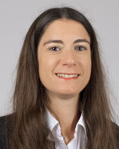
Anne Bonninمشاهده پروفایل
پژوهشگر
- Bioimaging
- Cardiac Imaging
- X-ray Tomography
- +۴ مورد دیگر
Dr. Anne Bonnin serves as a Beamline Scientist at the Paul Scherrer Institute (PSI) in Switzerland, where she has been instrumental in X-ray imaging research since joining the X-ray Tomography Group in 2014 and assuming her current role at the TOMCAT Beamline in 2016. Affiliated with PSI's Center for Photon Science and Laboratory for Macromolecules and Bioimaging, she operates at the forefront of synchrotron-based imaging techniques. Her academic foundation includes a PhD from INSA de Lyon focused on material properties for explosive detection, followed by postdoctoral work at the European Synchrotron Radiation Facility (ESRF) in X-ray diffraction and phase contrast tomography, and an NSF Research Fellowship for paleontology research at Harvard University and ESRF. Specializing in X-ray imaging (micro/nano-tomography, phase-retrieval) and powder diffraction, Dr. Bonnin leads the bioimaging program at TOMCAT with particular emphasis on the international Heart Imaging Project. Her research develops novel methodologies for materials characterization across diverse domains including cardiac microstructure analysis, paleontology, and neurodegenerative disease modeling, with significant contributions to understanding material behavior at microscopic scales. Her recent publications (2019-2021) demonstrate strong interdisciplinary impact, advancing X-ray imaging applications in energy storage (battery materials), biomedical research (cardiac/auditory systems), and materials engineering (aerogels). A defining trend is the integration of machine learning for image analysis, alongside methodological innovations like non-rigid image stitching and Fourier ptychography. These works reflect extensive international collaboration and address critical challenges in healthcare, energy, and fundamental material science. Dr. Bonnin leads the Heart Imaging Project to quantify cardiac microstructure using contrast-agent-free X-ray phase-contrast imaging, while actively contributing to the SLS2.0 upgrade project preparing TOMCAT for multiscale, multimodal, and dynamic tomographic capabilities. Her collaborative framework spans global researchers in materials science, paleontology, and biomedical engineering. As manager of the TOMCAT nanoscope—a full-field imaging setup achieving 150 nm 3D resolution—she enables cutting-edge research in absorption and phase-contrast imaging. Her team within the X-Ray Tomography Group drives the bioimaging program forward, particularly through the Heart Imaging Project's dynamic cardiac studies using modified Langendorff setups.