معرفی
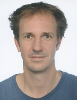
Prof. Dr. Christian Stigloher is a Professor for Microscopy at the Biocenter of the Julius-Maximilians-University Würzburg, where he serves as Group Leader of the Central Department of Microscopy (Imaging Core Facility). His research focuses on the ultrastructural analysis of neuronal synapses using advanced electron microscopy techniques.
His educational background includes:
- Studied biology at the University of Würzburg and Duke University, North Carolina, USA
- Doctoral thesis in the laboratory of Laure Bally-Cuif at the Helmholtz Center Munich and the Technical University Munich
- Postdoctoral fellowship in Jean-Louis Bessereau's laboratory at the Ecole Normale Superieure in Paris, France
Prof. Stigloher's research centers on synaptic architecture and function, with particular focus on how synaptic vesicles are retained close to the presynaptic active zone to maintain efficient signaling. He employs a synergistic combination of two model organisms: C. elegans neuromuscular junctions for efficient candidate identification and manipulation, and zebrafish larva neuromuscular junctions as a vertebrate model to test for evolutionary conservation of function. His lab specializes in electron tomography as an ultra-high 3D resolution method to dissect components of synaptic architecture, and in correlative light and electron microscopy (CLEM) approaches that combine the advantages of both techniques.
His recent publications demonstrate a strong focus on ultrastructural analysis across diverse biological systems, from neural synapses to cellular clearance mechanisms and host-pathogen interactions. The work spans multiple model organisms including C. elegans, zebrafish, and mammalian cell systems, with consistent application of advanced microscopy techniques. There's a clear trajectory toward increasingly sophisticated correlative microscopy methods that bridge molecular localization with ultrastructural context.
Prof. Stigloher leads the Central Department of Microscopy at the Biocenter, which functions as an Imaging Core Facility providing electron microscopy, high-content microscopy, light microscopy, and sample preparation services to researchers. His team includes several researchers and technical staff who collaborate on various projects related to cellular architecture and ultrastructural analysis.