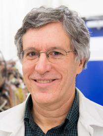
Menghua Xia is a Research Fellow in the Department of Radiology & Biomedical Imaging at Yale School of Medicine, Yale University. Their research focuses on advancing medical imaging techniques through deep learning and artificial intelligence, particularly in PET, SPECT, and CT imaging. Key areas include low-dose imaging, image denoising, and federated learning applications for clinical settings. Research interests emphasize improving diagnostic accuracy through computational methods, such as cross-tracer generalizability in PET attenuation correction, dynamic cardiac imaging optimization, and anatomically informed diffusion models. Collaborations involve multi-institutional validation and real-world low-dose data analysis to enhance clinical workflows. Prominent contributions include frameworks like Fed-NDIF for federated low-count PET denoising and anatomically/metabolically informed diffusion models. Their work bridges machine learning innovation with practical medical imaging challenges, aiming to reduce radiation exposure while maintaining diagnostic quality.
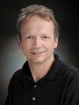
Dr. Peter Kench is an Associate Professor at the University of Sydney , affiliated with the Sydney School of Health Sciences and the Discipline of Medical Imaging Science . His work focuses on optimizing medical imaging techniques for cancer patients, particularly children, through radiation dose reduction and enhanced image quality. PhD, GradCertHlthScEd, BAppSc(MRT)Hons Research spans transmission/emission computed tomography (CT/SPECT/PET) innovations, with emphasis on pediatric imaging safety , spectral CT radiomics , and dynamic SPECT liver kinetics . His educational leadership includes developing simulation-based learning and computer-assisted instruction. Recent publications address radiation dose management , infection control in imaging , and patient-driven imaging requests . Awards include the Young Investigator Award (2021) and Top Cited Article (2020). Supervises research higher degree students and serves on multiple medical imaging societies .
Prof. Charalampos Tsoumpas is a Full Professor in Quantification in Molecular Diagnostics & Radionuclide Therapy at the University Medical Centre Groningen (UMCG), affiliated with the Faculty of Medical Sciences. His research focuses on advancing quantitative PET and SPECT imaging, including novel reconstruction algorithms and motion correction techniques. He holds academic roles including team leader in the Crystal Clear Collaboration (CERN), editorial board memberships in journals like European Journal of Nuclear Medicine , and leadership in initiatives such as the Collaborative Computational Project Synergistic Reconstruction in Biomedical Imaging (CCP SyneRBI) . Education includes a BSc in Physics (National University of Athens), MSc in Biomedical Engineering (National Technical University of Athens), and a PhD from Imperial College London. His career includes roles at King’s College London and the University of Leeds, where he led research projects funded by the Leverhulme Trust, EPSRC, and EU grants. Research Highlights: Pioneered methods for parametric image reconstruction, developed open-source STIR software, and contributed to PET-MRI integration. His work on motion correction and scatter correction has influenced commercial systems. Notable achievements include the Rotblat Medal (2017) for impactful research in medical physics. Leadership and Mentorship: Supervised 23 PhD students, fostering open science through collaborative coding and educational resources. Maintained industrial partnerships with companies like GE Healthcare and Bruker. Current projects include the SAFIR PET-MRI preclinical scanner and LAFOV PET/CT advancements. Labs/Teams: Leads the Quantification in Molecular Diagnostics lab at UMCG, collaborates on the SAFIR project (ETH Zurich), and contributes to CCP SyneRBI for synergistic reconstruction frameworks.
Steven Beyea is a Professor at Dalhousie University, holding joint appointments in the Department of Physics and Atmospheric Science, Department of Diagnostic Radiology, and School of Biomedical Engineering. He leads the Biomedical Translational Imaging Centre (BIOTIC) , focusing on developing and clinically translating novel diagnostic imaging technologies. His work integrates MRI, MEG, and multimodal imaging to advance pre-surgical functional neuroimaging, abdominal/pelvic cancer diagnostics, and biomarker-driven patient stratification. Research Interests : His interdisciplinary research spans compressed sensing algorithms for parametric mapping, automated analysis of functional neuroimaging data, and machine learning applications in healthcare. Projects include high-resolution liver iron/fat quantification without a priori assumptions and enhancing reliability in pre-surgical brain mapping. Infrastructure includes clinical 3T MRI/MEG and preclinical PET/SPECT/CT systems strategically located in Halifax’s major hospitals. Key Projects : Compressed Sensing for High-Temporal-Resolution Parametric Mapping Algorithms for Functional Neuroimaging Reliability in Pre-Surgical Mapping Novel MRI Pulse Sequences for Iron/Fat Quantification Machine Learning for Patient Stratification using MRI/MEG Grants & Labs : As head of BIOTIC, he oversees translational research infrastructure. Ongoing work explores imaging biomarkers for neurological diseases and cognitive impairment in systemic lupus erythematosus.
Grant Gullberg, PhD, is an Adjunct Professor in the School of Medicine at the University of California, San Francisco (UCSF). His primary affiliation is with the Department of Radiology, where he focuses on advanced imaging technologies. He holds significant expertise in dynamic cardiac SPECT and PET imaging, myocardial blood flow quantification, and novel hardware designs for medical imaging systems. Research Interests: His work spans biomedical engineering, nuclear medicine, and radiology, with a focus on developing and optimizing imaging techniques for cardiovascular applications. Key areas include: Dynamic PET/SPECT imaging methodologies Quantitative myocardial perfusion analysis Advanced reconstruction algorithms (e.g., MLEM, factorization methods) Hardware innovations like CZT detectors and collimatorless systems Grants & Funding: Dr. Gullberg has led or co-led multiple NIH-funded projects, including: Dynamic Cardiac SPECT Imaging (R01HL050663, 1995–2015) Molecular Imaging of Cardiac Hypertrophy (R01EB007219, 2008–2013) PET imaging of glucose and fatty acid metabolism (R01HL135490, 2017–2021) Labs/Teams: Collaborates actively with multidisciplinary teams in UCSF’s Radiology department, focusing on translational research in cardiac imaging and medical physics. His work integrates computational methods, deep learning, and clinical validation for diagnostic advancements.
Yuni Dewaraja, PhD is the William Martel Research Professor in the Department of Radiology at the University of Michigan Medical School. She is affiliated with the Center for Positron Emission Tomography and has made significant contributions to the field of nuclear medicine physics and radiopharmaceutical therapy dosimetry. Dr. Dewaraja earned her PhD in 1994 from the University of Michigan, following an MS from Kansas State University in 1990 and a BS from the University of Western Australia in 1986. Her academic journey has positioned her as a leading expert in quantitative imaging and radiation dosimetry. Dr. Dewaraja's research focuses on estimating radiation absorbed dose in radionuclide therapy , quantitative SPECT imaging , image reconstruction techniques , and correction methods for scatter, attenuation and partial volume effects . She extensively employs Monte Carlo methods for image reconstruction and patient-specific absorbed dose estimation. Her work bridges physics, engineering, and clinical applications to improve the precision of targeted radionuclide therapies. Her recent publications demonstrate a strong emphasis on Lutetium-177 and Yttrium-90 therapies , with research spanning dosimetry standardization , predictive modeling of treatment response , optimization of imaging protocols , and integration of multiple therapeutic modalities . She has been instrumental in international efforts to reduce variability in dosimetry calculations and establish best practices in the field. Can 177Lu-DOTATATE Kidney Absorbed Doses be Predicted from Pretherapy SSTR PET? Multicycle Dosimetric Behavior and Dose–Effect Relationships in [177Lu]Lu-DOTATATE Therapy Shorter SPECT scans using self-supervised coordinate learning techniques Sequential 90Y SIRT and SBRT using PET-based Absorbed Dose Maps 177Lu-PSMA-617 SPECT/CT Dosimetry and Radiobiological Models Dr. Dewaraja has received recognition through numerous citations of her work, with over 3,000 total citations according to the Michigan Research Experts profile. Her research has been featured in leading journals including the Journal of Nuclear Medicine (93 publications), European Journal of Nuclear Medicine and Molecular Imaging (30 publications), and Medical Physics (12 publications). As a dedicated educator and mentor, Dr. Dewaraja collaborates extensively with colleagues across multiple institutions on research projects focused on advancing precision in nuclear medicine therapy. Her work has significant implications for improving the safety and efficacy of targeted radionuclide treatments for cancer patients.
Justin Mikell, PhD is an Associate Professor of Radiation Oncology and Associate Professor of Radiology at Washington University School of Medicine, where he is part of the Division of Medical Physics within the Siteman Cancer Center. He joined Washington University in 2022 after previously holding a faculty position at the University of Michigan, where he was part of the brachytherapy and machine QA teams. Dr. Mikell earned his dual BS degrees in Engineering and Computer Science from the University of Illinois, Urbana-Champaign (2002), followed by a PhD in Medical Physics from the University of Texas at Houston Graduate School of Biomedical Sciences and UT MD Anderson Cancer Center (2015). He completed his residency in Medical Physics at the University of Michigan, Ann Arbor (2018). His research focuses on combination radiotherapy, quantitative emission imaging, and absorbed dose calculations for both microspheres and unsealed sources, particularly Y-90 microspheres. His work has significantly advanced the field of radioembolization for liver cancer treatment, with a special emphasis on precise dosimetry modeling and treatment planning. Dr. Mikell's expertise spans medical physics, radiation oncology, and nuclear medicine, allowing him to bridge these disciplines for improved patient outcomes. Dr. Mikell's publication record demonstrates consistent contributions to the field, with research spanning from technical aspects of dosimetry calculation to clinical applications in liver cancer treatment. His recent work shows increasing focus on integrating advanced imaging techniques like PET/CT with precise dose calculation methods, and exploring combination therapies involving radioembolization and stereotactic body radiation therapy. With approximately 1,200 citations across his 47 research outputs, Dr. Mikell has established himself as a significant contributor to the field of radiation oncology physics, particularly in the area of radioembolization dosimetry and treatment planning. He continues to collaborate extensively with nuclear medicine colleagues and radiation oncologists, advancing research in quantitative imaging for radiation therapy and developing more precise methods for calculating and delivering therapeutic radiation doses to cancer patients.
Fredrik Hedeer is a Consultant and Researcher affiliated with Lund University and the Lund Cardiac MR Group . His clinical and research focus lies in cardiovascular imaging , particularly comparing diagnostic modalities like myocardial perfusion SPECT (MPS) and cardiac magnetic resonance imaging (CMR) for assessing myocardial injury and perfusion in patients with suspected ischemic heart disease. Specializes in digital SPECT techniques and CMR correlation Active in UN SDG-related research (Radiology, Cardiology) Collaborates internationally on myocardial perfusion studies His work emphasizes diagnostic accuracy improvement and understanding perfusion dynamics under stress/rest conditions. Current research explores sex-based differences and comorbidity impacts in chronic coronary syndrome patients. Key collaborations include teams working on hybrid imaging modalities and radiotracer development . Publications demonstrate expertise in cardiovascular magnetic resonance, positron emission tomography, and multimodal imaging validation.
Sang-Geon Cho is a Visiting Assistant Professor at the Yale School of Medicine, Department of Internal Medicine. His research focuses on advanced cardiovascular imaging techniques, particularly in nuclear cardiology and PET applications. He has contributed to studies on coronary artery disease, myocardial ischemia, and cardiac amyloidosis. His work integrates artificial intelligence into medical imaging for improved diagnostic accuracy and patient prognosis. Education: Not explicitly stated in provided text. Affiliations: Yale School of Medicine, collaborating with departments such as Internal Medicine and Radiology. Research Interests: Dr. Cho specializes in translational imaging research, including: Development of AI-driven algorithms for coronary artery calcium scoring and myocardial blood flow analysis. Evaluation of PET and SPECT imaging as biomarkers for pulmonary fibrosis, cardiac amyloidosis, and thyrotoxicosis. Prognostic applications of phase analysis in heart failure and bone scintigraphy in cardiac amyloidosis. Publications: His recent work (2023-2025) emphasizes AI integration in imaging, validation of coronary stenosis models, and clinical utility of cardiac biomarkers. Key themes include optimizing imaging protocols for non-invasive diagnostics and improving risk stratification in cardiovascular diseases. Awards: No specific awards listed in the provided information. Grants/Advising: Not explicitly detailed, but his publications suggest involvement in multicenter studies and clinical trial protocols. Labs/Teams: Collaborates with the Stephen & Denise Adams Center for Parkinson’s Disease Research and other interdisciplinary teams at Yale.
Yongyi Yang is the Harris Perlstein Professor of Electrical and Computer Engineering and Professor of Biomedical Engineering at Illinois Institute of Technology (IIT), part of the Armour College of Engineering. He holds dual roles in both departments and is a Fellow of IEEE and AIMBE. His research focuses on medical imaging, machine learning, and image processing, with over 300 publications. Education: Ph.D. (1994), M.S. (1992), and M.S.E.E. (1988) from IIT, and B.S.E.E. (1985) from Northern Jiaotong University, Beijing. Research interests include tomographic image reconstruction, computer-aided diagnosis (CAD), and medical imaging technologies. Current projects involve cardiac and pediatric kidney imaging, breast cancer detection in mammography, and brain atlas construction. He has served as an editor for multiple IEEE journals and on NIH and NSF review panels. Key contributions include advancing SPECT reconstruction techniques, developing machine learning models for microcalcification detection, and authoring the textbook Vector Space Projections (1998). Recent work leverages deep learning for medical image analysis, including context-sensitive detection models and respiratory motion correction in SPECT. Awards include the IEEE Fellow (2021) and AIMBE Fellow (2018), alongside recognition for student paper awards and impactful publications. His research bridges applied mathematics, signal processing, and medical applications, with a focus on improving diagnostic accuracy and reducing radiation exposure in imaging.
Dr. Thomas Humphries is an Associate Professor in the Division of Engineering and Mathematics at the University of Washington Bothell since 2022. He earned his Ph.D. in Applied and Computational Mathematics from Simon Fraser University and holds a B.Math from the University of Waterloo. His research focuses on tomographic image reconstruction and mathematical optimization techniques. Ph.D., Applied and Computational Mathematics, Simon Fraser University (2011) M.Sc., Applied and Computational Mathematics, Simon Fraser University (2007) B.Math, Joint Honours Applied Math and Computer Science, University of Waterloo (2005) His work in Medical Imaging addresses challenges in CT and SPECT reconstruction, particularly for polyenergetic/sparse data. He also explores derivative-free optimization in oil field operations and has developed open-source MATLAB code for polyenergetic CT reconstruction available on GitHub. Recent publications focus on superiorization methodology and machine learning integration. Key research trends include iterative reconstruction algorithms, metal artifact reduction, dynamic SPECT imaging, and regularization techniques. No formal scientific awards are listed in the provided text. Dr. Humphries teaches mathematics courses including calculus, linear algebra, and numerical analysis. His professional journey includes postdoctoral work at Memorial University (2011-2013) and Oregon State University (2013-2015) before joining UW Bothell in 2015.
Joel Karp is a Professor of Radiologic Physics in Radiology and holds a secondary appointment in Physics and Astronomy at the University of Pennsylvania. He leads the PET Imaging/Cyclotron Facility and directs the Nuclear Medicine Core in the Medical School's Small Animal Imaging Facility. His work focuses on advancing PET technology, including time-of-flight imaging, detector design, and 3D reconstruction algorithms. He has pioneered the Philips Gemini TF scanner and developed novel scintillator-based instruments. Dr. Karp has been instrumental in establishing imaging standards like the NEMA NU-2 protocol and has contributed to multi-center trial accuracy initiatives. Education: PhD in Nuclear Physics from MIT (1980) Affiliations: Penn Medicine, IEEE Nuclear Medical Imaging Sciences Council, SNM, and ACRIN PET Core Lab. His research interests span PET/SPECT instrumentation, quantitative imaging, and clinical translation. He has received the Ed Hoffman Memorial Award (2007) and led national committees, including the NAS Nuclear Medicine Science Committee. Dr. Karp's lab collaborates extensively on preclinical imaging, deep learning denoising, and low-dose protocols, advancing both clinical and research imaging capabilities. Notable contributions include the PennPET Explorer total-body PET system and collaborations on dedicated breast-PET/DBT systems. His work bridges physics, engineering, and medicine to enhance diagnostic precision and reduce radiation exposure.
Manfred Trummer is a Professor in the Department of Mathematics at Simon Fraser University (SFU), part of the Faculty of Science. His research focuses on computational methods in medical imaging, particularly dynamic single photon emission computed tomography (SPECT), and numerical methods for differential equations, including spectral collocation and radial basis function techniques. He emphasizes solving ill-posed problems and improving stability, accuracy, and adaptivity in numerical solutions. Trummer holds a Ph.D. in Mathematics from ETH Zurich (1983). His work bridges applied mathematics and computational science, addressing challenges in medical imaging reconstruction and high-order numerical methods for differential equations. Notable contributions include advancements in spectral methods, iterative reconstruction techniques for SPECT, and preconditioning strategies for ill-conditioned systems. His research spans dynamic imaging applications, such as kidney imaging and 4D SPECT reconstruction, alongside foundational numerical analysis topics like matrix factorization, boundary layer resolution, and RBF stability. Recent publications highlight innovations in spectral collocation for mixed functional differential equations and conformal mapping using Szegő kernels. Trummer teaches graduate courses such as APMA 923 (Numerical Methods in Continuous Optimization). While no specific grants or labs are detailed in the text, his work reflects sustained engagement with interdisciplinary computational challenges in mathematics and medical imaging.
Benjamin H. Brinkmann, PhD, is a Professor of Neurology and Associate Professor of Biomedical Engineering at Mayo Clinic. He serves as a Consultant in the Division of Epilepsy within the Department of Neurology. His research focuses on developing noninvasive seizure detection and prediction technologies using biosensors, with a strong emphasis on improving pre-surgical evaluations and epilepsy management. Education: PhD in Biophysics/Biophysical Sciences (Mayo Graduate School, Mayo Clinic College of Medicine) and BS in Physics and Biology (North Park College). Research Interests: Noninvasive wearable devices for seizure forecasting, machine learning applications in neurophysiological data analysis, EEG and MEG source reconstruction, ictal SPECT imaging, and volumetric neuroimaging techniques. His work aims to translate technologies into therapies that prevent seizures and address comorbidities like depression and cognitive impairment. Key Projects: Optimizing Pharmacotherapy with Noninvasive Wearable Sensors and Subscalp EEG (NIH-funded grant, 2022–2025) Leadership roles: Technical co-chair of Neurotechnology in Epilepsy Section (International League Against Epilepsy), EEG project lead in Mayo Clinic Neurology AI Program Laboratory Affiliation: Collaborates with the Bioelectronics Neurophysiology and Engineering Lab at Mayo Clinic.