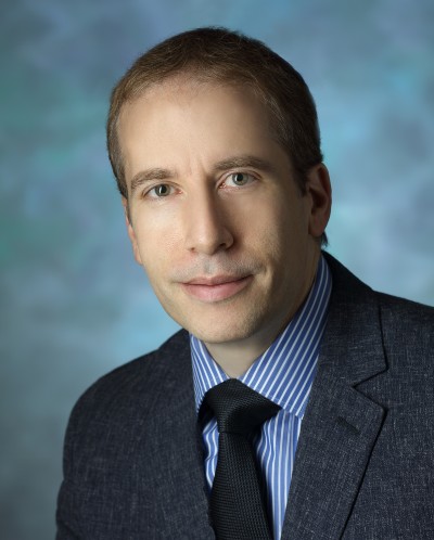
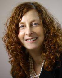
Professor Guy Williams is a leading academic at the University of Cambridge with a focus on imaging science and clinical neurosciences, affiliated with Downing College and the Wolfson Brain Imaging Centre . Holding a PhD in Physics from his initial Natural Sciences degree, he specializes in nuclear magnetic resonance (NMR) and MRI techniques for brain imaging. Education: BA, PhD in Physics His research centers on non-invasive imaging of brain structure and function, particularly in traumatic brain injury (TBI) and dementia. His work involves developing novel MRI pulse sequences and advanced data analysis algorithms, including AI-based diagnostic tools. He leads studies on white matter integrity post-trauma, longitudinal dementia assessment, and applications of MRI in disorders of consciousness and addiction. Recent publications highlight collaborations in traumatic brain injury outcomes, AI-guided dementia prediction, and neuroimaging of post-COVID cognitive deficits. His team's work on ultra-high field laminar fMRI and distortion correction methods has advanced clinical neuroscience applications. Key techniques include diffusion tensor imaging (DTI), 7 Tesla MRI, and positron emission tomography (PET/MR). His research spans from basic NMR physics to clinical translation, with a strong emphasis on multi-site studies and real-world diagnostic implementation.
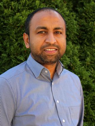
Dr. Tanzil M. Arefin is an Assistant Professor of Neuroscience at the University of Rochester School of Medicine and Dentistry and Associate Director of the Preclinical Imaging Core at the Center for Advanced Brain Imaging and Neurophysiology (CABIN). His research focuses on developing neuroimaging techniques to study brain functions and microstructures in animal models of human disorders, including neurodegenerative and psychiatric illnesses. He holds affiliations with the Del Monte Institute for Neuroscience and the Neuroscience Ph.D. Program. **Education**: Ph.D., Neuroscience, University of Freiburg and University of Strasbourg (2017) M.Sc., Biomedical Engineering, Czech Technical University and University of Groningen (2012) B.Sc., Electrical and Electronic Engineering, Islamic University of Technology (2007) **Research Interests**: Dr. Arefin's lab employs multimodal MRI methodologies (resting-state fMRI, diffusion MRI, ASL perfusion MRI, MR spectroscopy) alongside optogenetics and chemogenetics to elucidate molecular mechanisms impairing brain plasticity. Current projects include studying cerebellar connectivity's role in non-motor behaviors and developing interventions for alcohol-dependent brains. **Awards**: Magna cum Laude, Summa Cum Laude, Erasmus Mundus Fellowships (both Doctoral and Masters). **Grants & Advising**: Not explicitly listed in texts, but lab activities suggest involvement in NIH-funded projects. Advising details are pending explicit student listings. **Lab & Affiliations**: Arefin Lab focuses on translational imaging tools. Affiliated with UR CABIN and URMC's Neuroscience programs. Location: 430 Elmwood Ave, Rochester, NY.
Charles A. Greer is Professor of Neurosurgery and Neuroscience at Yale School of Medicine, where he serves as Co-Vice Chair of Research for Neurosurgery and Director of the Yale Interdepartmental Neuroscience Graduate Program. He maintains joint appointments in both the Department of Neurosurgery and Department of Neuroscience, with additional affiliations through the Wu Tsai Institute, Yale Combined Program in the Biological and Biomedical Sciences, and Yale Stem Cell Center. Dr. Greer's research focuses on understanding the fundamental mechanisms that establish orderly topographic maps within the central nervous system during development and regeneration. His laboratory primarily investigates the complex olfactory system, studying how axons sort into functional subsets and target specific regions in the olfactory bulb. His work has significant implications for understanding neural regeneration following injury or disease, particularly through his research on ensheathing cells that support axon growth and can promote remyelination in damaged spinal cord tissue. His research spans multiple disciplines including neuroanatomy, developmental neuroscience, and neural regeneration, with particular emphasis on the molecular and cellular mechanisms of axon targeting, glial cell function, and neural circuit formation. Dr. Greer has established extensive collaborations across Yale departments including Neurology, Neurobiology, Anesthesiology, and Ophthalmology, as well as with institutions including Columbia University, Emory University, The Rockefeller University, University of Maryland, and University of Colorado. Dr. Greer has received numerous prestigious awards including the Max Mozell Award for Outstanding Achievements in the Chemical Senses, the R.H. Wright Award for Outstanding Research in Olfaction, and recognition as Distinguished Professor from Universite Pierre et Marie Curie in Paris. He has served as President of the Association for Chemoreception Sciences and as a member of NIH study sections and the Advisory Council for the National Institute on Deafness and Other Communication Disorders. As an educator, Dr. Greer has directed the Yale Interdepartmental Neuroscience Graduate Program and mentored numerous students and postdoctoral fellows. He serves as Associate Editor for The Journal of Comparative Neurology and Journal of Neuroscience, and sits on the editorial boards of several other neuroscience journals. His extensive publication record demonstrates sustained contributions to understanding the organization and development of neural systems, particularly the olfactory pathway.
John G Georgiadis is the Interim Chair and R. A. Pritzker Professor of Biomedical Engineering at Illinois Institute of Technology's Armour College of Engineering. He holds affiliations with the Illinois Tech Digital Medical Engineering and Technology (IDMET) Research and Education Center. His academic journey includes a Ph.D. (1987) and M.S. (1984) in Mechanical Engineering from UCLA, and a Diploma in Mechanical Engineering from the National Technical University of Athens (1983). Georgiadis’ research focuses on aging-related changes in the brain and skeletal muscle, leveraging MRI and computational models. Key projects include intramyocellular biotransport, cerebral microvasculature imaging, and multiscale brain mechanics. He has pioneered advancements in magnetic resonance elastography (MRE) for non-invasive tissue stiffness measurement, contributing to clinical applications in neurology and cardiology. His awards include the NSF Presidential Young Investigator Award (1991–1997) and Fellow status in the American Institute for Medical and Biological Engineering. Georgiadis has authored over 150 peer-reviewed publications and holds multiple patents in medical device technology and imaging techniques. His work bridges biomechanical engineering, computational imaging, and clinical diagnostics, with implications for aging populations and chronic disease management. Professional memberships include the Biomedical Engineering Society, IEEE, and AIMBE. His labs focus on translational research, integrating advanced imaging modalities with biomechanical principles to address complex biomedical challenges.
Jessica D. Rosarda PhD is an Assistant Professor in the Department of Anatomy, Physiology and Genetics at the Uniformed Services University (USU) of the Health Sciences School of Medicine in Bethesda, MD. She leads a research laboratory focused on cellular stress mechanisms in military-relevant disorders. PhD in Chemical and Biological Sciences, The Scripps Research Institute (2023) MSc in Pharmacy, University of Florida (2013) BSc in Biology, Washington and Lee University (2010) Dr. Rosarda's research centers on cellular stress response pathways, particularly how cells respond to stress through signaling mechanisms that can either protect or damage tissues. Her work spans chemical biology, molecular medicine, neuroscience, and molecular/cell biology with a focus on proteostasis, unfolded protein response, and stress signaling dynamics in disease contexts. She investigates how imbalances in these pathways contribute to conditions ranging from retinal degeneration to traumatic brain injury, with particular relevance to military medicine. Analysis of Dr. Rosarda's publication record reveals a strong focus on stress response pathways, particularly the unfolded protein response and integrated stress response. Her work demonstrates how perturbations in proteostasis contribute to diverse pathologies including neurodegeneration, amyloidosis, and inflammatory conditions. She employs chemical biology approaches to develop therapeutic strategies targeting these pathways, with several publications on pharmacological modulators of stress responses. Dr. Rosarda maintains an active research program with numerous publications in high-impact journals including Nature Communications, Cell Chemical Biology, and ACS Chemical Biology. Her work frequently involves collaborations with the Wiseman laboratory and other researchers in the fields of proteostasis and stress response. Dr. Rosarda's laboratory at USU focuses on defining stress pathway signaling dynamics in military-relevant disorders, determining the metabolic factors that govern these stress responses, and identifying novel approaches for resolving toxic stress involved in both acute and chronic conditions.
Associate Professor Anna Leonard is part of the School of Biomedicine at the University of Adelaide, within the Faculty of Health and Medical Sciences. She leads the Spinal Cord Injury Research Group, focusing on understanding secondary injury processes post-SCI, particularly oedema, haemorrhage, and neuroinflammation. Her research utilizes porcine and rodent models to explore translational therapies and chronic outcomes like cognition and neuropathic pain. Dr. Leonard completed her PhD in 2012 (awarded Dean’s commendation and University Doctoral Medal) and has held postdoctoral fellowships at the University of Adelaide and the University of Alabama at Birmingham. She currently supervises research students at all levels and holds national recognition in neurotrauma research. Educational background: PhD (2012, University of Adelaide), postdoctoral training at UoA and UAB. Research interests include: neuroinflammation, spinal cord trauma mechanisms, neuroprotective interventions, and translational clinical models. Scientific achievements: Developed Australia’s first clinically relevant large-animal spinal cord injury model. Awards include the University Doctoral Medal (2012). Active in mentoring HDR students and grant acquisition. Currently investigates peripheral stimulation therapies, aging effects on SCI recovery, and concomitant brain injury impacts.
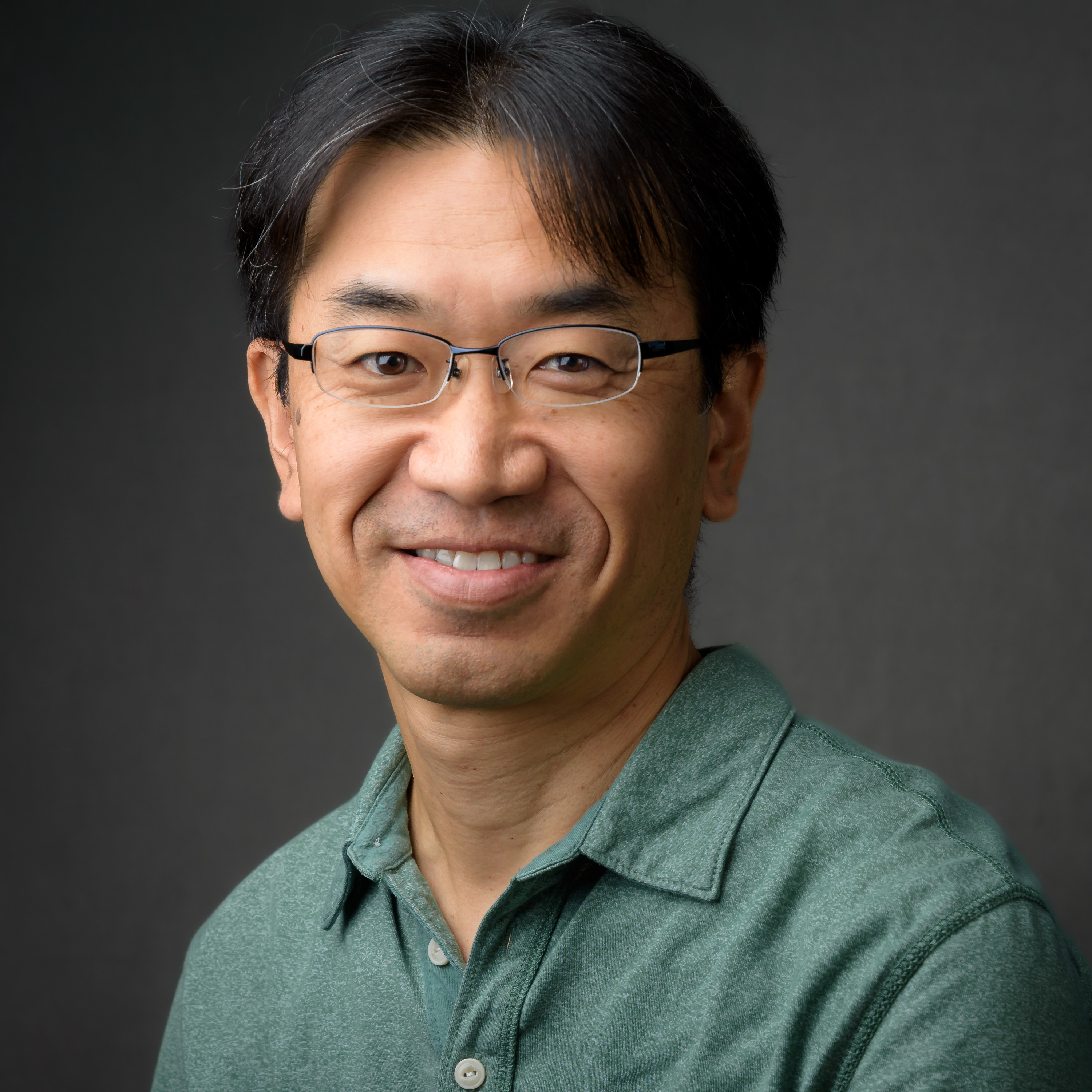
Hiroshi Nishiyama is an Associate Professor in the Department of Neuroscience at the University of Texas at Austin. His research focuses on synaptic plasticity and circuit reorganization in the mammalian cerebellum, with implications for learning, memory, and brain injury recovery. Education: B.S., Kyoto University (1992–1996) M.S., Kyoto University (1996–1998) Ph.D., Kyoto University & RIKEN Brain Science Institute (1998–2002) Postdoctoral Research, Johns Hopkins University (2002–2008) His work explores how cerebellar circuits are refined during development, reorganized in adulthood through learning, and rewired after injury. Techniques include electrophysiology , optogenetics , and long-term in vivo imaging of synaptic dynamics. Recent publications highlight mechanisms of Purkinje cell ablation , lesion-induced axonal sprouting , and dendritic translocation in synaptic competition. These studies span developmental neuroscience , neurotrauma , and neurodegenerative models . The Nishiyama Lab investigates cerebellar circuits as critical nodes in brain-wide functional architecture, bridging motor control and broader neurological disorders.
Professor Mark Lythgoe is a distinguished academic at University College London (UCL), where he serves as Professor of Biomedical Imaging in the Department of Imaging within the Faculty of Medical Sciences. He is the Founder and Director of the Centre for Advanced Biomedical Imaging (CABI) at UCL, a multidisciplinary research center hosting 12 state-of-the-art imaging modalities and 50 researchers. Additionally, he is Co-Director of the UCL Department of Imaging and Director of Biomedical Imaging Research at the Francis Crick Institute. Mark Lythgoe earned his Doctor of Philosophy from University College London in 1999 and his Master of Science from the University of Surrey in 1993. Professor Lythgoe has a long-standing track record in the development and application of biomedical imaging techniques, with research spanning from fundamental imaging science to clinical applications. His work focuses on advancing imaging technologies for biomedical research and clinical practice, with particular emphasis on neuroimaging, molecular imaging, and the application of imaging to understand neurological disorders and cancer. He has translated his research findings into clinical radiological practice and established training programs in biomedical imaging. His extensive publication record demonstrates consistent innovation across imaging modalities, with recent work increasingly focused on the glymphatic system in neurological disorders, advanced tumor imaging techniques, and the development of novel imaging technologies for both preclinical and clinical applications. His research bridges engineering, physics, biology, and clinical medicine to solve complex biomedical challenges. Professor Lythgoe's significant contributions to the field have been recognized with numerous prestigious awards: IET Achievement Medal (2023) - for major and distinguished contribution in Medical Imaging Royal Society of Medicine Ellison-Cliffe Award (2021) - for contribution of fundamental science to the advancement of medicine Davies Medal from the Royal Photographic Society (2013) - for significant contribution to imaging science Alumni Achievement Award from the University of Salford Neuroscience Prize for Public Understanding from the British Neuroscience Association Dorothy Hodgkin Award Biosciences Federation Science Communication Award Fellow of the British Science Association Professor Lythgoe has secured substantial research funding, with £45 million awarded for his collaborative imaging research program. He is deeply committed to training the next generation of imaging scientists, serving as Co-Director of the MSc in Advanced Biomedical Imaging and Co-Founder of the UCL Centre for Doctoral Training in Medical Imaging. His public engagement efforts include directing the Cheltenham Science Festival, which has become one of the largest science festivals in the world. As Director of the Centre for Advanced Biomedical Imaging, Professor Lythgoe leads a vibrant research team of 50 researchers working across 12 state-of-the-art imaging modalities. His center fosters interdisciplinary collaboration between engineers, physicists, biologists, and clinicians to develop and apply cutting-edge imaging technologies. He has published over 300 papers including publications in Nature, Nature Photonics, Nature Medicine and The Lancet, demonstrating the high impact of his research across multiple disciplines.
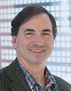
Peter J. Basser is a leading research scientist at the National Institutes of Health (NIH), specifically within the Eunice Kennedy Shriver National Institute of Child Health and Human Development (NICHD), where he heads the Section on Quantitative Imaging and Tissue Sciences (SQITS). His work bridges physics, engineering, and neuroscience to develop non-invasive MRI methods for probing tissue microstructure, particularly in the brain. His educational background is not explicitly mentioned, but his scientific achievements reflect deep training in biophysics and medical imaging. He earned his Ph.D. and has built a career at NIH as a principal inventor of key neuroimaging technologies. Basser's research focuses on quantitative imaging and tissue sciences , especially using diffusion MRI to study brain structure and function. He pioneered Diffusion Tensor MRI (DTI) , Streamline Tractography , and advanced methods like MAP MRI , CHARMED , and AxCaliber , enabling in vivo measurement of axon diameters and microstructural features previously accessible only through histology. His work aims to translate these tools into clinical use for diagnosing developmental disorders, trauma, and neurodegeneration. The 15 most recent articles reflect a consistent focus on developing novel MRI biomarkers, particularly through diffusion and relaxometry methods. They explore water exchange, restriction, glymphatic clearance, latency connectomes, and cortical microstructure, demonstrating a trajectory toward in vivo MRI histology and precision imaging for pediatric and neurological applications. Scientific Awards: National Academy of Engineering (NAE), Inducted 2020 National Academy of Inventors (NAI) Fellow, 2024 Eduard Rhein Technology Award, 2021 ISMRM Gold Medal, 2008 ISMRM Lauterbur Lecturer, 2020 American Society of Neuroradiology Honorary Member, 2019 Victor M. Haughton Award, 2017 ISMRM Fellow, 2010 AIMBE Fellow Best Paper Award, Frontiers in Physics, 2023 Basser leads a dynamic research group that mentors postdoctoral fellows and trainees, many of whom have received prestigious awards. His lab has secured significant grants from the NIH BRAIN Initiative, NICHD, USUHS, and the Bill & Melinda Gates Foundation. The SQITS lab develops open-source software tools like TORTOISE , dmritool , and HI-SPEED , which are widely used in the neuroimaging community. The lab collaborates with institutions such as Uniformed Services University and participates in major initiatives like the Human Placenta Project and the Human Connectome Project. Basser’s vision is to transform clinical MRI scanners into quantitative scientific instruments for precision medicine and large-scale brain mapping. Labs and Teams: Section on Quantitative Imaging and Tissue Sciences (SQITS), NICHD, NIH Neuropathology-Neuroradiology Integration Core (with USUHS) Advanced Translational Neuroimaging Research & Development Core Diffusion – Data Processing Center (DPC)
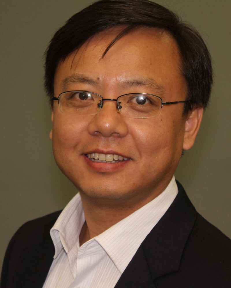
Junzhong Xu is an Associate Professor of Radiology and Radiological Sciences, Biomedical Engineering, and Physics and Astronomy at Vanderbilt University. His research focuses on developing advanced quantitative MRI methods for applications in neurodegenerative diseases and cancer, including MRI cell size imaging and tumor treatment response assessment. He holds affiliations with the Vanderbilt University Institute of Imaging Science (VUIIS) and has expertise in microstructural diffusion MRI and GPU-accelerated imaging tools. Xu’s work spans interdisciplinary collaborations in biomedical engineering and physics. Education: B.S., University of Science and Technology of China M.S., University of Science and Technology of China Ph.D., Vanderbilt University Research Interests: Development of MRI cell size imaging for cancer and neurodegenerative diseases Quantitative assessment of tumor microstructure and treatment response Applications of microstructural diffusion MRI in multiple sclerosis and oncology Key Contributions: Advancement of MRI cytometry for non-invasive disease assessment GPU-accelerated tools for diffusion MRI simulation (MATI) Imaging biomarkers for early detection of neurodegenerative pathology Labs/Teams: Active member of the Vanderbilt University Institute of Imaging Science (VUIIS), collaborating on interdisciplinary imaging projects.
Bruce Wasserman, MD is a Professor in the Department of Diagnostic Radiology and Nuclear Medicine at the University of Maryland School of Medicine. He serves as Vice Chair of Clinical Research and Director of the Neurovascular Imaging Center. His expertise spans neuroradiology, vascular imaging, and MRI techniques development for studying neurovascular diseases. Dr. Wasserman has held significant leadership roles including Director of the Diagnostic Neurovascular Imaging Center at Johns Hopkins for 16 years. His educational background includes a BS in Biomedical Engineering from Rensselaer Polytechnic Institute, Internal Medicine internship at University of Rochester School of Medicine, Residency at Mount Auburn Hospital in Harvard Medical School, and Neuroradiology Fellowship and MD from Johns Hopkins University School of Medicine. Dr. Wasserman's research focuses on innovative MRI techniques to study blood vessel diseases in the head and neck, particularly atherosclerotic plaque formation and instability. His work aims to reduce stroke risk through better understanding of plaque development and vulnerability. He has made significant contributions to the field of intracranial atherosclerotic disease and its relationship to cognitive decline and dementia. His research also encompasses vessel wall imaging for characterizing plaque features and assessing stroke risk. His publication record shows consistent research output from 2002 through 2025, with recent emphasis on carotid plaque classification systems, intracranial atherosclerosis in dementia development, and vessel wall imaging techniques. His work bridges neuroradiology, cardiology, and neurology with applications in stroke prevention and risk assessment. Dr. Wasserman has been continuously funded by the NIH as a PI or co-PI since 2002. He leads several major multi-institutional studies including the Multi-Ethnic Study of Atherosclerosis (MESA) carotid MRI study, the MRI Reading Center for the Atherosclerosis Risk in Communities (ARIC) study, and the Intracranial Atherosclerotic Disease and Cognitive Impairment Study. He chairs the American Society of Neuroradiology Cervical and Intracranial Vessel Wall Imaging Study Group. His laboratory includes multiple postdoctoral fellows and research staff working on neurovascular imaging projects. Dr. Wasserman collaborates extensively with researchers across institutions, particularly through large cohort studies that follow thousands of participants to study imaging biomarkers of cardiovascular events.
Gwenn Smith, PhD, is a Professor of Psychiatry and Behavioral Sciences at Johns Hopkins University School of Medicine. She serves as Director of the Division of Geriatric Psychiatry and Neuropsychiatry and Co-Director of the Center for Translational Molecular Imaging at Johns Hopkins Bayview Medical Center. Her primary research facility is located at the Department of Psychiatry, 5300 Alpha Commons Drive, Baltimore. Dr. Smith leads a research program focused on molecular neuroimaging of neurodegenerative and mood disorders. For over two decades, her work has centered on: Developing PET imaging methods to study neurobiological mechanisms of cognitive decline Investigating monoaminergic system dysfunction in late-life depression and dementia Mapping treatment response mechanisms for brain stimulation therapies Identifying early biomarkers for Alzheimer's, Parkinson's, and related disorders Her publication portfolio demonstrates consistent focus on multimodal neuroimaging approaches integrating PET, MRI, and proteomic analyses. Recent work emphasizes: Interactions between serotonin degeneration and amyloid deposition Optimization of deep brain stimulation for Alzheimer's disease Neurobiological signatures of psychosis across neurodegenerative disorders Advanced 7T MR spectroscopy applications Dr. Smith directs several neuroimaging initiatives including studies on fornix deep brain stimulation (ADvance trial), blood-brain barrier opening using focused ultrasound, and traumatic brain injury in athletes. She maintains active collaborations across neurology, neurosurgery, and radiology departments to advance translational applications of neuroimaging.
Mattias Sköld serves as an Adjunct Lecturer at Karolinska Institutet's Department of Neuroscience and Associate Professor of Neurosurgery at Uppsala University, concurrently functioning as Senior Consultant Neurosurgeon at Akademiska Sjukhuset Uppsala and Senior Researcher in Neuroscience. His academic credentials include: M.D. from Karolinska Institutet (2003) Ph.D. in Neuroscience from Karolinska Institutet (2004) Medical Internship at Karolinska University Hospital (2004-2006) Neurosurgery residency at Akademiska Sjukhuset Uppsala (2006, 2012-2019) Sköld's research investigates traumatic nervous system injuries through methodologies ranging from in vitro cavitation models to animal studies, with military medicine applications forming a critical focus. His work examines blast-induced trauma mechanisms, neural regeneration pathways, and molecular responses to high-energy injuries, particularly emphasizing VEGF signaling and inflammatory cascades in neural repair processes. Analysis of his 15 most recent publications reveals consistent exploration of blast injury modeling frameworks, peripheral nerve regeneration techniques using biomaterials, and temporal gene expression patterns following neural trauma across military and civilian contexts. No scientific awards are documented in the provided materials. He supervises Ph.D. students in both basic neuroscience and clinical neurosurgery domains. Current research is supported by Swedish Research Council grants including "Novel strategies for nervous tissue regeneration" (2020-2023) and previously "Targeted anti-inflammatory treatment in traumatic brain injuries" (2009-2010). Sköld leads Karolinska Institutet's Experimental Traumatology Research Unit and contributes to multiple NATO human factors and medicine task groups focused on brain injury modeling standards and combat casualty care performance metrics.
Dr. Jacqueline Rushby is a Conjoint Senior Lecturer at the School of Psychology, University of New South Wales. Her position involves part-time academic engagement in psychophysiological research and neuropsychological rehabilitation. Her research investigates disorders of social cognition and emotional processing in clinical populations, with emphasis on traumatic brain injury (TBI), autism spectrum disorder (ASD), and frontotemporal dementia. Key methodologies include EEG/ERP analysis, transcranial direct current stimulation (tDCS), and autonomic arousal measurement to explore neural correlates of empathy deficits and emotion regulation. Recent publications demonstrate a consistent focus on neurorehabilitation mechanisms, with frequent examination of tDCS efficacy, emotion recognition impairments, and autonomic dysregulation in TBI populations. Studies increasingly integrate multimodal physiological measures (e.g., HRV biofeedback, facial EMG) with neurocognitive assessments. No scientific awards or prizes are documented in the provided materials. Information regarding student advising, research grants, laboratory affiliations, or team collaborations is unavailable in the source text.
Audny Gabriele Wagner Anke serves as Head of Department of Physical Medicine and Rehabilitation, Senior Consultant, and Professor II at the Department of Clinical Medicine, UiT The Arctic University of Norway in Tromsø. She is affiliated with both the Rheumatology and Rehabilitation research group and the Neuromuscular Diseases Research Group. Her research expertise spans multiple critical areas in rehabilitation medicine: Long-term stroke rehabilitation outcomes (particularly at 4-year follow-up) Traumatic brain injury and spinal cord injury recovery Rehabilitation approaches for individuals with intellectual disabilities Chronic pain management, especially neck and back complaints Quality of life and functional outcomes following various injuries Professor Wagner Anke's recent publications reveal a strong emphasis on longitudinal studies examining recovery trajectories over extended periods. Her work frequently bridges physical and cognitive rehabilitation domains, with growing interest in technology-assisted interventions like augmented reality exergames. She contributes significantly to large-scale registry studies such as the Norwegian Neck and Back Registry (NNRR) and international collaborations including the CENTER-TBI study. Her most notable research contributions address: Metacognitive factors influencing stroke recovery Barriers to physical activity for adults with intellectual disabilities Unmet rehabilitation needs in trauma populations Vocational rehabilitation and return-to-work outcomes Family-centered care following pediatric trauma As Head of Department, Professor Wagner Anke maintains active clinical responsibilities while leading research initiatives and mentoring junior researchers. Her interdisciplinary collaborations span multiple institutions in Norway and internationally, demonstrating her commitment to advancing rehabilitation science through evidence-based practice.