Manuel Antonio Burgos Olmos is a Professor specializing in Computational Fluid Dynamics (CFD) applied to nasal airway function and surgical optimization. His interdisciplinary work bridges biomedical engineering, oto-rhino-laryngology, and anthropology, with significant contributions to virtual surgery tools like DigBody®, MeComLand®, and Noseland® for objective airflow analysis and surgical planning. His research focuses on nasal airflow simulation across diverse contexts: human climate adaptation studies in anthropology, veterinary applications for big cats (e.g., lions using Flowgy software), and clinical conditions including empty nose syndrome and nasoseptal perforations. Key interests include 3D modeling of nasal cavities, vorticity analysis, and reducing surgical outcome variability through computational approaches. Analysis of his 46 publications (1998-2025) reveals a consistent trajectory toward integrated computational modeling, with recent work (2016-2025) emphasizing cross-species comparisons (humans/chimpanzees), sexual dimorphism in airways, and clinical-veterinary translational applications. His publications in high-impact journals demonstrate methodological innovation in CFD-based surgical prediction and anthropological morphology assessment.
Danail Stoyanov is a Professor of Robot Vision at University College London's Department of Computer Science, holding the prestigious Royal Academy of Engineering Chair in Emerging Technologies. He serves as Co-Director of the UCL Hawkes Institute and previously directed the Wellcome/EPSRC Centre for Interventional and Surgical Sciences until September 2024. His academic journey at UCL progressed from Lecturer (2014) to Senior Lecturer (2015), Associate Professor (2016), and ultimately Professor of Robot Vision (2018). His educational background includes a Bachelor of Engineering in Computer Systems and Electronics from King's College London (2001) and a PhD in Computer Science specializing in medical image computing from Imperial College London (2006). His progression through Royal Academy of Engineering Research Fellowships at both Imperial College and UCL demonstrates his exceptional research trajectory. Professor Stoyanov's research focuses on developing surgical robotics and artificial intelligence to improve minimally invasive interventions. His work spans vision-based surgical guidance systems, computational biophotonic imaging modalities, and quantitative measurements during robotic-assisted procedures. His research bridges computer vision, medical robotics, and clinical applications with emphasis on gastrointestinal surgery, pituitary surgery, and ophthalmic interventions. Analysis of his recent publications reveals a strong trend toward real-time AI applications in surgical contexts, with particular emphasis on stereo vision, depth estimation, surgical workflow recognition, and error detection systems. His work increasingly incorporates foundation models and self-supervised learning approaches while maintaining clinical relevance across multiple surgical specialties including neurosurgery, ophthalmology, and gastrointestinal procedures. His distinguished recognition includes multiple prestigious fellowships: Fellow of the Royal Academy of Engineering (2022) Fellow of the Institute of Engineering and Technology (2020) Fellow of the MICCAI Society (2023) Fellow of the Institute of Electrical and Electronics Engineers (2024) Fellow of the Academy of Medical Sciences (2025) As an educator, Stoyanov founded and directs the MSc Robotics and Computation program while teaching the advanced Robotic Systems Engineering course. His leadership extends to conference organization including roles as Programme Chair for IPCAI (2014), General Chair for IPCAI (2017-2020), and Programme Co-Chair for MICCAI (2018, 2020). He maintains an active industry collaboration as Chief Scientist for Digital Technologies at Medtronic since 2016. His SRV Group at UCL serves as the primary research hub driving innovation in surgical robotics and vision, with strong connections to the Wellcome/EPSRC Centre for Interventional and Surgical Sciences and the UCL Hawkes Institute which he now co-directs. The group maintains active collaborations with clinical partners and industry including Medtronic, facilitating rapid translation of research into clinical practice.
Lars Lönn is a Clinical Professor in the Department of Clinical Medicine at the University of Copenhagen's Faculty of Health and Medical Sciences, based at Rigshospitalet in Copenhagen. His clinical and research work focuses on interventional radiology techniques and medical education. His research centers on expanding endovascular treatment applications, including percutaneous transluminal angioplasty (PTA), stent placement, embolization techniques, and endovascular prosthetic insertion. He evaluates these against conventional diagnostic methods like duplex ultrasound, arteriography, and 3D CT scanning. A major emphasis is medical simulation training for endovascular procedures, with projects examining simulator-based training effectiveness across clinical scenarios in collaboration with CEKU, vascular surgery, cardiology, and radiology departments. Recent publications reveal expertise in prostatic artery embolization for benign prostatic hyperplasia, genicular artery embolization for knee osteoarthritis, and AI-driven lung perfusion quantification in pulmonary embolism, demonstrating strong trends in vascular interventions, embolization therapies, and advanced imaging analytics integration. Professor Lönn leads key collaborative initiatives including the Copenhagen Mesenteric stent study (COMESS) comparing stent treatments for mesenteric ischemia and the TUMT-PAE-1 trial evaluating alternatives for urinary symptoms. His work emphasizes interdisciplinary coordination across trauma surgery, anesthesiology, and specialty departments to optimize acute care protocols and patient outcomes.
Anupama Grandhi, BDS, DDS, serves as Associate Professor in Oral and Maxillofacial Surgery at Loma Linda University School of Dentistry and Assistant Professor in Pathology and Human Anatomy at the School of Medicine. Her dual appointment bridges dental and medical pathology through interdisciplinary research and clinical education. Her educational foundation includes: Bachelor of Dental Surgery (BDS) from Rajiv Gandhi University of Health Sciences (2002) Doctor of Dental Surgery (DDS) from University of Southern California (2006) Dr. Grandhi's research pioneers AI-integrated approaches to oral cancer and dysplasia, focusing on epigenomic/transcriptomic signatures and noninvasive diagnostics. She investigates perineural invasion mechanisms and biomaterial effects on neural tissues, with recent work developing brush swab techniques as alternatives to tissue biopsies. Her methodology combines computational analysis with molecular biology to advance early detection and precision oncology in dentistry. Analysis of her 2021-2024 publications reveals accelerating interdisciplinary output (3 papers in 2024 alone), with dominant themes in AI-driven diagnostics, multi-omics integration for tobacco-related cancer signatures, and clinical translation of noninvasive methods. This trajectory positions her at the forefront of computational oral pathology. Her professional recognitions include: Diplomate, American Board of Oral and Maxillofacial Pathology (2012-present) Fellow, American Academy of Oral and Maxillofacial Pathology (2012-present) Fellow, Southern California Academy of Oral Pathology (2017-present) Dr. Grandhi actively secures research funding through industry partnerships, currently leading two Vigilant Biosciences clinical studies: the BEVIGILANT™ ORAFUSION SYSTEM trial (2023) and OncAlert RAPID evaluation (2024). While specific student names aren't published, her collaborative publications indicate active mentorship of trainees across dental and medical disciplines. Her teaching portfolio demonstrates consistent commitment to clinical oral pathology education through courses like GRDN 535 and DENT 880. Though no formal lab name is designated, her research group operates at the convergence of oral oncology, computational pathology, and translational diagnostics, driving innovation in cancer detection methodologies and neural-tumor interaction studies.
Nuno Malta dos Santos is a Researcher at the Center for Interdisciplinary Research in Health (CIIS) within the Faculty of Dental Medicine at Universidade Católica Portuguesa. His academic career focuses on periodontal surgery, particularly in the areas of gingival recession treatment, root coverage procedures, and implantology. He actively participates in multiple research projects including the InDig project on digital teaching technologies in dental medicine and the CIIS research center. His research interests span Gingival Recession , Connective Tissue Graft techniques, Systematic Review methodologies, Platelet-Rich Fibrin applications, and advanced Incision Techniques in periodontal surgery. Dr. dos Santos has developed expertise in minimally invasive approaches for root coverage procedures and the assessment of various biomaterials in periodontal regeneration. Analysis of his recent publications reveals a strong focus on evidence-based dentistry through systematic reviews, particularly examining the relationship between root anatomy and gingival recession, bisphosphonate effects on implant success, and innovative surgical techniques like the Mixed-Thickness Tunnel Access approach. His work consistently bridges clinical practice with scientific validation, contributing significantly to advancements in periodontal plastic surgery. Dr. dos Santos has supervised multiple students including Rebelo, C. G. on bisphosphonate research and Júnior, L. S. D. F. on longitudinal studies of gingival recession in dental students. His research has been supported by projects funded through Portugal2020 and Fundação para a Ciência e a Tecnologia. He is an active contributor to the dental research community through conference presentations and collaborations within the CIIS center, focusing on improving clinical outcomes in periodontal surgery and implant dentistry through evidence-based approaches and technological innovation.
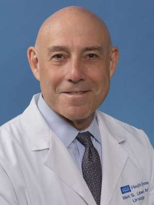
Mark S. Litwin is Professor and Chair of the Department of Urology at the David Geffen School of Medicine at UCLA. He also holds appointments in health policy & management and nursing, reflecting his interdisciplinary approach to healthcare. Trained at Harvard, Dr. Litwin specializes in testicular, bladder, prostate and kidney cancer, with a particular focus on improving quality of care, quality of life, and equitable access to care in urologic oncology. Dr. Litwin's research spans multiple domains including Health Care Access, Health Care Quality, Quality-of-life Measurement, Medical Outcomes Research, Health Policy, Resource Utilization, Medical Decision Making, and the Epidemiologic Burden of Illness of Urologic Diseases. His work has significant implications for patient care, particularly in prostate cancer treatment and LGBTQ+ health services. He created and directs IMPACT, a state-funded program providing free medical care for low-income, uninsured men with prostate cancer in California. His publication portfolio demonstrates expertise across urologic oncology, healthcare policy, and quality improvement. Recent articles focus on prostate cancer diagnostics (including AI applications), global treatment variations, graphical representations of symptoms for underserved populations, and the impact of the COVID-19 pandemic on gender-affirming care. His work consistently emphasizes patient-centered outcomes and healthcare accessibility. Super Doctors® Southern California, 2013-2025 Sherman M. Mellinkoff Faculty Award, David Geffen School of Medicine at UCLA, 2021 Top Doctors, Los Angeles Magazine, 2018-2021 UCLA Community Service and Praxis Award for Diversity, Equity and Inclusion, 2017 Barringer Medal, American Association of Genitourinary Surgeons, 2016 As Founder and Co-Director of the UCLA Gender Health Program, Dr. Litwin has expanded his leadership beyond traditional urology into gender-affirming care. He directs an NIH-funded fellowship training program in outcomes research in urologic oncology and previously led Urologic Diseases in America, the NIH's largest effort documenting the burden of urologic diseases. His clinical work spans multiple UCLA-affiliated medical centers including Ronald Reagan UCLA Medical Center, Harbor-UCLA Medical Center, and UCLA Santa Monica Medical Center.
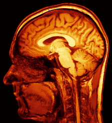
Professor Franklyn Howe serves as Professor of Magnetic Resonance Imaging and Director of the Neuroscience & Cell Biology Research Institute at City St George's, University of London (formed from the August 2024 merger of St George's, University of London and City University of London). With a career spanning since 1988 at St George's, he has established himself as a leading expert in multimodal MRI applications for disease diagnosis and treatment monitoring, particularly in brain tumor analysis. His work bridges physics, medical imaging, and clinical applications across multiple disease areas. Professor Howe earned his BA in Physics from the University of Oxford, followed by a DPhil on 'Magnetisation and microwave absorption in rare earth metal alloys' from the same institution in 1984. He completed postdoctoral research evaluating MRI contrast agents at St Bartholomew's Hospital and in MR image analysis at the Royal Postgraduate Medical School (now part of Imperial College School of Medicine) before joining St George's. Howe's research centers on developing automated analysis of multimodal MRI to aid diagnosis of brain tumors, using various MRI methods including perfusion and diffusion imaging for structural and functional information, and magnetic resonance spectroscopy (MRS) for biochemical data. He applies pattern recognition techniques to optimally combine data from different MRI methods, aiming to develop biomarkers for diagnosis, prognosis assessment, and treatment response monitoring. His work extends beyond brain tumors to dementia, Tourette syndrome, cerebrovascular disease, chronic obstructive pulmonary disease, lupus, lymphedema, osteoarthritis, and normal aging. In 1992, he was instrumental in developing MR Neurography at St George's, a non-invasive method for detailed nerve imaging. Analysis of Professor Howe's recent publications (2021-2024) reveals consistent focus on advancing MRI techniques across multiple clinical applications. His work demonstrates strong interdisciplinary collaboration, with significant contributions to lymphatic imaging, brain tumor characterization, neurodegenerative disorders, and quantitative MRI methodology. The publications show progression from fundamental technical improvements in MRI acquisition and processing toward increasingly sophisticated clinical applications, particularly in neuroimaging and vascular disorders. Chair of ISMRM MRS Study Group Co-founder and Chair of Pediatric MR Study Group Long-standing member of International Society of Magnetic Resonance in Medicine Professor Howe actively supervises lab-based MRes and BSc student research projects and library-based Special Study Courses on Biomedical Applications of Magnetic Resonance. He has secured substantial research funding from diverse sources including the Medical Research Council, Cancer Research UK, Alzheimer's Research UK, and St George's Hospital Charity. His collaborative approach is evident in the wide range of clinical research studies he participates in across multiple departments. As Director of the Neuroscience & Cell Biology Research Institute, Howe leads a research team including Ian Storey (Image Analyst) and Mike Mills (MRI physicist). He maintains extensive collaborations within St George's with Dr Thomas Barrick, Dr Atticus Hainsworth, Mr Timothy Jones (Neurosurgery), Dr Jeremy Isaacs (Neurology), Dr Pia Ostergaard, Prof Sahar Mansour (Clinical Genetics), Prof Nidhi Sofat (Rheumatology), and Mr Michael Hart (Neurosurgery), creating a robust interdisciplinary research environment focused on translating MRI advances into clinical practice.
Dr. Kurt Busuttil Naudi is a Clinical Senior University Lecturer and Honorary Consultant in the Dental School at the University of Glasgow's School of Medicine, Dentistry & Nursing. He serves as coordinator of the 4th Year BDS undergraduate course and co-coordinator of the postgraduate MSc in Oral and Maxillofacial Surgery, while also managing international elective visits to Glasgow Dental School. His educational background includes: BChD from the University of Malta (2001) MFDS from the Royal College of Physicians & Surgeons of Glasgow MSurgDent from the Royal College of Surgeons of Edinburgh (2008) Doctorate in Dental Surgery from the University of Glasgow (2011) Masters in Education in Learning and Teaching in Higher Education from the University of Glasgow (2014) Dr. Busuttil Naudi's primary research focuses on two main areas: Bone Biology and Dental Education. In Bone Biology, he specializes in bone bioengineering, particularly investigating growth factors such as Bone Morphogenetic Protein and various synthetic injectable, solid and 3D printed scaffolds for repairing critical-size craniofacial defects. His Dental Education research examines longitudinal data for competence assessment in dentistry, aiming to improve educational outcomes and assessment methodologies for dental students. His publication record shows a consistent trajectory in oral and maxillofacial surgery research, with particular emphasis on bone regeneration techniques, surgical planning using 3D technologies, and dental education assessment. Over the past two decades, his work has evolved from basic case reports to sophisticated tissue engineering approaches and educational research methodologies. Professional recognition includes: Fellow of the Higher Education Academy Dr. Busuttil Naudi actively supervises PhD students in both his research domains, guiding projects related to bone bioengineering applications and innovative dental education assessment techniques. His work bridges clinical practice with academic research, contributing significantly to advancements in oral surgery techniques and dental education methodologies. He leads research efforts in the Glasgow Dental Hospital & School laboratory environment, focusing on translational research that connects laboratory findings with clinical applications in oral and maxillofacial surgery.
Vesna Zeljkovic serves as a Professor in the Chemistry & Physics Department at Lincoln University, where she applies her expertise in signal and image processing to develop mathematical models and novel algorithms for medical applications. Her office is located in the Ivory V. Nelson Science Center Room 334, and she can be reached at vzeljkovic@lincoln.edu or by phone at 484-365-7258. Professor Zeljkovic's research spans multiple interdisciplinary domains with a strong focus on medical diagnostics. Her work integrates advanced signal processing techniques with clinical applications, particularly in cardiopulmonary analysis and dermatological imaging. She has developed numerous algorithms for medical image analysis, sound signal processing, and diagnostic classification systems that bridge physics, engineering, and medical science. Her publication record demonstrates a consistent research trajectory from 2003 through 2025, with recent work focusing on vaccination effectiveness quantification, dermatological condition assessment, and advanced cardiopulmonary signal analysis. The articles reveal a methodological trend toward increasingly sophisticated machine learning applications while maintaining strong foundations in mathematical modeling and signal processing theory. While specific awards are not documented in the available materials, her extensive publication record across medical and engineering disciplines indicates significant scholarly contributions. Her research appears to focus on practical applications of signal and image processing to solve real-world medical diagnostic challenges across multiple specialties. Professor Zeljkovic's work demonstrates strong interdisciplinary collaboration, particularly between physics, engineering, and medical fields. Her research group appears to focus on developing computational tools for medical diagnostics, with particular emphasis on non-invasive assessment techniques using signal and image analysis.
Mustafa Bulut serves as Clinical Associate Professor at the University of Copenhagen's Faculty of Health and Medical Sciences within the Department of Clinical Medicine. His clinical and research activities are centered at Regionshospitalet Køge and Rigshospitalet in Copenhagen, with affiliations to both surgical and gastrointestinal research units. His research focuses on innovative approaches in colorectal cancer management, including electroporation therapy, AI-assisted endoscopic diagnostics, and hybrid surgical techniques. Key areas include neoadjuvant immunotherapy trials, mortality prediction models using data-driven approaches, and development of novel endoscopic procedures for high-risk patients. His work bridges clinical practice with technological innovation in gastrointestinal medicine. Recent publications demonstrate strong emphasis on translational research, with multiple studies on calcium electroporation for tumor ablation and AI applications in colonoscopy. His collaborative network includes prominent Danish surgical oncology centers and international research groups focused on improving colorectal cancer outcomes through technological integration. Professor Bulut maintains active clinical practice while contributing to medical literature through high-impact publications in surgical oncology and endoscopy journals. His current research trajectory shows increasing focus on data science applications in surgical decision-making and minimally invasive cancer treatment modalities.
Flemming Jannik Vind Bjerrum holds a dual appointment as Clinical Associate Professor at the University of Copenhagen's Department of Clinical Medicine and as a surgeon at the Capital Region of Denmark, primarily affiliated with Amager and Hvidovre Hospital and Copenhagen Academy for Medical Education and Simulation. His work bridges clinical practice and academic research in surgical innovation. His research focuses on simulation-based training and competency assessment for minimally invasive and robotic surgery. Key areas include development of AI algorithms for surgical gesture annotation, virtual reality simulation modules, and evidence-based training protocols. His fingerprint analysis reveals dominant expertise in surgeon training , systematic reviews , laparoscopy , and simulation training , with significant contributions to surgical AI applications. Recent publications (2024-2025) show a strong trend toward artificial intelligence integration in surgical training and assessment, particularly in robotic surgery gesture analysis, bronchoscopy, and bowel preparation evaluation. Over 80 research outputs demonstrate consistent productivity, with 17 publications in 2024 alone indicating active current research. No scientific awards are publicly documented in the provided materials. His advising activities and grant funding are not explicitly detailed in available sources, though his leadership in multicenter trials (e.g., robotic cardiac surgery simulation assessment) suggests collaborative grant involvement. His work with the Copenhagen Academy for Medical Education and Simulation indicates institutional leadership in surgical education infrastructure. Bjerrum co-develops simulation technologies including virtual reality modules for canine surgery and AI-driven assessment tools, operating within interdisciplinary teams at Copenhagen Academy for Medical Education and Simulation. His research network shows extensive international collaboration, particularly in surgical AI standardization efforts.