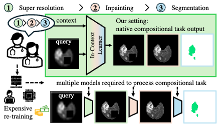
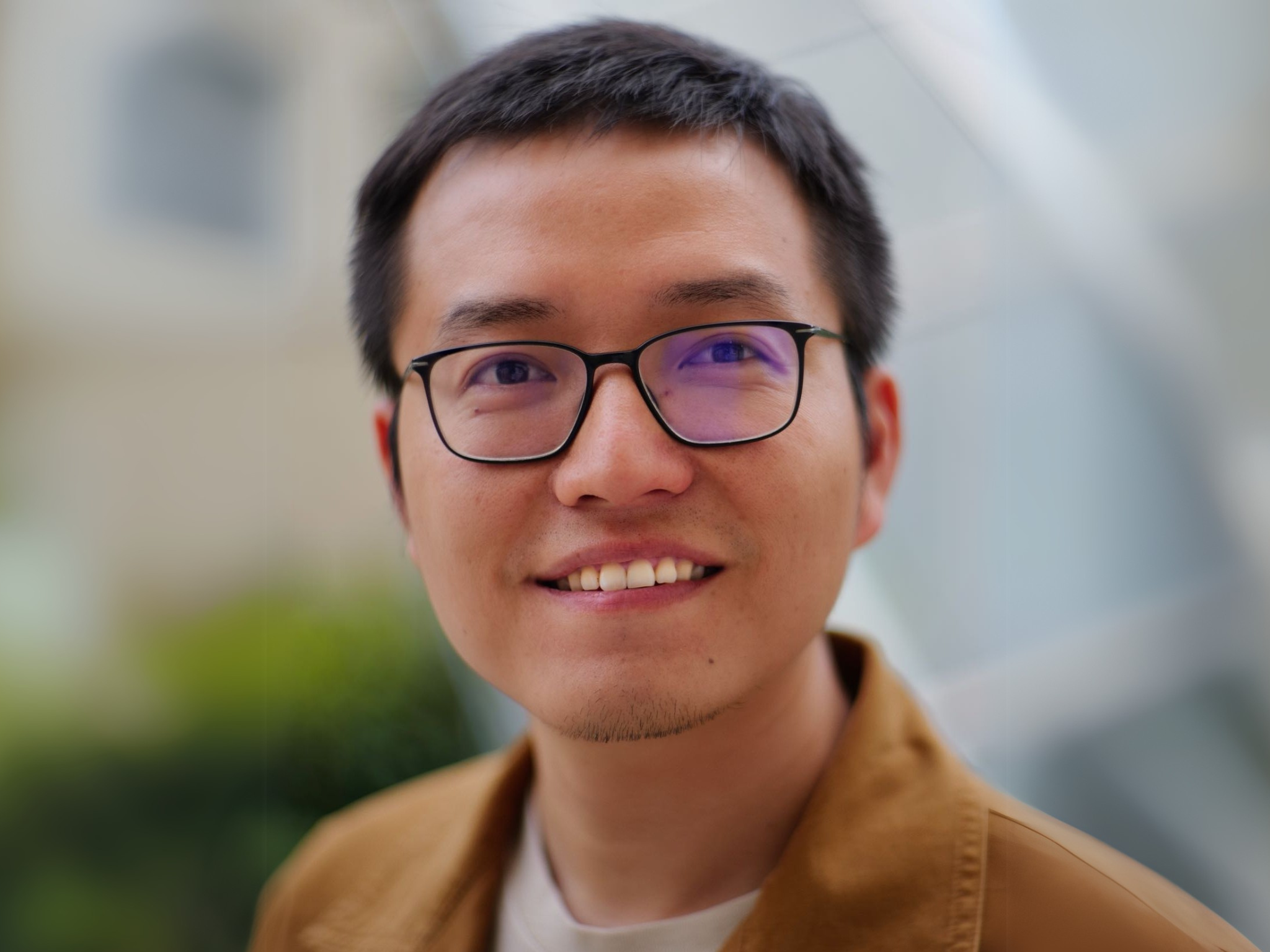
Prof. Dr. Erik Rodner is a faculty member at the University of Applied Sciences Berlin (HTW Berlin), where he serves as a Professor for Machine Learning and Data Science. He also contributes to the School of Engineering Sciences - Technology and Life. His research spans computer vision, machine learning, and biomedical applications, with a focus on learning with limited data, robust visual recognition models, and medical image analysis. He has developed innovative methods for medical diagnostics, industrial classification, and anomaly detection. Recent publications (2025-2016) highlight his expertise in visual in-context learning, semi-weakly segmentation, and active learning frameworks. He has collaborated with institutions such as ZEISS Group, Friedrich Schiller University Jena, and UC Berkeley. Scientific Awards: Award for Excellent Teaching (2023)
Bjoern Menze is a Professor and Rudolf Mößbauer Tenure Track Chair at the Technical University of Munich (TUM), leading the Image-based Biomedical Modeling Group within the Munich School of Bioengineering. His research focuses on medical image computing, tumor growth modeling, and computational physiology, with applications in clinical neuroimaging and personalized radiotherapy design. He holds a Ph.D. in Computer Science from Heidelberg University and has held positions at ETH Zurich, INRIA Sophia Antipolis, MIT, and Harvard Medical School. His academic journey includes a postdoc at MIT’s CSAIL and Harvard Medical School, followed by roles at ETH Zurich and INRIA. His work bridges biomedical imaging with machine learning, emphasizing model-driven analysis of physiological processes. He has been a visiting professor at Maastricht University and contributes to initiatives like the Center for Translational Cancer Research at TUM. Key research areas include tumor growth modeling, quantitative imaging biomarkers, and integrating mathematical models with clinical data. His awards include the MICCAI Young Scientist Award (2014), Leopoldina Fellowship (2009), and DFG Research Fellowship (2008). He advises on medical AI, leads interdisciplinary projects, and publishes extensively in top journals like Nature Neuroscience and IEEE Transactions on Medical Imaging. His lab’s work spans applications such as glioblastoma radiotherapy optimization, whole-body bone lesion detection, and neural connectivity imaging. Collaborations include institutions like Harvard, MIT, and ETH Zurich. He emphasizes translating computational methods into clinical practice for personalized healthcare solutions.
Paul J. Kennedy is a Professor at the University of Technology Sydney's Centre for Artificial Intelligence. He holds a PhD from the same institution (1999). His research focuses on machine learning applications in healthcare, bioinformatics, medical imaging, and data mining. Key areas include developing algorithms for genomic data analysis, healthcare pathway modeling, and edge-cloud frameworks for omics data. Education: PhD in Artificial Intelligence (1999, UTS). Research interests span machine learning, health informatics, and data compression. Notable work includes studies on administrative health records, lung nodule detection, and virtual reality-based cancer cohort analysis. He has co-authored over 100 publications across journals like BMC Bioinformatics, IEEE Transactions, and Artificial Intelligence in Medicine. Advising: Collaborates extensively with students/researchers but no explicit student list provided. Grants and labs: Active in interdisciplinary projects involving medical and computational teams, though specific grants are not detailed here.
Muhammad Waqas is a researcher affiliated with COMSATS University Islamabad , where he holds a position in the Department of Meteorology under the School of Applied Sciences and Humanities . His academic collaborations span institutions like Bahria University, National University of Technology, and University of Bahrain, indicating a multidisciplinary approach. Research interests include Mechanisms for integrating fuzzy logic and machine learning in health monitoring Application of deep learning to medical imaging and clinical diagnostics Development of smart sensors for wearable technology in biomechanics Analysis of social media data for public health surveillance and sentiment analysis Investigation of digital citizenship and ICT leadership in educational contexts Trends in his 15 most recent publications (2025-2024) reveal a focus on medical diagnostics (e.g., monkeypox, breast cancer), smart infrastructure (e.g., sensor placement, structural health monitoring), and social media analytics for health and behavioral insights. These works leverage machine learning , fuzzy systems , and multi-objective optimization .
Andrea Volkamer is a computational chemist and active principal investigator in the field of computer-aided drug design (CADD), with a focus on kinase targets, druggability prediction, and machine learning applications. She has published extensively in journals such as the Journal of Chemical Information and Modeling and Journal of Medicinal Chemistry , with recent work up to 2025 indicating an ongoing academic research program. Her research group, referred to as 'volkamerlab,' develops open-source tools including DoGSite, KiSSim, KinFragLib, and TeachOpenCADD, which are widely used in both academic and industrial drug discovery settings. Her research interests span computational drug discovery , structural bioinformatics , kinase inhibitor design , off-target and polypharmacology prediction , and educational platforms for CADD . She emphasizes open science and reproducibility, particularly through the TeachOpenCADD initiative, which provides interactive Jupyter Notebooks and KNIME workflows for teaching cheminformatics concepts. The 15 most recent publications reflect a strong trend toward integrating machine learning and deep learning (e.g., transformers, graph neural networks) with structure-based methods such as molecular docking, free energy calculations, and binding site comparison. Her work increasingly addresses real-world challenges in drug discovery, including kinase mutation resistance, selectivity optimization, and in vivo toxicity prediction using conformal and hybrid models. Scientific Contributions and Awards: Development of key computational tools: DoGSite, KiSSim, KinFragLib, TeachOpenCADD. Leadership in open-source and open-education initiatives in cheminformatics. Active publication record in top-tier journals with interdisciplinary impact. Advising and Grants: While specific student names and grant details are not mentioned in the provided text, her role as a corresponding author on numerous publications and the existence of a dedicated research lab ('volkamerlab') imply that she mentors students and postdoctoral researchers. She likely secures competitive funding to support her research in computational drug discovery and method development. Labs and Teams: She leads the Volkamer Lab ('volkamerlab'), which focuses on developing and applying computational methods for drug discovery. The lab collaborates with both academic and pharmaceutical partners and emphasizes open-source software development and educational outreach.
Li Wei is a distinguished academic affiliated with Tsinghua University, with a focus on interdisciplinary research spanning artificial intelligence, machine learning, and computer vision. His work often intersects with medical informatics, remote sensing, and signal processing, demonstrating a commitment to advancing technological solutions in healthcare, environmental monitoring, and engineering systems. Research interests include deep learning applications in clinical diagnostics, satellite data analysis for climate modeling, and optimization of energy storage systems. He has contributed to innovative solutions in areas such as UAV-enabled edge computing, privacy-preserving blockchain protocols, and thermal-based surveillance systems. His collaborative projects often involve multidisciplinary teams across institutions. Publications reflect a strong emphasis on practical applications, such as mobile health tools for tumor recognition, transformer-based super-resolution techniques for oceanography, and AI-driven risk classification models for respiratory diseases. While no specific awards or grants are listed, his prolific output across top-tier journals indicates sustained research impact. Professional activities include contributions to conferences like RecSys, MICCAI, and AAAI, and editorial roles are implied through his extensive publication record. Collaborations with industry partners (e.g., in energy systems and medical imaging) suggest engagement with real-world problem-solving.
Despina Kontos, PhD is the Herbert and Florence Irving Professor of Radiological Sciences at Columbia University Irving Medical Center (CUIMC), with appointments in the Department of Radiology and the Herbert Irving Comprehensive Cancer Center. She serves as the Chief Research Information Officer for CUIMC, Vice Chair of Artificial Intelligence and Data Science Research in the Department of Radiology, and Director of Biomarker Imaging at NewYork-Presbyterian Hospital. Additionally, she holds appointments in the Departments of Biomedical Informatics and Biomedical Engineering. Dr. Kontos received her educational training from prestigious institutions: BS in Engineering from the University of Patras, Greece MSc and PhD in Computer and Information Sciences from Temple University Postdoctoral training in Radiology at the University of Pennsylvania Certificates in Biostatistics and Epidemiology from UPenn, Cancer Biology from Harvard, and AI for Decision Making from Wharton As a computer scientist with expertise in artificial intelligence and machine learning, Dr. Kontos focuses on developing computational methodologies to leverage imaging as quantitative biomarkers for personalized disease prediction, particularly in cancer. Her research program investigates how imaging data can be mined to extract sophisticated phenotypic signatures with diagnostic, prognostic, and predictive value. While her primary focus has been on breast cancer, her lab also pursues related research in lung cancers, evaluating the integration of CT radiomic features with liquid biopsy data to characterize tumor heterogeneity. Dr. Kontos founded and directs Columbia University's Center for Innovation in Imaging Biomarkers and Integrated Diagnostics (CIMBID), a multidisciplinary center dedicated to developing and integrating quantitative imaging and non-imaging biomarkers for personalized disease prediction. Through CIMBID, she has built a vibrant scientific ecosystem that brings together expertise across Columbia's campuses, linking basic science, engineering, clinical medicine, public health, and health services research. Analysis of Dr. Kontos's publication record reveals a strong focus on applying AI and machine learning to biomedical imaging, particularly for cancer risk prediction and personalized treatment. Her work demonstrates a progression from foundational methodological development to clinical translation, with increasing emphasis on multi-modal biomarker integration. Recent publications show expansion into new disease areas including Alzheimer's disease prediction, while maintaining her strong focus on breast and lung cancer applications. Dr. Kontos has received significant recognition for her contributions to the field: Academy for Radiology and Biomedical Imaging Research Distinguished Investigator Award (2020) Eastern Cooperative Oncology Group - American College of Radiology Imaging Network ECOG-ACRIN Young Investigator Award of Distinction for Translational Research (2014) Dr. Kontos has been highly successful in securing research funding, with numerous grants from federal agencies including the National Institutes of Health (NIH) and the Department of Defense (DOD), as well as private foundations such as the American Cancer Society (ACS) and the Radiological Society of North America (RSNA). Her leadership extends to mentoring students and postdoctoral researchers through her roles at CIMBID and the Department of Radiology. As the founding director of CIMBID, Dr. Kontos leads a multidisciplinary team that includes the Computational Imaging Biomarker Group (CBIG), the Laboratory of AI and Biomedical Science (LABS), and several other affiliated research labs. The center leverages Columbia's institutional strengths in engineering, data science, and clinical medicine to advance personalized healthcare through AI and imaging technologies.
Prof. Dr. Julia Schnabel is the TUM Liesel Beckmann Distinguished Professor and Helmholtz Distinguished Professor at TUM's TUM School of Computation, Information and Technology. Her research focuses on computational imaging and AI in medicine, including medical image processing, machine learning, motion modeling, and quantitative imaging. She holds IEEE, Ellis, and MICCAI Society fellowships, and has pioneered work in image reconstruction, artifact correction, and AI-based diagnostics. Educations: Bachelor/Master from TU Berlin (1993) PhD from University College London (1998) Postdocs at UMC Utrecht, King's College London, and UCL Her research interests span medical AI, deep learning for medical imaging, and clinical evaluation methodologies. Key contributions include frameworks for motion artifact correction in MRI, physics-informed neural networks, and benchmark datasets like NOVA for anomaly detection in brain MRI. She has authored over 100 publications, with recent work advancing unsupervised anomaly detection and federated learning in healthcare. Prof. Schnabel leads interdisciplinary projects at TUM and Helmholtz Zentrum München, focusing on AI-driven solutions for diagnostic and therapeutic challenges. Her labs develop tools for real-time cardiac imaging, histopathology segmentation, and trustworthy AI guidelines (FUTURE-AI initiative).
Prof. Wolfgang Weber is a Professor and Director of the Department of Nuclear Medicine at the Klinikum rechts der Isar, Technical University of Munich (TUM). His academic career includes roles such as Associate Professor at UCLA (2003–2007) and Chair of Nuclear Medicine at Albert Ludwig University of Freiburg (2007–2013). He specializes in molecular imaging and targeted radionuclide therapy, with a focus on cancer theranostics. His research addresses precision oncology through advanced imaging techniques for diagnosis and treatment monitoring. Education: Doctorate in Medicine from TUM (1995). Key prior appointments include Director of the Molecular Imaging and Therapy Service at Memorial Sloan Kettering Cancer Center (2013–2017) and Professor at Weill-Cornell Medical College. His work spans prostate cancer imaging, neuroendocrine tumors, and radiopharmaceutical development. Research interests emphasize integrating imaging modalities like PET/CT and SPECT for cancer staging, therapy response prediction, and novel radiotracer development. Notable contributions include PSMA-ligand PET imaging for prostate cancer recurrence detection and PARP inhibitor engagement studies in lung cancer. Publications highlight innovations in theranostic agents, such as 18F-rhPSMA-7 and Ga-68-based tracers. His work frequently addresses clinical trial validation of imaging biomarkers and dosimetry safety in radiolabeled therapies.
Tina Dorosti is a researcher at the Technical University of Munich , affiliated with the TUM Faculty of Medicine and the Department of Physics . Her work focuses on applying artificial intelligence to medical imaging, particularly in CT and X-ray technologies. Research Interests: Tina specializes in AI-driven medical imaging solutions, with emphasis on machine learning for disease detection, dark-field X-ray imaging, and spectral X-ray imaging. Her projects address challenges in low-dose imaging, artifact reduction, and lung volume quantification. Publications: Her recent work (2025) includes optimizing CNNs for COPD detection in CT scans, enhancing lung tumor imaging with sparse sampling, and developing deep learning methods for lung volume estimation from chest radiographs. Earlier studies (2024-2021) explore hemorrhage detection, artifact correction, and bone segmentation in clinical imaging. Awards: Cover image of the Radiology: Artificial Intelligence July 2025 issue Collaborations: Tina collaborates with Prof. Franz Pfeiffer and colleagues at the Chair of Biomedical Physics, contributing to interdisciplinary projects in radiology, oncology, and respiratory disease diagnostics.
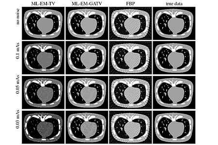
Jürgen Hesser is a Professor at the Mannheim Medical Faculty , Heidelberg University, specializing in Experimental Radiotherapy and Medical Imaging . His research focuses on solving inverse problems in imaging, particularly for CT reconstruction , brachytherapy planning , and low-dose imaging . Current affiliations: Clinic for Radiotherapy and Radiooncology, Mannheim University Hospital Collaborative ties: Interdisciplinary Center for Scientific Computing (IWR) and Center for Bioinformatics (ZITI) at Heidelberg University Research interests center on anisotropic total variation techniques for medical and industrial applications, including MR-guided interventions and real-time radiation therapy . His work has led to a 1000x speed improvement in brachytherapy planning algorithms. Recent publications highlight expertise in image reconstruction (CT/X-ray), noise optimization , machine learning for cancer classification, and big data management solutions. His methods are applied to both clinical and industrial imaging challenges. Additional contributions include scientific data infrastructure development and variance stabilization techniques for medical sensors. The research group maintains strong interdisciplinary links with Physics, Mathematics, and Computer Science faculties.
Rizwan Qureshi is an active researcher and academic specializing in artificial intelligence, machine learning, and their applications in medical imaging and bioinformatics. With a robust publication record spanning from 2017 to 2025, he has established himself as a significant contributor to the fields of computer vision and biomedical AI. His research interests focus on Artificial Intelligence , Machine Learning , Medical Imaging , Computer Vision , and Biomedical Engineering . Qureshi's work demonstrates particular expertise in object detection systems (especially YOLO variants), medical image segmentation, vision-language models, and applications of AI to healthcare problems including lung cancer research and diabetic retinopathy detection. Analysis of his recent publications (2023-2025) reveals a strong trend toward medical applications of AI, with approximately 60% of his work focusing on healthcare-related problems. His research shows increasing emphasis on model robustness, explainability, and handling distribution shifts in real-world applications. The publications span top venues including IEEE Access, IEEE Transactions on Medical Imaging, CVPR, and BIBM. Qureshi maintains extensive collaborations with researchers across multiple institutions, with frequent co-authorship with Hong Yan, Tanvir Alam, Jia Wu, and Sheheryar Khan. His work demonstrates both technical depth in machine learning methodologies and practical application to significant healthcare challenges. While specific details about his academic advising are not evident from the publication record alone, his numerous publications with multiple co-authors suggest active participation in research teams and likely supervision of graduate students. His work shows consistent funding support through publication in reputable journals and conferences.
Mostafa Mayar is a researcher affiliated with the Chair of Computer Science Applications in Medicine at the Technical University of Munich . His work bridges artificial intelligence and medical imaging, with a focus on improving surgical precision and tumor segmentation. His research interests include: Hyperspectral imaging for medical diagnostics Deep learning in surgical oncology Graph neural networks for tumor analysis Computer vision in healthcare applications AI-driven margin assessment in head and neck cancer Orthopedic fracture classification using machine learning Recent publications highlight his contributions to hyperspectral imaging integration with AI for ex vivo studies in squamous cell carcinoma and femur fracture classification. He also explores masked autoencoders for surgical event recognition.
Chang Liu is a Research Associate at NHR@FAU (Center for National High Performance Computing Erlangen) at Friedrich-Alexander-Universität Erlangen-Nürnberg (FAU), where he joined the AI group in April 2025 to support AI-oriented projects across diverse research fields. Prior to this position, he was a doctoral researcher at the Pattern Recognition Lab at FAU until March 2025. Chang Liu earned his degree in Medical Engineering at FAU. His academic journey at FAU began in September 2016 as a student, progressed to a researcher at the Pattern Recognition Lab starting in March 2020, and culminated in his doctoral research until March 2025. Dr. Liu's research focuses on medical image processing and analysis, with particular emphasis on the automated segmentation of computed tomography (CT) images and the generation of high-quality CT images. His work bridges computer science and medical applications through artificial intelligence to solve complex healthcare problems. Beyond his core research, he has contributed to applying AI technologies in diverse fields including second language education and nail disease diagnosis, demonstrating his interdisciplinary approach to problem-solving. His expertise spans data augmentation techniques, multi-organ segmentation, CT reconstruction, and radiation dose optimization. His publication record reveals consistent advancement in medical image analysis techniques, particularly in CT imaging and segmentation. His work shows a progression from foundational deep learning applications to more sophisticated approaches incorporating anatomical knowledge and addressing practical clinical constraints like limited annotations and radiation safety. Dr. Liu has mentored numerous students through their thesis work, guiding them in cutting-edge research at the intersection of AI and medical imaging. His advisees have completed projects on breast cancer risk stratification, medical segmentation annotation, U-Net architecture configuration, and other innovative topics in medical image analysis. As part of NHR@FAU, Dr. Liu works with the AI group to enhance research projects using modern high-performance computing systems, applying his expertise in medical image analysis to support diverse research fields across FAU.
Mario Cesarelli is a Professor at the University of Naples Federico II's Department of Biomedical Engineering, with an extensive publication record spanning over three decades. His research bridges engineering and clinical medicine, focusing on developing computational methods for disease diagnosis and patient monitoring through biomedical signal processing and artificial intelligence. Dr. Cesarelli's research spans multiple domains of biomedical engineering with particular emphasis on: Medical imaging analysis and radiomics for neurodegenerative disorders Explainable AI for cancer detection and diagnosis Biomechanics and motion analysis for neurological conditions Cardiac signal processing and analysis Generative models for medical image synthesis and authentication His recent publications demonstrate a strong trend toward explainable deep learning applications in healthcare, particularly for neurodegenerative diseases like Parkinson's and Alzheimer's, as well as various cancer diagnostics. The work increasingly focuses on model interpretability to build clinician trust in AI-assisted diagnosis. Dr. Cesarelli maintains extensive collaborations with a core research group including Paolo Bifulco (46 joint publications), Maria Romano (41), Gianni D'Addio (34), Antonella Santone (27), and Francesco Mercaldo (26), forming interdisciplinary teams that combine engineering expertise with clinical knowledge.