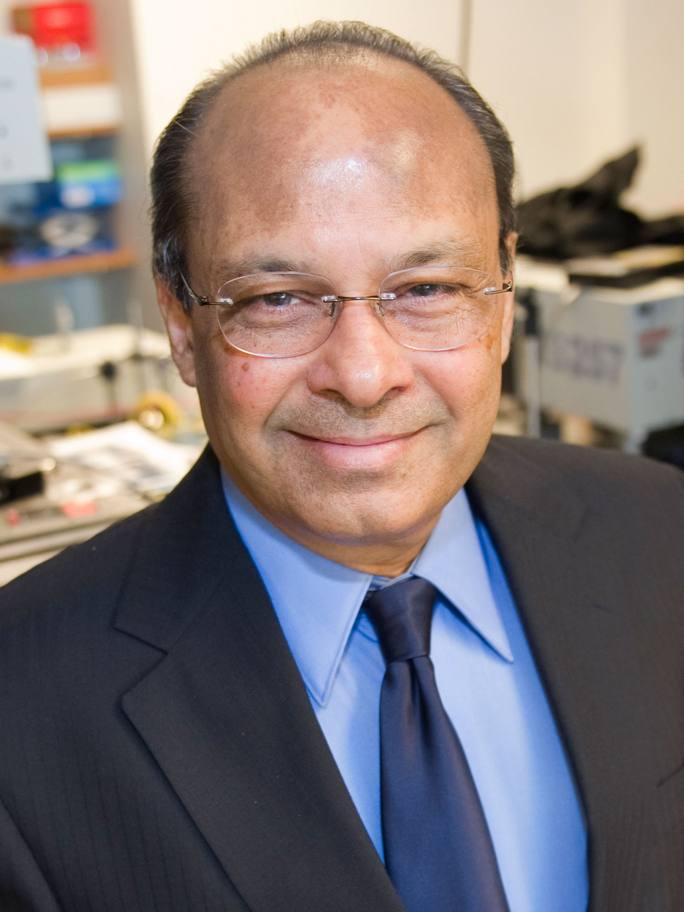
Srinivas Sridhar is a University Distinguished Professor of Physics, Biomedical Engineering, and Chemical Engineering at Northeastern University, with a secondary appointment as Lecturer on Radiation Oncology at Harvard Medical School. He previously served as Vice Provost for Research at Northeastern University (2004–2008), overseeing its research portfolio. As an elected Fellow of the American Physical Society and the American Institute of Medical and Biological Engineering, his research spans nanomedicine, neurotechnology, drug delivery, and quantitative MRI, with over 450 publications and patents. He founded the Nanomedicine Innovation Center and directs major NIH/NSF programs like CaNCURE and IGERT, focusing on undergraduate and graduate training in nanomedicine, particularly for underrepresented communities. His research interests include Nanomedicine Neurotechnology Quantitative MRI Drug Delivery Systems Metamaterials and Nanophotonics Quantum Chaos Superconductivity . Recent work involves machine learning-enhanced diagnostics for glaucoma, engineered nanoparticles for BRCA-deficient cancers, and portable neuro-ophthalmic devices. His publications from 2025–2017 reflect interdisciplinary applications in oncology, neurology, and materials science, with a focus on therapeutic and diagnostic innovation. Scientific accolades include the 2016 Biomedical Engineering Society Diversity Award University Distinguished Professorship . As an educator and entrepreneur, he has trained over 120 researchers, developed first-of-their-kind nanomedicine courses, and founded companies commercializing technologies like QUTE-CE MRI. His lab leads projects on cancer nanomedicine, quantitative imaging, and nanoscale magnetism, supported by grants from NIH, NSF, DoD, and private foundations.
Associate Professor Kai-Hsiang Chuang is a Principal Research Fellow at the School of Biomedical Sciences within the Faculty of Health, Medicine and Behavioural Sciences at the University of Queensland. He is also affiliated with the Queensland Brain Institute and the Centre for Advanced Imaging. His research focuses on understanding brain networks, developing advanced imaging techniques, and translating these findings to improve diagnosis and intervention for neurological disorders. Dr. Chuang received his Ph.D. in electrical and biomedical engineering from the National Taiwan University, Taiwan, in 2001. His doctoral research focused on improving the detection of brain activity using functional magnetic resonance imaging (fMRI). Ph.D. in Electrical and Biomedical Engineering, National Taiwan University (2001) Dr. Chuang's research spans multiple areas of brain imaging and neuroscience. His primary focus is on functional brain mapping , where he develops in vivo imaging techniques including functional MRI and multimodal integration with optogenetics, calcium imaging, and electrophysiology. He applies these techniques in both humans and animal models to improve understanding and intervention of brain function, disease processes, and treatment effects. Another key area is brain networks in learning, memory, and dementia . His work explores how brain network wiring and activity underpin cognition and behavior, with particular focus on understanding the causal relationship between brain network activity and memory formation. He develops techniques to modulate behavior by manipulating brain network activity. More recently, Dr. Chuang has expanded into brain waste clearance research, studying the brain's fluid drainage system that clears waste and toxic molecules like amyloid plaques. His lab is developing imaging techniques to track this system's function and understand its regulatory mechanisms, which could provide new treatment targets for dementia. Analysis of Dr. Chuang's recent publications reveals a strong focus on advancing functional MRI techniques for brain network analysis, particularly in rodent models. His work consistently bridges basic neuroscience with clinical applications, especially in understanding memory formation and dementia. A notable trend is the development of multimodal approaches that combine fMRI with optogenetics, calcium imaging, and electrophysiology to establish causal relationships in brain networks. His research increasingly addresses the translation of preclinical findings to human applications, with growing emphasis on Alzheimer's disease mechanisms and potential interventions. Dr. Chuang serves on the editorial boards of multiple prestigious journals including Frontiers in Neuroscience: Brain Imaging Methods , Imaging Neuroscience , and Scientific Reports , reflecting his standing in the field. Editorial Board Member, Frontiers in Neuroscience: Brain Imaging Methods Editorial Board Member, Imaging Neuroscience Editorial Board Member, Scientific Reports Dr. Chuang is actively involved in research supervision, currently serving as Principal Advisor for one PhD student working on "Developing imaging and neuro-technologies for decoding memory formation" and Associate Advisor for two other PhD projects. He has successfully completed supervision of three PhD students on topics related to resting-state networks, memory consolidation, and functional MRI. ARC Discovery Projects (2024-2028): "Decoding the brain network of memory formation" ARC Training Centre for Innovation in Biomedical Imaging Technology (2017-2024) NHMRC-NIH BRAIN Initiative Collaborative Research Grants (2016-2023) Universities Australia - Germany Joint Research Co-operation Scheme (2017-2018) Mater Medical Research Institute Limited grant for mindfulness-based cognitive therapy research (2017-2020) Dr. Chuang leads the Functional and Molecular Neuroimaging Group at the Queensland Brain Institute. His laboratory focuses on understanding the functional connectome of the brain and developing functional and molecular imaging techniques to study brain connectivity associated with behavior. The group has developed various MRI techniques to track neuronal connections, map large-scale brain synchrony, and quantify cerebral blood flow and metabolism in vivo. His research team collaborates extensively with other experts at UQ and internationally, including collaborations with Associate Professor Darryl Eyles, Professor Jürgen Götz, Professor Tianzi Jiang, Dr. Fatima Nasrallah, Professor Linda J. Richards, Professor Pankaj Sah, Professor Elizabeth Coulson, Dr. Patricio Opazo, Professor Feng Liu, and Professor Markus Barth.
Professor Jianhui Zhong is a distinguished physicist and imaging scientist at the University of Rochester, holding dual appointments as Professor in the Department of Imaging Sciences and Department of Biomedical Engineering within the School of Medicine and Dentistry. He currently serves as Associate Director of the MRI Research Group at the University Medical Center and has been a leading figure in MRI physics and applications for over 25 years. Dr. Zhong's research focuses on Biological Physics and advanced MRI techniques, particularly in the areas of diffusion-weighted imaging, functional MRI, and intermolecular multiple-quantum coherence (iMQC) methods. His work has led to significant advancements in understanding brain imaging biomarkers for neurological disorders, HIV-related cognitive impairment, stroke detection, and tissue characterization. He has pioneered novel MRI techniques for measuring diffusion behaviors in complex biological systems and developing quantitative imaging methods for clinical applications. His extensive publication record includes over 130 papers, with recent work emphasizing quantitative mapping techniques like MR fingerprinting, diffusion tensor imaging, and advanced reconstruction methods using deep learning. Dr. Zhong has received numerous prestigious awards including the Dean's Research Award from University of Rochester and Yale School of Medicine, and has contributed significantly to the field through his service on the Editorial Board of Magnetic Resonance Imaging since 2001. Dean's Award for Excellency in Research, University of Rochester (2004) Summa Cum Laude Citation, American Society of Neuroradiology (2000) Certificate of Merit Citation, Radiological Society of North America (1999) The Dean's Award, Yale University School of Medicine (1994) Dr. Zhong has been instrumental in developing advanced MRI techniques for clinical applications, particularly in neurological disorders and HIV-related brain changes. His research group has made significant contributions to understanding the relationship between physical processes in tissues and MRI signal changes, leading to improved diagnostic methods for conditions like acute stroke and cognitive impairment.
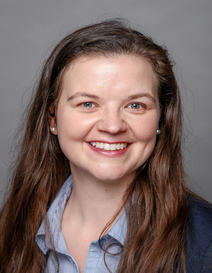
Laura Walkup is Associate Professor in Pediatric Pulmonary Medicine at University of Cincinnati College of Medicine. Her research focuses on advanced pulmonary imaging techniques including hyperpolarized 129Xe MRI and ultrashort echo time MRI for pediatric respiratory conditions. Research applications include cystic fibrosis, pediatric asthma, bronchopulmonary dysplasia, and lymphatic anomalies. Walkup develops quantitative methods for assessing lung structure-function relationships and ventilation heterogeneity in clinical populations.
Uzay Emir is a Joint Associate Professor in Radiology at UNC-Chapel Hill, with cross-institutional roles including Principal Investigator at the University of Oxford and prior experience as an Assistant Professor at Purdue University. His research focuses on advancing MRI/MRS methodologies for neurodegenerative disease biomarker discovery, particularly using ultra-short echo time (UTE) and Rosette trajectory-based imaging techniques. Emir's work emphasizes translational applications across preclinical and clinical settings, including 3T to 9.4T field strengths. He pioneered the PETALUTE sequence for accelerated phosphorus spectroscopic imaging and led the multicenter 'Repeat it with me challenge' for test-retest reproducibility in Rosette MRI(S)I. His innovations include density-weighted concentric ring trajectories and 3D Rosette-based methods for brain iron content mapping and myelin fraction analysis. Research interests span neurochemical profiling, metabolic imaging, and functional MRI-fMRS integration at 7T. Education: PhD in imaging modalities (fMRI signal transients), postdoctoral training in MRS methods at the University of Minnesota's Center for Magnetic Resonance Research Key Methods: UTE MRI/MRSI, Rosette trajectory, 31P-MRSI, PETALUTE sequence Key Projects: ME/CFS metabolic studies, lead toxicity neuroimaging, sodium cartilage quantification Research trends in his articles highlight development of novel imaging sequences (e.g., ZTE fMRI, accelerated J-resolved spectroscopy) and their application to neurological disorders. Emphasis on clinical feasibility of 31P-MRS after decades of technical challenges underscores his translational impact. Recent work includes simultaneous multi-slice MRSI and NIfTI-MRS data standardization efforts. His contributions bridge preclinical-clinical research through standardized protocols enabling biomarker validation. Current efforts explore spatiotemporal dynamics of neural networks using integrated fMRI-fMRS approaches.
Dr. Catherine Johnson is the Robert H. Quenon Associate Professor of Mining & Explosives Engineering at Missouri University of Science and Technology. She serves as Director of the Explosives Research Facility and Associate Director of the PRECISE-TBI Education Core for the VA Interagency Resource Center (I50). Her research focuses on shock physics, blast-induced traumatic brain injury (TBI), and mining safety. Education: Ph.D. in Mining Engineering (Explosives Emphasis), University of Kentucky (2014) M.Eng. in Mining and Quarry Engineering, University of Leeds (2012) B.Eng. in Mining and Quarry Engineering, University of Leeds (2012) Her research explores the adverse effects of high explosives and blasting agents, including coal dust explosions, air overpressure from quarry blasting, and shock wave injuries such as traumatic brain and lung injuries. She develops policy, training, and protective equipment improvements to mitigate these risks. Recent publications highlight trends in blast wave propagation , TBI biomarker discovery , protective armor design , and explosive material characterization , with applications in neuroscience , materials science , and mining safety . Scientific Awards: 2018 President’s Award, International Society of Explosives Engineers 2018 Inaugural Member, University of Missouri System Presidential Engagement Fellows 2019-2021, 2023 Mining Engineering Department Research Award 2020, 2022 Department Teaching Award 2021 Dean’s Scholar, College of Engineering and Computing 2020-2021 University Research Award 2023 Missouri S&T Woman of the Year Dr. Johnson has graduated 6 PhD and 4 MS students and currently advises 10 PhD and 1 MS student. She has secured $11.7M in collaborative research funding and authored 39 peer-reviewed journal articles, 41 conference proceedings, and 3 book chapters.
Prof. Dr. Shaukat Khan is a faculty member in the Department of Physics at the Technical University of Dortmund, where he conducts research in accelerator physics at the Center for Synchrotron Radiation. His work is centered on the DELTA accelerator facility, a 1.5 GeV electron storage ring used for both user experiments and advanced accelerator research. His research focuses on modern subfields of accelerator physics, particularly the interaction between electrons and femtosecond laser pulses to generate ultrashort pulses in the extreme ultraviolet and terahertz ranges. His group develops electronic and optical beam diagnostic techniques and applies machine learning methods to optimize accelerator parameters. A key ongoing activity involves Compton scattering of laser photons by electrons for precise determination of electron beam energy and other characteristics. Future projects include the design of an electron source for time-resolved electron diffraction. The research group collaborates with other major accelerator centers on topics such as the development of synchrotron radiation sources and free-electron lasers, temporal and spectral shaping of terahertz pulses, and control of beam parameters in electron and proton machines. Students are trained in an interdisciplinary environment encompassing condensed matter research, laser physics, and artificial intelligence applications in accelerator science. Prof. Khan's work contributes to cutting-edge advancements in accelerator technology, with recent milestones including the world's first observation of EEHG (echo-enabled harmonic generation) signal at a storage ring, achieved at DELTA. Scientific Affiliations and Activities: Faculty of Physics, Department of Physics, Technical University of Dortmund Center for Synchrotron Radiation (DELTA facility) Research Group Leader in Accelerator Physics Member of Condensed Matter and Accelerator Physics research groups Conducts lectures, seminars, and supervises student theses on accelerator-related topics Organizes educational excursions (e.g., to DESY in Hamburg) Research Collaborations: German Electron Synchrotron (DESY) Other international accelerator centers
Misung Han, Ph.D., is a Researcher in the Department of Radiology and Biomedical Imaging at the University of California, San Francisco (UCSF). She specializes in developing advanced MRI techniques for musculoskeletal, breast, and brain applications. Her work focuses on improving diagnostic capabilities through novel imaging methods, such as high-resolution structure depiction and tissue parameter quantification in musculoskeletal tissues. She collaborates across departments, including Neuro Imaging and Focused Ultrasound Lab, to advance clinical applications like accelerated perfusion imaging and temperature mapping. Education: B.S., Electrical Engineering, Korea Advanced Institute of Science and Technology (2002) M.S. and Ph.D., Electrical Engineering, Stanford University (2004 & 2010) Postdoctoral Training in Radiology and Biomedical Imaging, UCSF (2014) Research Interests: Misung Han's research emphasizes MRI innovation for musculoskeletal disorders, including rotator cuff pathology, osteoarthritis, and bone marrow analysis. Her techniques, such as UTE temperature mapping and deep learning reconstruction, aim to enhance diagnostic precision and quantify disease biomarkers. She also explores accelerated imaging protocols to improve clinical workflows. Grants & Awards: NIAMS K01 Grant (2020–2025): Developing MRI techniques for rotator cuff pathology GE Healthcare ISA (2023–2025): Deep-learning reconstruction for UTE imaging Multiple Distinguished Reviewer Awards from ISMRM and NMR journals Labs & Teams: She leads the Musculoskeletal and Quantitative Imaging Research Group at UCSF, collaborating with interdisciplinary teams to translate technical innovations into clinical practice. Her work bridges engineering, radiology, and clinical medicine to advance imaging science.
Douglas Kelley, PhD holds the title of Research Associate and Adjunct Professor at the University of California, San Francisco (UCSF). His work focuses on advanced MRI techniques, particularly in ultra-high field (7T) imaging, quantitative susceptibility mapping, and diffusion MRI. He has contributed to innovations in RF coil design, image reconstruction algorithms, and applications in neuroimaging, oncology, and musculoskeletal imaging. Key research areas include: Development of high-resolution MRI methods for brain tumor characterization and neurodegenerative diseases Optimization of 7T MRI protocols for phase imaging, T1-weighted imaging, and diffusion tensor imaging Advancements in RF coil architectures and parallel imaging strategies Applications of quantitative susceptibility mapping in clinical diagnostics His publications span over 30 years, with a focus on advancing imaging hardware, software algorithms, and clinical applications of MRI technology. Notable contributions include studies on hippocampal atrophy in Alzheimer's disease, multiple sclerosis lesion characterization, and the use of ultrashort echo time (UTE) and zero echo time (ZTE) imaging. Dr. Kelley has collaborated extensively with researchers in radiology, biomedical engineering, and neurology, contributing to both technical innovations and translational medical research.
Peder Larson is a Professor in Residence at the University of California, San Francisco (UCSF) Department of Radiology and Biomedical Imaging, where he serves as Principal Investigator for The Larson Advanced Imaging Technologies Research Group. Based at the UCSF Mission Bay campus in Byers Hall as part of the Quantitative Biosciences Institute, his research group takes an engineering-driven approach to develop advanced medical imaging methods, primarily focusing on MRI with some work in CT and PET. Dr. Larson's research interests span radio frequency pulse design, pulse sequence development, novel imaging strategies, and optimized reconstruction methods for MRI, with particular emphasis on applications in Hyperpolarized carbon-13 agents and semi-solid tissue imaging with ultrashort echo time (UTE) methods. His group works on metabolic imaging with hyperpolarized MRI for cancer imaging, pediatric lung MRI methods, myelin MRI with ultrashort echo time techniques, quantitative imaging on PET/MRI systems, and AI-based analysis of prostate and kidney cancer imaging data. The team frequently draws on backgrounds in engineering, physics, biology, and chemistry to analyze complex imaging data and provide novel information about tissue structure and function. His research publications and educational presentations from 2017-2019 demonstrate a strong focus on hyperpolarized MRI technology, covering everything from fundamental physics to specific clinical applications in cancer, cardiac imaging, and neurology. His work shows a progression from theoretical foundations to practical implementations across multiple organ systems. As an educator, Dr. Larson has developed significant educational resources including the 'Introduction to Principles of MRI' eBook, edited 'Hyperpolarized Carbon-13 Magnetic Resonance Imaging and Spectroscopy,' and created numerous educational materials, courses, and software tools for MRI education. He teaches UCSF Biomedical Imaging 201: Principles of Magnetic Resonance Imaging and Bioengineering 297: Hyperpolarized MR Seminar. Dr. Larson joined UCSF as a post-doctoral scholar in 2007 under Dan Vigneron, PhD, and became faculty in 2011. He completed his undergraduate and graduate studies at Stanford University with Dwight Nishimura, conducting doctoral research on 'MRI of Semi-solid Tissues.' He emphasizes the importance of understanding patient experiences by having his team volunteer for medical and research imaging themselves, stating 'It is important to understand what your patient and research subject experience is going to be.'
Stephanie Merhar, MD, is an Associate Professor of Pediatrics at the University of Cincinnati and attending neonatologist at Cincinnati Children’s Hospital Medical Center. Her clinical and research portfolio centers on neonatal neurology, advanced neuro-imaging in the NICU, and the developmental consequences of extremely preterm birth and prenatal opioid exposure. Education & Training: While specific degree dates are not listed, Dr Merhar’s extensive peer-reviewed output and faculty rank indicate subspecialty fellowship training in Neonatal–Perinatal Medicine following pediatric residency. Research Interests: Neonatal brain development and injury in extremely preterm infants Functional and structural connectivity MRI applied to high-risk neonates Neonatal opioid withdrawal syndrome (NOWS) – pathophysiology, care pathways, and long-term neurodevelopment Advanced MRI techniques (diffusion tensor imaging, ultrashort echo-time pulmonary MRI, on-site NICU scanners) Early biomarkers of cerebral palsy and cognitive/ language impairment Feeding intolerance and post-hemorrhagic hydrocephalus biomarkers Publication Trends: Between 2021-2023 Dr Merhar has led or co-authored >25 high-impact papers. Work clusters into three major domains: (1) prospective multicenter trials of NOWS management (Eat-Sleep-Console, OBOE, ESC-NOW), (2) longitudinal imaging studies correlating neonatal MRI metrics with 2-year neurodevelopmental outcomes, and (3) technical innovations enabling safe, quiet, on-site MRI of unstable neonates. These publications appear in The New England Journal of Medicine , JAMA , Pediatric Research , NeuroImage: Clinical , and American Journal of Neuroradiology . Scientific Awards & Honors: None explicitly listed in the provided text. Grants & Collaborative Networks: Dr Merhar is an active investigator within the NICHD Neonatal Research Network, leading neuroimaging components of several multi-site trials. She collaborates closely with the Outcomes of Babies with Opioid Exposure (OBOE) consortium and serves as site PI for Cincinnati Children’s in the ESC-NOW stepped-wedge RCT. Laboratories & Teams: She conducts imaging research within the neonatal neuro-imaging program at Cincinnati Children’s, utilizing the on-site 1.5-T small-footprint MRI scanner and the hospital’s 3-T research systems. Her team includes MRI physicists, neonatal neuro-radiologists, developmental neuropsychologists and biostatisticians.