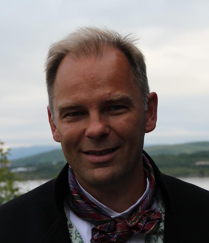
Sven Hermann is a Senior Lecturer at the European Institute for Molecular Imaging (EIMI) within the University of Münster, leading the Preclinical Imaging Group. His research focuses on developing molecular imaging strategies for inflammation and infection, including PET, SPECT, fluorescence reflectance imaging, and optoacoustic imaging. Key targets include matrix metalloproteinases (MMPs) and alarmins (S100A8/S100A9), with applications in cardiovascular diseases, rheumatoid arthritis, and bacterial imaging. He participates in the Cells in Motion Cluster of Excellence and leads DFG-funded projects TRR332-C07 and CRC1450-C03, investigating immune regulation and imaging techniques. Affiliations: EIMI, University of Münster; Core Unit Pix (preclinical imaging). Research Themes: Bacterial imaging via maltodextrin transporters, MMP activity visualization in atherosclerosis, and S100A8/A9-driven immune modulation. His work integrates engineering and biology to translate imaging innovations into clinical applications, emphasizing multimodal approaches for disease monitoring.
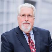
Albert J. Sinusas, M.D., is a Professor of Medicine (Cardiovascular Medicine and Radiology & Biomedical Imaging) at Yale University School of Medicine and holds adjunct roles in Biomedical Engineering. He leads the Yale Translational Research Imaging Center (Y-TRIC) and directs Advanced Cardiovascular Imaging at Yale New Haven Hospital. His roles include Chairman of multiple committees (Yale Radioactive Drug Research Committee, Radiation Safety Committee) and Board Member of the Intersocietal Accreditation Commission (IAC) for Nuclear/PET imaging. Dr. Sinusas earned his MD from the University of Vermont and completed training in Internal Medicine at the University of Oklahoma and Cardiology/Nuclear Cardiology at the University of Virginia. His research focuses on cardiovascular imaging innovations, including molecular imaging of myocardial injury, angiogenesis, and peripheral artery disease. His translational work employs multidisciplinary modalities such as PET/CT, SPECT/CT, MRI, and echocardiography. He leads NIH-funded grants and trains researchers through a T32 grant. With over 250 peer-reviewed publications, he co-edited textbooks on cardiovascular molecular imaging and hybrid imaging in cardiovascular medicine. Key research interests include developing novel imaging tracers, AI-enhanced diagnostic tools, and non-invasive assessment of cardiovascular pathophysiology. Notable contributions include advancements in PET-derived myocardial blood flow quantification and SPECT imaging optimization using deep learning. Dr. Sinusas has received prestigious awards like the SNMMI Hermann Blumgart Award (2008) and is frequently recognized in 'Best Doctors in America.' He serves on editorial boards for Journal of Nuclear Medicine , Journal of Nuclear Cardiology , and JACC: Cardiovascular Imaging . His clinical expertise includes ECG, stress testing, and interpretation of SPECT/PET imaging studies. He oversees Y-TRIC’s mission to bridge preclinical and clinical imaging research, fostering collaboration across engineering, radiology, and cardiology.
Mary Rusckowski, PhD, is an Associate Professor in the Department of Radiology at UMass Chan Medical School and holds an additional appointment at the T.H. Chan School of Medicine. Her research focuses on developing novel radiopharmaceuticals and molecular imaging agents for cancer, infection, and other diseases. Education: BA in Biology, Albertus Magnus College PhD in Biochemistry, Rutgers University Research Interests: Rusckowski's work integrates chemistry, biology, and imaging to create targeted diagnostic and therapeutic agents. Her major areas include: Design of radiolabeled antisense oligonucleotides for infection and cancer imaging Pretargeting strategies using morpholino oligomers and complementary chemistry Nanoparticle-based delivery systems for theranostics Development of novel chelators and radiolabeling techniques for technetium, indium, and lutetium isotopes Publication Trends: Her extensive publication record (1997–2024) reveals a consistent focus on advancing nuclear medicine and molecular imaging. Recent works emphasize infection-specific imaging agents, VEGF-targeted radiotherapy, and precision delivery systems for dermatological and neurological applications. Labs and Collaborations: While specific lab names are not provided, her co-authorship network includes prominent researchers such as Dale Greiner, John Harris, and Robert Brown, indicating active collaboration within UMass Chan and beyond.
Dr. Albert J. Sinusas is a Professor of Medicine (Cardiology) , Radiology & Biomedical Imaging , and Biomedical Engineering at Yale University . He serves as Director of the Yale Translational Research Imaging Center (Y-TRIC) and Advanced Cardiovascular Imaging at Yale New Haven Hospital. Education: BS from Rensselaer Polytechnic Institute (1979), MD from University of Vermont (1983), Internal Medicine training at University of Oklahoma (1986), Cardiology/Nuclear Cardiology at University of Virginia (1989) Dr. Sinusas specializes in non-invasive cardiovascular imaging with expertise in PET/CT, SPECT/CT, echocardiography, and MR imaging . His research focuses on molecular imaging of myocardial injury , angiogenesis , post-infarction remodeling , and deep learning applications in cardiac diagnostics. He has pioneered multimodality imaging approaches for cardiovascular pathophysiology assessment. Recent publications highlight his work in AI-driven cardiac imaging , novel PET tracers , and medical robotics . His team's 15 most recent articles (2024-2025) span topics from ARDS diagnostics to cardiovascular risk stratification using CT and PET technologies. Scientific Awards: SNMMI Hermann Blumgart Award (2008) Best Doctor in America (2001-2002, 2005-2015) M.A. Privatim from Yale (2006) Robert Wilkinson Lectureship (2014) Interurban Clinical Club membership (2017) As Principal Investigator on multiple NIH grants, Dr. Sinusas directs the NHLBI-funded T32 training program in multimodality cardiovascular imaging. His lab (Y-TRIC) houses state-of-the-art imaging resources including hybrid SPECT/CT , microCT , and 3D ultrasound systems for translational research from animal models to clinical applications.
Kosta Popovic serves as Associate Department Head and Associate Professor in the Department of Physics and Optical Engineering at Rose-Hulman Institute of Technology. His expertise lies in the design and development of Multimodal Medical Imaging Systems for Surgical Guidance and Diagnostics, with active supervision of undergraduate research projects spanning X-ray imaging phantoms and CNN-based medical image analysis. His educational background includes: PhD in Physics from the University of Virginia (2013) BA in Physics and Mathematics from Hamilton College (2006) Dr. Popovic's research integrates medical imaging device engineering with computational analysis techniques, focusing on gamma camera development, surgical guidance systems, and multimodal imaging integration. His teaching philosophy emphasizes hands-on laboratory experiences, covering Introductory Physics, Advanced Physics Labs, Physics of Medical Imaging, and Nuclear Physics curricula. His publication trends reveal consistent contributions to medical imaging hardware (gamma cameras, SPECT systems) and educational pedagogy, with recent work emphasizing machine learning applications in surgical diagnostics and adaptable course design for engineering education. His notable recognitions include: APS Career Mentoring Fellow (2024-2025) Engineering Unleashed Fellow (2021) As an active APS and ALPhA member, he serves as a reviewer for Physics in Medicine and Biology and ASEE Annual Conference. His academic service extends to co-advising the institute's Volleyball and SCUBA clubs, demonstrating commitment to holistic student development through both research mentorship and extracurricular engagement.
Paul O'Callaghan is a Researcher at Uppsala University's Department of Medical Cell Biology within the Faculty of Medicine. His work spans multiple disciplines at the intersection of cell biology, neuroscience, and biomedical engineering. Based at the BMC campus in Uppsala, he maintains an active research program with publications spanning from 2008 to the present, demonstrating sustained scholarly productivity. Dr. O'Callaghan's research interests focus on cellular mechanisms relevant to neurodegenerative diseases, particularly Alzheimer's disease, with emphasis on amyloid-beta pathology, heparan sulfate proteoglycans, and glial cell biology. His recent work has expanded into biomedical engineering applications including bioprinting, microfluidics, and biomaterials for neural applications. His publication record reveals a trajectory from fundamental cell biology toward translational applications, with consistent focus on cellular signaling mechanisms. O'Callaghan's recent publications (2021-2024) demonstrate a shift toward more engineering-focused approaches to biological problems, including 3D bioprinting, microfluidic cell culture systems, and biomaterial testing. His work maintains strong connections to both fundamental cell biology questions and potential clinical applications, particularly in neurodegenerative disease and cancer research. He leads his own research group within the Department of Medical Cell Biology, collaborating extensively with colleagues across Uppsala University including Johan Kreuger, Olle Eriksson, and other researchers in related fields. His work appears in high-impact journals across cell biology, neuroscience, and biomaterials disciplines.
Dr. Elahe Alizadeh is an Assistant Professor (Adjunct) at Queen's University and a Research Scientist at the Queen's Centre for Particle Studies (QCPU). She holds a BSc in Applied Physics from Amirkabir University of Technology (Iran), an MSc in Medical-Radiation Physics from the same institution, and a PhD in Nano-Bio-Physics from the University of Innsbruck (Austria). Her career includes a postdoctoral fellowship at Université de Sherbrooke (Canada) and roles at the University of Saskatchewan, where she developed pre-clinical radiopharmaceutical programs and managed advanced imaging facilities. Currently, she oversees nuclear imaging projects using tri-modality µPET/SPECT/CT scanners and maintains radiation safety certifications (RSO-1). Research Focus : Radiation physics, medical imaging, radiobiology, and pre-clinical theranostics Awards : Radiation Research Society Jack Fowler Award (2013) Committees : Canadian Radiation Protection Association (CRPA), Queen’s Radiation Safety Committee Her work bridges radiation safety, imaging technology, and translational medicine. Key research areas include radiation damage mechanisms, targeted cancer therapies, and oxygen-sensing pathways in pulmonary diseases. Recent studies span pulmonary hypertension treatments, PET imaging biomarkers, and plasma-based cancer therapies.
Dr. Ying-hui Chou is an Associate Professor in the Department of Psychology and Director of the Brain Imaging and Transcranial Magnetic Stimulation (TMS) Laboratory at The University of Arizona. Her research integrates advanced neuroimaging techniques, TMS, and behavioral assessments to address cognitive aging, mild cognitive impairment (MCI), and Alzheimer’s disease (AD). Education: Sc.D. in Biomedical Engineering Dr. Chou’s work focuses on developing image-guided TMS protocols for memory enhancement, investigating brain-behavior relationships through region-to-region interactions, and identifying TMS-derived/imaging-based biomarkers for early diagnosis and therapeutic outcome prediction. Her studies span neurodegenerative disorders, with emphasis on the aging brain and its plasticity. The 15 most recent publications highlight her lab’s expertise in TMS-fMRI integration, biomarker discovery, and neuromodulation for cognitive decline. Key themes include hippocampal plasticity, default mode network targeting, and multi-modal approaches combining EEG, MRI, and gut microbiome analysis. Dr. Chou teaches courses such as PSY 300 (Mind & Brain), PSY 321 (Brain Rehabilitation), and PSY 422 (Brain Connectivity). Her lab actively recruits participants for TMS/MRI studies on memory preservation in aging. Current funding includes: NIH/NIA R01: Enhancing hippocampal plasticity via TMS DoD grant for TMS sleep improvement VA Merit Grant: Carotid disease and dementia Past grants from NIH, University of Arizona, Arizona Alzheimer’s Consortium, and Cancer Center support her ongoing contributions to neurostimulation methodology and clinical applications.
Prof. Wolfgang Weber is a Professor and Director of the Department of Nuclear Medicine at the Klinikum rechts der Isar, Technical University of Munich (TUM). His academic career includes roles such as Associate Professor at UCLA (2003–2007) and Chair of Nuclear Medicine at Albert Ludwig University of Freiburg (2007–2013). He specializes in molecular imaging and targeted radionuclide therapy, with a focus on cancer theranostics. His research addresses precision oncology through advanced imaging techniques for diagnosis and treatment monitoring. Education: Doctorate in Medicine from TUM (1995). Key prior appointments include Director of the Molecular Imaging and Therapy Service at Memorial Sloan Kettering Cancer Center (2013–2017) and Professor at Weill-Cornell Medical College. His work spans prostate cancer imaging, neuroendocrine tumors, and radiopharmaceutical development. Research interests emphasize integrating imaging modalities like PET/CT and SPECT for cancer staging, therapy response prediction, and novel radiotracer development. Notable contributions include PSMA-ligand PET imaging for prostate cancer recurrence detection and PARP inhibitor engagement studies in lung cancer. Publications highlight innovations in theranostic agents, such as 18F-rhPSMA-7 and Ga-68-based tracers. His work frequently addresses clinical trial validation of imaging biomarkers and dosimetry safety in radiolabeled therapies.
Kaylena Ehgoetz Martens serves as an Associate Professor in the Department of Kinesiology and Health Sciences at the University of Waterloo, where she directs the Neurocognition and Mobility Lab. Her research program integrates movement kinematics, functional neuroimaging, psychophysiology, and cognitive neuroscience to investigate the neural basis of gait control and its disruption in neurodegenerative conditions, with particular emphasis on Parkinson's disease, dementia with Lewy bodies, and isolated REM sleep behavior disorder. She focuses on the complex interplay between cognition, emotion, and motor function to develop translational approaches for early diagnosis and intervention in mobility disorders. Dr. Martens' academic training includes a BSc in Kinesiology & Physical Education from Wilfrid Laurier University, an MA in Psychology from the University of Waterloo, a PhD in Cognitive Neuroscience from the University of Waterloo, and postdoctoral training at the Medicine, Brain and Mind Centre, University of Sydney, Australia. Her educational background established the foundation for her multidisciplinary approach to movement neuroscience. Her research program centers on three interconnected aims: (1) investigating cognitive-emotional interactions in gait and balance control; (2) leveraging gait complexity to identify subclinical predictors of neurodegeneration; and (3) developing technology-enhanced diagnostic and intervention tools using virtual reality and mobile recording devices. This work addresses critical gaps in understanding how anxiety, threat processing, and autonomic dysfunction contribute to movement impairments in aging and neurodegenerative diseases. Analysis of her recent publications (2023-2025) reveals a strong trajectory in subtype-specific characterization of freezing of gait, identification of sex-specific neurodegeneration patterns, and development of AI-driven detection methods. Her work increasingly incorporates machine learning for gait analysis while maintaining clinical relevance through biomarker discovery and therapeutic innovation, particularly in the prodromal phases of synucleinopathies. Scientific Awards: No specific awards were documented in the provided source material. Dr. Martens actively supervises graduate students across all levels including undergraduate theses, MSc, PhD, and postdoctoral fellows within her Neurocognition and Mobility Lab. She provides research opportunities for volunteers, coursework interns, and research coordinators, with a focus on translating laboratory findings to clinical applications. While specific grant details weren't provided, her extensive use of advanced neuroimaging, wearable sensors, and virtual reality technologies indicates substantial research funding supporting her program. The Neurocognition and Mobility Lab operates as a collaborative hub bridging basic neuroscience with clinical practice, working closely with healthcare providers to develop practical tools for early mobility impairment detection. Current projects emphasize translating gait complexity metrics into clinical biomarkers and developing anxiety-targeted interventions to prevent falls in neurodegenerative populations, with particular attention to preserving functional independence throughout the lifespan.
Rune Sundset is an Associate Professor at UiT The Arctic University of Norway, affiliated with the Nuclear Medicine and Radiation Biology research group. His work focuses on radiopharmaceutical development, molecular imaging, and liposomal drug delivery systems. Research themes: Radioligand design, PET/SPECT imaging optimization, and nanoparticle-biology interactions Key collaborations: European Journal of Nuclear Medicine and Molecular Imaging, BMC Research Notes, and Acta Physiologica Recent publications highlight his contributions to: Development of Cu-67 and Ga-68 radiotracers for tumor imaging Liposomal formulations for arginase inhibition in cancer therapy Machine learning applications in dynamic PET imaging Sphingolipid effects on liposome uptake by human phagocytes In the SECURE project, he investigates radioligand therapy and imaging biomarkers. His research group (NMRB) spans nuclear medicine, radiation biology, and biomedical engineering, with applications in glioblastoma, prostate cancer, and neurodegenerative diseases.
Henry VanBrocklin, PhD, is a Professor in Residence and Director of the Radiopharmaceutical Research Program in the Center for Molecular and Functional Imaging (CMFI) at the University of California, San Francisco. He also serves as a Joint Faculty Scientist at the Lawrence Berkeley National Laboratory and is a core faculty member for the Master's in Biomedical Imaging (MSBI) program. Dr. VanBrocklin received his educational training from: Doctor of Philosophy: Washington University, St. Louis - Radiopharmaceutical Chemistry Postdoctoral Fellow: University of Illinois, Urbana Dr. VanBrocklin's research focuses on radiopharmaceutical chemistry and molecular imaging. His work centers on developing novel radiotracers for PET and SPECT imaging applications, with particular emphasis on cancer imaging (prostate, pancreatic, and breast cancer) and neurological imaging. His laboratory specializes in short-lived radioisotope production and the creation of fluorine-18 and carbon-11 labeling chemistry strategies for new radiotracer preparations. His recent publications demonstrate a strong focus on prostate cancer imaging and therapy using PSMA-targeted agents, CD46-targeted theranostics, and novel nanocarrier systems for enhanced tumor penetration. There's also significant work on T-cell imaging using [18F]F-AraG for applications in infectious disease, autoimmune disorders, and cancer immunotherapy monitoring. Dr. VanBrocklin has received numerous prestigious awards: Paul C. Aebersold Award, SNMMI, 2023 Western Pioneer Award, Western Regional SNMMI, 2022 Fellow, Society of Radiopharmaceutical Sciences, SRS, 2021 Michael J. Welch Award, RPSC, SNMMI, 2020 MSBI Outstanding Teacher Award (2019, 2021, 2023) Distinguished Investigator, Acad Radiol & Biomed Imag Res, 2018 Fellow, Society of Nuclear Medicine and Molecular Imaging, SNMMI, 2018 As Director of the Radiopharmaceutical Research Program at UCSF's CMFI, Dr. VanBrocklin oversees a major research operation that develops and produces radiotracers for both basic science and clinical research. His laboratory has secured significant funding for developing novel imaging agents and therapeutic approaches, particularly in the area of targeted alpha therapy for prostate cancer. He serves as Editor-in-Chief of Molecular Imaging (2011-2025) and has been instrumental in advancing regulatory frameworks for PET drug development. Dr. VanBrocklin leads the Radiopharmaceutical Research Program at UCSF's Center for Molecular and Functional Imaging, which includes radiochemistry laboratories, imaging facilities, and a team of researchers focused on developing novel radiotracers and imaging methodologies. His program collaborates extensively with the Lawrence Berkeley National Laboratory, where he holds a joint appointment, facilitating access to advanced isotope production capabilities.
Professor Hosen Kiat is a Professor of Cardiology at Macquarie University's Department of Clinical Medicine and holds adjunct roles at ANU College of Health and Medicine, Charles Stuart University, and Western Sydney University. He contributed to founding Macquarie University Hospital and was Head of Medicine (2007-2015). His research focuses on medical imaging, preventative medicine, and cardiovascular disease, with over 200 publications. He leads projects on MRI advancements, heart disease risk factors, and imaging techniques for cardiac patients. Notable collaborations include work on photobiomodulation for Parkinson's disease and computational vascular modeling. Education: MBBS (Monash), DMedSc (UNSW), specialized training in cardiology and nuclear medicine in Australia and the US. Research highlights include SPECT imaging efficacy studies, aortic stiffness computational models, and randomized trials of transcranial photobiomodulation devices. He maintains a clinical practice in Sydney and is fluent in Mandarin/Bahasa Indonesia.
Roger Fulton is a Professor of Medical and Preclinical Imaging at the University of Sydney and a Principal Nuclear Medicine Physics Specialist at Westmead Hospital's Department of Medical Physics. He is part of the Brain and Mind Centre and leads the Physics and Biomodelling team. His roles include Conjoint Professor in Medical Radiation Sciences, RHD student supervision, and coordination of the MRTY5134 (Computed Tomography Theory) and MRTY2107 (Imaging Technology 2) units of study. He holds a PhD in Applied Physics. His career has spanned collaborations with institutions like the Research Center Julich (Germany), Massachusetts General Hospital (USA), and the International Atomic Energy Agency (Austria). Professional recognitions include Fellow of ACPSEM, Senior IEEE Membership, and Chair of the Nuclear Medical Imaging Sciences Council (IEEE). Research interests focus on motion tracking/correction for SPECT, PET, and CT imaging, including patent innovations and leadership in radiation dose reduction techniques. He has secured $9.2M in competitive grants since 2004 and contributed to global standards through IAEA initiatives. Grants and advising: Over $9.2M in grants since 2004, advising PhD student Bader ALOUFI on liver imaging kinetics. His work bridges hardware engineering (e.g., Intel RealSense depth cameras) with software solutions (neural networks, Bayesian methods) to enhance imaging accuracy and reduce patient motion artifacts. Scientific Awards: US Patent (2020) ARC College of Experts (2016–2018) ACPSEM Fellowship IEEE Senior Membership IEEE Nuclear and Plasma Sciences Society leadership Labs/Teams: Physics and Biomodelling team at the Brain and Mind Centre, collaborating with international groups on preclinical imaging systems and awake-animal PET methodologies.
Dale Bailey is a Professor in the Faculty of Medicine & Health at the University of Sydney, with clinical appointments at Royal North Shore Hospital's Department of Nuclear Medicine. He previously directed the Sydney Vital Northern Translational Cancer Research Centre and is a member of the Centre for Drug Discovery Innovation, the University of Sydney Nano Institute, and the Charles Perkins Centre. Nuclear Medicine Physicist Theranostics Researcher Clinical Imaging Expert His research focuses on quantitative functional imaging using SPECT and PET, hybrid imaging modalities, radiation dosimetry for personalized cancer therapy, and the biological effects of radiation. He pioneered combined structure/function imaging and advanced radionuclide therapy applications. Recent publications emphasize PET/CT with novel radionuclides, metabolic tumor volume as a prognostic biomarker, and dosimetry validation methods . Key themes include cancer, nuclear medicine, and translational research. PhD in Physics Fellow of the Institute of Physics and Engineering in Medicine (FIPEM) Chartered Scientist (CSci) in the UK International Consensus Guidelines Contributor He supervises research students in radiotheranostics and collaborates on clinical trials for neuroendocrine tumors and glioblastoma. Grants and projects involve hybrid imaging optimization, radiation safety, and personalized treatment planning.