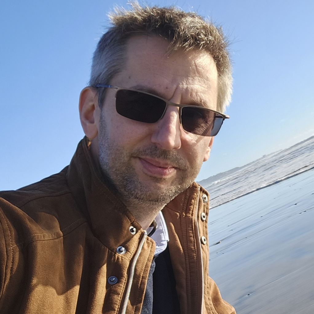
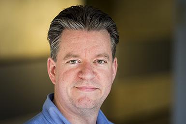
Uwe Himmelreich is a Full Professor at the Faculty of Medicine, KU Leuven , leading the Biomedical MRI unit. He is actively involved in the Medical Imaging Division , KU Leuven Brain Institute , KU Leuven Institute for Integration of Micro- and Nano-scale Technologies , and KU Leuven Cancer Institute . Role: Full Professor and Head of Biomedical MRI Affiliations: Faculty of Medicine, Medical Imaging Division, LBI, LIMNI, LKI His research spans neuroscience , cardiovascular imaging , and nano/micro-scale technologies . Key projects include: MindMAP: Radiotherapy-induced neurotoxicity in juvenile brains Preclinical cancer models for oral tumors Quantitative T2 mapping of lung disease in murine models Neuroinflammation and cognitive decline in cryptococcosis Resistance training effects on cortical thickness in aging cohorts His work integrates MRI , multi-photon microscopy , and novel contrast agents for longitudinal in vivo studies. Methodologies include vascular density mapping , proton therapy verification , and preclinical radiotherapy evaluation . Notable contributions include: 2025: JAK/STAT inhibition in malaria-induced inflammation 2025: Manganese-enhanced MRI for cardiac injury 2025: Quantitative lung imaging at 9.4T 2024: IVIM as vascular density marker in rat brain 2024: Phase-change ultrasound agents for proton therapy
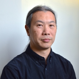
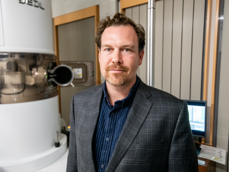
Cheuk Wai Tai is a Senior Staff Researcher at Stockholm University's Department of Environmental and Materials Chemistry since 2009. He manages the transmission electron microscopes and sample preparation equipment at the Electron Microscopy Center and serves as Section Editor for the Journal of Electronic Materials. His work focuses on quantitative structure characterization in functional materials research, particularly within nanoscience and nanotechnology contexts. Education: Ph.D. in Applied Physics, The Hong Kong Polytechnic University, 2004 M.Phil. in Applied Physics, The Hong Kong Polytechnic University, 2001 M.Sc. in Physics, The Chinese University of Hong Kong, 1998 B.Sc. (Hons) in Engineering Physics, The Hong Kong Polytechnic University, 1997 Dip. in Mechanical Engineering (Computer Aided Engineering), Institute of Vocational Education (formerly Haking Wong Technical Institute), Hong Kong, 1992 His research centers on structure-property relationships in functional materials through advanced electron microscopy techniques. Current specializations include Pair Distribution Function (ePDF) & Diffuse Scattering, Energy Materials characterization, and EM sample preparation methodology development. The group maintains strong focus on translating structural data into functional performance metrics for nanomaterials. Recent publications (2013-2019) demonstrate consistent emphasis on electron microscopy applications for energy storage materials (batteries, photocatalysts) and functional ceramics. Key trends include structural disorder analysis in piezoelectrics, development of quantitative TEM methods like SUePDF, and nanoscale characterization of electrocatalyst surface phases. His work bridges materials chemistry with advanced imaging techniques. Scientific recognition includes: Fellow of The Royal Microscopical Society (U.K.) Senior Member of IEEE Marie Curie Fellowship (2007-2009) from European Commission Sir Edward Youde Memorial Fellowship (2003/2004) from Hong Kong S.A.R. Government He teaches Solid State Chemistry (KZ7003) and leads Introduction to Analytical Electron Microscopy (KZ8009), having previously taught Advanced Transmission Electron Microscopy (KZ8010) before 2011. Major grants supporting his work include: "Quantitative structural characterisation using 3D electron-based pair distribution function" (Swedish Research Council) "A Multidimensional Toolkit for Modern Electron Microscopy" (Swedish Foundation for Strategic Research) "Mitigating Ni-rich Li-ion cathode side-reactions" (Swedish Energy Agency, Co-applicant) He leads the Cheuk-Wai Tai group within Stockholm University's chemistry department and oversees operations at the Electron Microscopy Center, where his team develops and applies advanced characterization techniques for functional materials research.
David A. Muller serves as the Samuel B. Eckert Professor of Engineering in the School of Applied and Engineering Physics at Cornell University and co-directs the Kavli Institute at Cornell for Nanoscale Science. His research group focuses on developing quantitative electron microscopy methods to understand materials properties at the atomic scale, with particular emphasis on sustainable energy applications and quantum materials. Muller's laboratory utilizes some of the world's highest resolution electron microscopes housed in specially designed, environmentally isolated rooms. Muller received his undergraduate education at the University of Sydney and earned his Ph.D. in Physics from Cornell University in 1996. Between 1997 and 2003, he was a member of the technical staff at Bell Laboratories, where he applied his expertise in imaging single atoms and atomic-scale spectroscopy to determine the physical limits of transistor miniaturization. In 2003, he returned to Cornell as a faculty member, where he has since established himself as a leader in advanced electron microscopy techniques. Muller's research spans multiple frontiers in materials science, with particular focus on understanding how electronic-structure changes at the atomic scale control macroscopic behavior in diverse systems like turbine blades, fuel cells, and transistors. His current work emphasizes the physics of renewable energy materials, atomic-scale control of materials to create electronic phases that cannot exist in bulk, and developing hardware and algorithms for 'big data' acquisition from high-bandwidth pixelated electron microscope detectors. His group's work bridges theoretical physics and experimental techniques, requiring researchers who can think in both real and reciprocal space while considering both fundamental principles and practical applications. Analysis of Muller's recent publications reveals a strong trend toward advancing electron ptychography and 4D-STEM techniques for atomic-scale imaging. His group has pioneered methods for 3D atomic-scale metrology, strain mapping, and imaging of radiation-sensitive materials. The research spans applications from semiconductor technology to quantum materials and energy storage systems, demonstrating the versatility of his microscopy approaches across multiple scientific domains. Top 100 Young Innovator by Tech Review Magazine (2003) Burton Medal from Microscopy Society of America (2006) Ernst Ruska Prize of German Society for Electron Microscopy (2021) John Cowley Medal from International Federation of Societies for Microscopy (2023) Fellow of American Physical Society Fellow of American Association for the Advancement of Science Fellow of Microscopy Society of America Muller has mentored an extensive group of students and postdocs who have gone on to successful careers in academia and industry. His former students hold faculty positions at institutions including Rice University, University of Southern California, Seoul National University, Colorado School of Mines, and the University of Michigan, among others. His research has been supported by substantial grants, including a $22.5M NSF grant that accelerates materials discovery. The Muller lab maintains close collaborations with the Kavli Institute at Cornell and PARADIM (Platform for the Accelerated Realization, Analysis, and Discovery of Interface Materials). The Muller lab operates at the forefront of electron microscopy, housing specialized instrumentation including high-resolution transmission electron microscopes in environmentally isolated rooms. The group collaborates extensively with other research teams at Cornell and worldwide, focusing on understanding materials atom by atom. Current research directions include applying machine learning to electron microscopy data analysis, developing cryogenic techniques for studying low-melting-point materials, and exploring quantum phenomena in engineered materials systems.
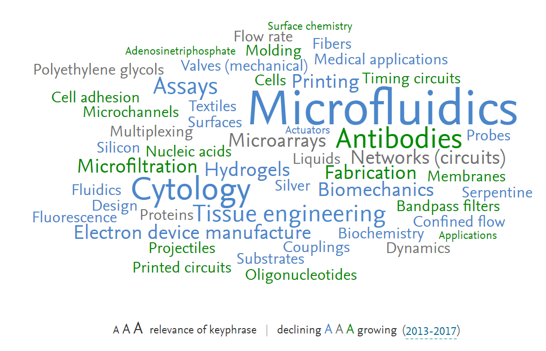
David Juncker is a Professor and Department Chair of the Department of Biomedical Engineering at McGill University. He serves as a Principal Investigator at the McGill University & Genome Quebec Innovation Centre and holds associate memberships in the Department of Neurology and Neurosurgery, Department of Electrical and Computer Engineering, Division of Experimental Medicine, Department of Surgery, and Goodman Cancer Research Centre. His research focuses on micro- and nano-bioengineering technologies for bioanalysis, precision medicine, and organs-on-chips. Key areas include microfluidics, lab-on-a-chip devices, biomedical sensors, medical diagnostics, biomaterials, tissue engineering, and cancer biomarker discovery. His lab develops scalable antibody microarrays, self-powered diagnostic platforms, microfluidic probes for brain tissue perfusion, and nanogradients for neuronal navigation, with applications in cancer diagnostics, global health, and neuroscience. Recent publications (2023-2025) reveal strong emphasis on extracellular vesicle analysis, single-cell proteomics, 3D-printed microfluidic/organ-on-a-chip systems, and capillary-driven circuits. Key trends include low-cost point-of-care diagnostics, advanced circulating tumor cell isolation methods, and biomimetic synthetic vesicles for drug delivery, demonstrating translational potential in early disease detection. Dr. Juncker leads a highly interdisciplinary team comprising undergraduate and graduate students, post-doctoral fellows, and staff from diverse scientific, engineering, and cultural backgrounds. His lab actively recruits Canadian/permanent resident graduate students for projects on single extracellular vesicle and protein detection in cancer and infectious diseases, leveraging microfluidics and wearables for biomarker discovery. The Juncker Lab operates from the McGill University & Genome Quebec Innovation Centre (740 Dr. Penfield Avenue, Room 6206). It maintains a collaborative, multicultural environment focused on developing transformative micro- and nano-bioengineering technologies with significant potential impact on human health diagnostics and treatment.
Thomas Grenier is an Associate Professor in the Department of Electrical Engineering at INSA Lyon and a member of the CREATIS laboratory (CNRS UMR 5220, INSERM U1294). He obtained his HDR (Habilitation à Diriger des Recherches) in 2023 and his Ph.D. in Image Processing from INSA Lyon in 2005. His research focuses on medical image segmentation, clustering, and filtering using feature space, scale-space, and deep learning approaches. Doctoral School: EEA (Electronics, Energy, and Automatics) Research Affiliation: CREATIS Lab (CNRS/INSERM/INSA Lyon/Université Lyon 1/Université Jean Monnet Saint-Etienne) He has contributed to 20 papers and co-supervised 5 PhD students, including Léo Dumortier and Florent Guépin. Grenier leads the annual Deep Learning for Medical Imaging (DLMI) school, which he co-founded, and has organized five editions across Lyon and Montreal since 2019. The school emphasizes practical deep learning applications in medical imaging for participants of all expertise levels. His work spans interdisciplinary domains such as medical imaging , deep learning , and image processing , with recent publications on generative AI for MRI synthesis, explainable networks, and segmentation of neurological pathologies in preclinical models. He manages pedagogical platforms, coordinates LabEx PRIMES project activities, and oversees lab room infrastructure for 200 hours/year across 10 training programs. Grenier also leads the MUSIC transversal project on Multiple Sclerosis since 2019.
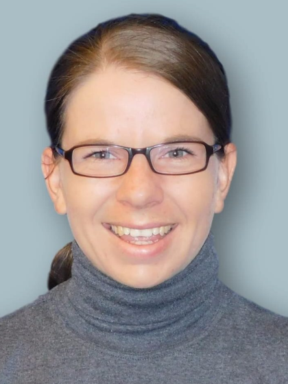
Dr. Julia Kamenz is an Assistant Professor (Rosalind Franklin fellow) at the University of Groningen's Faculty of Science and Engineering, where she leads research in the Molecular Systems Biology group within the Groningen Biomolecular Sciences and Biotechnology Institute (GBB). Her work focuses on understanding the molecular mechanisms that regulate cell cycle progression and cell division. Dr. Kamenz received her undergraduate training in Biochemistry at the University of Tuebingen, completed her PhD at the Friedrich Miescher Laboratory of the Max Planck Society under Dr. Silke Hauf (defended February 2015 with highest honors), and conducted postdoctoral research at Stanford University with Prof. James E. Ferrell. Her PhD work was supported by a Boehringer Ingelheim Fonds fellowship, and her postdoc was funded by a German Research Foundation (DFG) Postdoctoral Fellowship. Her research expertise spans cell cycle regulation and dynamics, post-translational modifications, Xenopus laevis model systems, and live cell microscopy. Dr. Kamenz investigates how kinases and phosphatases intricately regulate cell proliferation and division, with particular interest in the molecular mechanisms that ensure faithful chromosome segregation during mitosis. Her recent work has revealed novel insights into mitotic checkpoint signaling, particularly in early embryonic development where these checkpoints appear to function differently than in somatic cells. Dr. Kamenz's publication record demonstrates a strong focus on the dynamics of cell cycle transitions, with recent papers appearing in high-impact journals including Nature, The Journal of Biological Chemistry, and The Journal of Cell Biology. Her research integrates experimental biochemistry, live-cell imaging, and computational modeling approaches to understand complex regulatory networks. ERC Starting Grant (November 2022) NWO Vidi Grant (July 2021) Mansour Postdoctoral Travel Award (2019) Dr. Kamenz has secured significant research funding including an ERC Starting Grant (€1.5 million) and an NWO XS grant (€50,000) for her project "What limits mitotic checkpoint signaling in the early embryo?" Her research contributes to understanding fundamental biological processes with implications for developmental biology and cancer research. She collaborates extensively within the University of Groningen and with international partners, particularly in the areas of cell cycle research and biophysical approaches to biological problems. Dr. Kamenz leads a research group focused on cell cycle regulation within the Molecular Systems Biology division of the Groningen Biomolecular Sciences and Biotechnology Institute. Her lab combines biochemical approaches using Xenopus egg extracts with live-cell imaging and computational modeling to dissect the molecular mechanisms controlling cell division.
Suliana Manley is a Full Professor at the École Polytechnique Fédérale de Lausanne (EPFL) , affiliated with the School of Basic Sciences and the Laboratory of Experimental Biophysics . She also holds teaching and research roles in EPFL's School of Life Sciences and Swiss Plasma Center , focusing on interdisciplinary biophysical studies. Education : PhD in Physics (2004), Harvard University Bachelor's in Physics & Mathematics (1997), Rice University Manley's research centers on super-resolution fluorescence imaging , single-molecule tracking , and quantitative biophysics . Key themes include: Understanding protein assembly dynamics at cellular membranes Elucidating viral assembly mechanisms (e.g., HIV-Gag) Developing 3D imaging algorithms and high-density data reconstruction tools like PALMsiever and FALCON Quantifying nanoscale organization in systems like telomeres and centrioles Her work bridges optical physics , computational image analysis , and cellular biology , with notable Nature and PNAS publications. Collaborations span bioengineering , genetics , and medical research . Scientific Awards : Featured in Nature Methods Research Highlights (3x) Very Important Paper and Cover Article (ChemBioChem, 2012) Postdoctoral Fellow, NIH and MIT Advising & Collaborations : Current PhD students in biophysics, cellular biology, and bioengineering Former students: Anna Archetti, Aleksandr Benke, Andrea Callegari, and others Co-founder of tools for high-density super-resolution microscopy and live-cell imaging Labs & Teams : Leads the Laboratory of Experimental Biophysics at EPFL, integrating physics-based methods into biological questions. The lab focuses on quantitative imaging , computational modeling , and software development for nanoscale analysis.
Adam de la Zerda is an Associate Professor at Stanford University's Department of Structural Biology (School of Medicine) and Electrical Engineering (by courtesy). He develops advanced optical molecular imaging technologies combining nanoparticle contrast agents and adaptive OCT systems for cancer and ophthalmic disease research. Technion-Israel Institute of Technology (BSc, 2005) Stanford University (PhD, 2011) UC Berkeley (Postdoctoral Fellowship) Research Themes : Virtual biopsy using machine learning-enhanced OCT Gold nanorod-based molecular contrast agents Speckle noise reduction for cellular resolution Needle beam optical coherence tomography angiography His 15 most recent publications demonstrate technical innovations in: Metasurface optics for extended depth-of-field Spectral deconvolution of multiple contrast agents Speckle modulation for improved diagnostic clarity Noninvasive lymphatic system mapping Scientific Honors : Pew-Stewart Scholar for Cancer Research AFOSR Young Investigator NIH Early Independence Award Forbes 30 Under 30 (x2) Chan Zuckerberg BioHub Investigator His lab team has developed clinical prototypes including OcuBell Inc. 's ophthalmic imaging systems and Visby Medical 's diagnostic platforms. Current research spans from in vivo glycoprotein imaging to de novo biosensor development for real-time disease monitoring in awake animal models.
Prof. Casper Hoogenraad is a full professor in Molecular Neuroscience at the Department of Cell Biology, Faculty of Science, Utrecht University. His research focuses on understanding how intracellular protein trafficking underlies neuronal development and function, with particular emphasis on the microtubule cytoskeleton, synaptic cargo trafficking, and synaptic plasticity. He leads an active research group within Utrecht University's Cell Biology department and collaborates extensively with other neuroscience research groups. Education: PhD, Erasmus University Rotterdam (1996-2001) Postdoc, Massachusetts Institute of Technology (2002-2005) Hoogenraad's research spans three main themes: cytoskeleton dynamics during neurodevelopment and synaptic plasticity, motor proteins and adaptors as regulators of synaptic transport, and psychiatric and neurologic disease disorders linked to intracellular transport. His work combines genetics, biochemistry, molecular, and cellular biology methods in in vitro (neuron cultures), ex vivo (brain slices), and in vivo (mice) systems, along with advanced microscopy techniques including immunofluorescent confocal microscopy, high-resolution live cell imaging, and photo-activated localization microscopy (PALM). Analysis of Hoogenraad's recent publications reveals a strong focus on microtubule organization, neuronal polarity, and the molecular mechanisms underlying synaptic function and dysfunction. His work frequently explores how disruptions in intracellular transport contribute to neurological disorders including Alzheimer's disease, schizophrenia, and autism spectrum disorders, with particular attention to the relationship between cytoskeletal organization and cargo transport in neuronal compartments. Scientific Awards and Memberships: ZonMW-VIDI (2004) European Young Investigators (EURYI) award (2005) NWO-ALW VICI (2011) ERC Consolidator grants (2013) FENS-Kavli Network of Excellence (2014) European Molecular Biology Organization (EMBO) (2015) Young Academy of Europe (YAE) (2015) IBRO Kemali Prize (2016) Hoogenraad leads a research group studying neuronal development and function, with a particular focus on how intracellular transport mechanisms contribute to both normal brain function and neurological disorders. His laboratory employs a multidisciplinary approach combining molecular, cellular, and systems neuroscience techniques to investigate the molecular basis of neuronal polarity, synaptic plasticity, and the pathogenesis of neurological disorders. He has secured significant research funding through prestigious grants including ERC Consolidator grants. The Hoogenraad lab operates within the Cell Biology department at Utrecht University, collaborating with other research groups focusing on cellular dynamics, biophysics, and neurobiology. The lab utilizes advanced microscopy techniques including immunofluorescent confocal microscopy, high-resolution live cell imaging (spinning disc microscopy and total internal reflection fluorescence microscopy), and quantitative analysis using advanced high-resolution microscopy (photo-activated localization microscopy). Current lab technicians include Phebe Wulf and Bart de Haan.
Parvin Mousavi is a Professor at the School of Computing, Queen's University, and holds prestigious chairs including the Canada CIFAR AI Chair and Canada Research Chair in Medical Informatics. She directs the Medical Informatics (Med-i) Laboratory, focusing on computational approaches for biological process prediction and explanation. Education: Ph.D., University of British Columbia (2001) Research interests span machine learning in computer-assisted diagnosis, ultrasound imaging, medical image computing, bioinformatics, systems biology, and quantitative modeling of gene regulatory networks. The lab emphasizes interdisciplinary collaboration with medical professionals and institutions. Affiliations & Collaborations: Kingston General Hospital, Human Mobility Research Centre (HMRC), and University of California, San Francisco. The Med-i Laboratory is equipped with advanced parallel computing servers, clusters, and imaging devices. Awards: Canada CIFAR AI Chair Canada Research Chair in Medical Informatics Contact: Office: Goodwin 720 | Phone: 613 533-6070
Daniel Sage is a Lecturer and Scientific Advisor at École polytechnique fédérale de Lausanne (EPFL) , affiliated with the Biomedical Imaging Laboratory (LIB) under the College of Engineering (STI) and School of Life Sciences (SV) . He specializes in bioimage informatics , structured-illumination microscopy , and deep learning applications for biomedical imaging. His work spans algorithm development for single-molecule localization microscopy (SMLM) , fluorescence imaging , and 3D reconstruction . His research group has developed open-source tools like FlexSIM for light inhomogeneity correction, DeepImageJ for integrating deep learning in ImageJ, and Steer'n'Detect for orientation-accurate template detection. His publications focus on correcting multiple-blinking artifacts in PALM, optimal transport metrics for SMLM evaluation, and contextual feature analysis for xenograft cell classification. He mentors PhD students and contributes to interdisciplinary education through courses such as Bioimage Informatics and Fundamentals of Image Analysis , emphasizing practical software solutions and Java programming for bioimage processing. His collaborations include institutions like Howard Hughes Medical Institute and Centre National de la Recherche Scientifique (CNRS) .
Michael Vershinin is an Assistant Professor of Physics and Astronomy at the University of Utah, specializing in molecular motors and biophysics. He is also affiliated with the Biological Chemistry Program and leads a lab focused on understanding how molecular motors like kinesin and dynein drive intracellular transport and viral assembly. He earned his B.S. from Cooper Union College and Ph.D. from the University of Illinois, Urbana-Champaign. His research interests include: Molecular motor function and regulation Single-molecule biophysics Microtubule-based transport Viral particle assembly (especially SARS-CoV-2 and HIV) Optical trapping and fluorescence microscopy His lab uses in vitro reconstitution and optical trapping to dissect the biophysical properties of motor proteins and their regulation. He collaborates across disciplines, integrating biochemistry, molecular biology, physics, and computational modeling to explore how complex biological behaviors emerge from simpler components. His publications span a wide range of topics, from the structural stability of SARS-CoV-2 virus-like particles to the mechanical behavior of kinesin and dynein motors. A recurring theme is the use of quantitative biophysical tools to understand how motor proteins navigate complex cytoskeletal environments and how viruses hijack these systems for transport. He currently advises no listed students in the provided text and has not received any explicitly listed awards. His lab is located at the University of Utah and can be reached at vershinin@physics.utah.edu .
Rupert Huber is a Professor at the Department of Experimental and Applied Physics, University of Regensburg, where he has held a chair since 2010. His research focuses on ultrafast quantum phenomena, terahertz science, and lightwave electronics, with a strong emphasis on nanoscale imaging and quantum materials. He leads the Huber group, which has launched the ERC project 'Orbital Cinema' and produced numerous high-impact publications in journals like Nature and Nano Letters . Chair for Experimental and Applied Physics, University of Regensburg (2010–present) Emmy Noether Group Leader, University of Konstanz (2007–2010) Alexander von Humboldt Fellow, UC Berkeley/Lawrence Berkeley National Lab (2004–2006) His research explores terahertz spectroscopy , quantum materials , and ultrafast nanoscopy , often combining experimental innovation with theoretical insights. Recent work includes groundbreaking studies on exciton dynamics in van der Waals magnets and subcycle imaging of electron wave motion. The group’s publications frequently appear as coverstories in Nature Photonics and Nano Letters . Huber has received prestigious awards such as the Gottfried Wilhelm Leibniz Prize (2019) , ERC Starting Grant (2012) , and OSA Fellowship (2018) . He has supervised numerous Ph.D. and Master’s students, including recent awardees like Joshua Mornhinweg (faculty dissertation prize, 2024) and Josef Riepl (best tutor award, 2024).
Ueli Grossniklaus is an Ordinary Professor at the University of Zurich within the Faculty of Mathematical and Natural Sciences , affiliated with the Department of Plant and Microbiology . His work focuses on plant developmental biology, particularly epigenetic and genetic mechanisms governing reproduction and adaptation. Key Courses: Epigenetics, Plant Biology Workshop, Group Seminars on Current Research Laboratory Techniques: Advanced methods in plant cell mechanics, transcriptomics, and genome editing Research Interests span plant epigenetics, reproductive biology, and the interplay between environmental stress and genetic regulation. He investigates: Mechanistic control of gametogenesis and fertilization Epigenetic contributions to plant adaptation Evolutionary implications of asexual reproduction Biophysical forces in plant cell growth Publication Trends (2025–2018) reveal expertise in: Arabidopsis and fern model systems Epigenetic regulation (DNA methylation, histone dynamics) Apomixis and hybrid seed failure mechanisms Biomechanics of pollen tubes and carnivorous plants Genome editing tools (CRISPR) and long-read sequencing Scientific Collaborations include interdisciplinary projects on: Microfluidic devices for plant cell analysis Gene drive ecology and ethics 3D imaging of plant reproductive structures Advising and Grants focus on mentoring through research internships in developmental biology, genetics, and systems biology. His lab engages in: Epigenetic response to environmental stress Cell wall mechanics in reproduction Computational modeling of plant growth Laboratory Teams integrate plant biologists, bioengineers, and computational scientists to study: Mechanistic gene regulation Evolutionary developmental biology Microrobotics for cellular force measurement
Dr. Jamie Warner is a Professor and Temple Foundation Endowed Professor in the Walker Department of Mechanical Engineering at The University of Texas at Austin, leading the TMI Electron Microscopy Facility within the Cockrell School of Engineering. His research focuses on nanostructured materials, advanced transmission electron microscopy, and opto-electronic applications. Prior to UT Austin, he held a Full Professorship at the University of Oxford's Department of Materials, where he led the Nanostructured Materials Group and graduated 30 PhD students. Key roles include Director of the Texas Materials Institute and Visiting Professorships at MIT and Sungkyunkwan University. Education: PhD in Physics (University of Queensland, 2004), Postdoc (New Zealand/Australia, 2005-2006) Research Interests: Atomic-scale characterization of 2D materials (graphene, MoS₂, WS₂), electron microscopy techniques, nanoelectronic devices, and energy storage materials. His articles span advanced TEM techniques, 2D material synthesis, and opto-electronic device fabrication. Notable awards include the Royal Society University Research Fellowship (2010), ERC Consolidator Grant (2017), and ACS Nano Lectureship (2019). Awards: Fellow of the Royal Society of Chemistry (2019), Top 10 'Highly Prolific' ACS Nano Author (2018) Advising: 30+ PhD graduates, extensive postdoc and master's supervision Grants: ERC Consolidator Grant for opto-electronics, multiple industry and academic collaborations He leads the Warner Group, which operates cutting-edge facilities for electron microscopy and nanofabrication. Current projects include cryo-TEM for battery materials and single-atom catalysts.