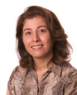
Dr. Kristin O'Grady is an Assistant Professor in the Department of Biomedical Engineering and Department of Radiology & Radiological Sciences at Vanderbilt University's School of Engineering. Her research focuses on developing quantitative MRI methodologies for the brain and spinal cord, particularly improving spinal cord MRI for neurological diseases like multiple sclerosis. She specializes in diffusion tensor imaging, functional connectivity analysis, and high-field MRI applications. Her work spans advanced imaging techniques including MP2RAGE, susceptibility-weighted MRI, and phase imaging, with a focus on clinical feasibility and disease markers. She has contributed to studies on spinal cord morphometry, paramagnetic rim lesions, and biological interactions affecting CNS structure. No scientific awards or grants are explicitly listed in the provided materials. Dr. O'Grady collaborates across interdisciplinary teams within the School of Engineering, focusing on translational research in neuroimaging technologies.
Professor Nellie Georgiou-Karistianis serves as the Pro Vice-Chancellor (Research Training) at Monash University within the Deputy Vice-Chancellor (Research) portfolio, where she leads and shapes the graduate research training agenda. She previously held significant leadership roles as Associate Dean (Graduate Affairs, 2015-2018) and Deputy Dean (Academic and Graduate Affairs, 2019-2021) within the Faculty of Medicine, Nursing and Health Science. She heads the Georgiou-Karistianis Experimental Neuropsychology Research Unit within the Turner Institute of Brain and Mental Health and serves as Director of the Huntington's Research Group of Victoria (HRGV). Her research focuses on neurodegenerative disorders, particularly Huntington's disease and Friedreich ataxia. She has pioneered work in discovering novel biomarkers for tracking disease progression that are now used in early drug development. Her research spans cognitive neuroscience, neuroimaging, biomarker development, and cognitive interventions. She has made significant contributions to understanding disease progression at brain, cognitive and motor levels, with work that has informed clinical trials and therapeutic approaches. Analysis of her recent publications reveals a strong focus on advanced neuroimaging techniques for tracking neurodegenerative processes, machine learning applications for biomarker development, and cognitive interventions for neurodegenerative conditions. Her work increasingly integrates multimodal data analysis and international collaboration to validate imaging biomarkers for clinical trial use. H-index of 57 with over 10,500 citations Editor-in-chief of Journal of Huntington's Disease since 2017 Editor-in-chief of Recent Patents on Biomarkers since 2017 Leadership certification from Harvard University Professor Georgiou-Karistianis has supervised 29 doctoral students to completion, seven of whom received NHMRC ECR Fellowships and one a Fulbright Fellowship. She has secured over $16 million in research funding from competitive government schemes (NHMRC, ARC Linkage) and philanthropic bodies. As Overall Coordinating Principal Investigator, she leads TRACK-FA, an international study with seven academic partners and six industry sponsors across four continents. She leads the Georgiou-Karistianis Experimental Neuropsychology Research Unit and chairs the TRACK-FA Neuroimaging Consortium. Her work involves close collaboration with international research networks including the Huntington's Disease Regulatory Science Consortium (HD-RSC), Critical Path Institute, and the Cure Huntington's Disease Initiative (CHDI, USA) Foundation.
Dale Bailey is a Professor in the Faculty of Medicine & Health at the University of Sydney, with clinical appointments at Royal North Shore Hospital's Department of Nuclear Medicine. He previously directed the Sydney Vital Northern Translational Cancer Research Centre and is a member of the Centre for Drug Discovery Innovation, the University of Sydney Nano Institute, and the Charles Perkins Centre. Nuclear Medicine Physicist Theranostics Researcher Clinical Imaging Expert His research focuses on quantitative functional imaging using SPECT and PET, hybrid imaging modalities, radiation dosimetry for personalized cancer therapy, and the biological effects of radiation. He pioneered combined structure/function imaging and advanced radionuclide therapy applications. Recent publications emphasize PET/CT with novel radionuclides, metabolic tumor volume as a prognostic biomarker, and dosimetry validation methods . Key themes include cancer, nuclear medicine, and translational research. PhD in Physics Fellow of the Institute of Physics and Engineering in Medicine (FIPEM) Chartered Scientist (CSci) in the UK International Consensus Guidelines Contributor He supervises research students in radiotheranostics and collaborates on clinical trials for neuroendocrine tumors and glioblastoma. Grants and projects involve hybrid imaging optimization, radiation safety, and personalized treatment planning.
Christian Büchel is a Professor in the Department of Neurology and Experimental Neurobiology at the University Medical Center Hamburg-Eppendorf (UKE). He holds a faculty position within the Medical Faculty of Hamburg University. His research focuses on understanding brain mechanisms underlying pain, placebo/nocebo effects, and psychiatric disorders using advanced neuroimaging techniques. He leads studies on brain-body interactions, developmental neuroscience, and translational neurology. Key research areas include structural and functional MRI investigations of pain modulation, addiction, and stress responses. He has contributed to multi-center neuroimaging studies and pioneered methods for spinal cord imaging. His work integrates cognitive neuroscience principles with clinical applications, particularly in chronic pain management and mental health. Büchel's recent studies explore how environmental factors (e.g., urban green spaces) influence brain structure, and he investigates neural correlates of resilience to substance use in adolescents. He collaborates internationally on projects like the IMAGEN study and the ENIGMA consortium, emphasizing reproducibility in neuroimaging research. His publications span over 20 years, with a focus on brain network dynamics, decision-making processes, and translational neuroscience. He actively contributes to developing open-access neuroimaging standards and has authored seminal papers on placebo mechanisms and brain plasticity.
Dr. Emmanuel Stamatakis is a Senior Research Associate at the University of Cambridge, affiliated with the School of Clinical Medicine and the Division of Anaesthesia. He maintains research ties with the Centre for Speech, Language, and the Brain (CSLB) while focusing on neuroimaging, consciousness studies, and traumatic brain injury (TBI) mechanisms. Institution: University of Cambridge Primary Affiliation: School of Clinical Medicine, Division of Anaesthesia Research Affiliation: Centre for Speech, Language, and the Brain (CSLB) Academic Rank: Research Fellow His research spans neuroscience, neuroimaging, and anaesthesia, with particular emphasis on: Consciousness dynamics under pharmacological and pathological conditions Functional and structural brain connectivity in TBI and delirium Neural mechanisms of anaesthetic agents Evolutionary and comparative neuroscience of brain states Long-term outcomes after neurological trauma Neurotechnology applications in clinical settings Recent publications reveal trends in: Quantifying consciousness through functional gradients and harmonic decomposition Linking anaesthesia-induced neural changes to clinical outcomes Exploring genetic and inflammatory factors in TBI recovery Multi-modal imaging approaches to neurological disorders Translational studies bridging animal models and human conditions Methodological advancements in brain network analysis
Jeffrey Hutsler is an Associate Professor in the Department of Psychology at the University of Nevada, Reno, affiliated with the Institute of Neuroscience. His research focuses on the microanatomical organization of the human brain, particularly in individuals with autism spectrum disorders. Ph.D. in Physiological Psychology, University of California, Davis (1993) M.A. in Physiological Psychology, University of California, Davis (1991) B.A. in Psychology, San Jose State University (1988) Hutsler's research explores cortical layering, dendritic spine densities, white matter diffusion, and subplate abnormalities in autism. He integrates histological assessments with neuroimaging techniques to study structural and functional brain differences. His recent publications (2025-2011) emphasize autism-related cortical anomalies, visual processing, hemispheric specialization, and computational modeling. Key trends include the use of fNIRS and diffusion MRI to link structural changes to behavioral symptoms. Slifka-Ritvo Award for Innovation in Autism Research (2010) Vada Trimble Outstanding Mentor Award (2013) Hutsler has mentored numerous students through courses like Human Neuropsychology, Developmental Neuropsychology, and the Neuropsychology of Autism. His work often involves collaborations in neuroimaging, developmental neuroscience, and autism research.
Mojtaba Zarei is a researcher at the Department of Clinical Research, Faculty of Health Sciences, University of Southern Denmark, with additional affiliations at Odense University Hospital (OUH) and Karolinska Institutet (KI). His primary research unit is the Neurology Research Unit in Odense, focusing on advanced neuroimaging techniques and their applications in neurological and sleep disorders. Dr. Zarei's research spans multiple domains within neuroscience, with particular expertise in Positron Emission Tomography (PET), Diffusion Tensor Imaging (DTI), and cognitive function assessment. His work frequently addresses Alzheimer's Disease, Parkinson's Disease, and insomnia disorders, utilizing both clinical and computational approaches. His fingerprint analysis shows strong activity in neuroscience (100% for PET), diffusion tensor imaging (66%), cognitive function (45%), and Alzheimer's Disease (40%). His recent publications reveal a clear trajectory toward integrating multimodal imaging techniques with machine learning approaches for improved diagnosis and understanding of neurological conditions. The work on OPETIA (Odense-Oxford PET Image Analysis) demonstrates his contribution to developing standardized tools for neuroimaging analysis. His research increasingly bridges computational methods with clinical neuroscience, as evidenced by his work on image stitching algorithms and machine learning applications for insomnia classification. Dr. Zarei actively collaborates with researchers across multiple institutions, with notable external collaborations visible on the international network map. His work has been mentioned by peer review sites, picked up by news outlets, and shared across social media platforms, indicating growing impact in his field. Within his research unit of Neurology in Odense, Dr. Zarei appears to be part of a multidisciplinary team working at the intersection of clinical neurology, advanced imaging, and computational analysis, contributing to both methodological development and clinical applications of neuroimaging techniques.
Susan A. Gauthier is Professor of Neuroscience at the Brain and Mind Research Institute, Professor of Neurology in the Department of Neurology, and Professor of Neurology in Radiology at Weill Cornell Medical College. She serves as Director of Clinical Research at the Judith Jaffe Multiple Sclerosis Center, where she leads a translational research program focused on uncovering the biological mechanisms of multiple sclerosis using advanced quantitative neuroimaging. Dr. Gauthier completed her undergraduate studies at the State University of New York at Buffalo, earned her D.O. from the Philadelphia College of Osteopathic Medicine, and obtained her MPH from Harvard School of Public Health. After completing neurology residency and chief residency at Boston University Medical Center, she was awarded a Clinical Trial Training Fellowship from the National Multiple Sclerosis Society at Brigham and Women's Hospital. Dr. Gauthier's research focuses on developing imaging biomarkers to track inflammation, demyelination, and clinical disability in multiple sclerosis. Her work spans quantitative susceptibility mapping (QSM), TSPO-PET imaging, brain connectivity analysis, and artificial intelligence applications in neuroimaging. She has made significant contributions to understanding chronic active lesions, paramagnetic rim lesions, and their relationship to disability progression in MS. Analysis of Dr. Gauthier's recent publications (2022-2025) reveals a strong focus on quantitative neuroimaging biomarkers for multiple sclerosis. Her work increasingly integrates advanced computational methods including artificial intelligence and machine learning to analyze structural and functional connectivity. A key theme across her recent work is the validation of imaging techniques against pathological findings, particularly regarding chronic active lesions and iron deposition. She has also contributed to several consensus statements that aim to translate imaging research into clinical practice for MS patients. Sylvia Lawry Fellowship in Clinical Trials, National Multiple Sclerosis Society (2002) Dr. Gauthier has served as Principal Investigator on multiple NIH and National Multiple Sclerosis Society grants, including studies on multi-scale imaging assessment of cognitive impairment in MS, quantification of innate immune activity within chronic lesions, and establishing the clinical relevance of chronic active MS lesions. She has built the MS Center's research infrastructure at Weill Cornell and forged strong collaborations with the Department of Radiology. As a dedicated mentor, she has trained numerous students, residents, fellows, and junior faculty who have gone on to academic and clinical leadership positions. Dr. Gauthier leads a research team within the Judith Jaffe Multiple Sclerosis Center that collaborates closely with the Department of Radiology at Weill Cornell. She serves on the Steering and Executive Committees of the North American Imaging in Multiple Sclerosis (NAIMS) Cooperative, which brings together experts from multiple institutions to advance imaging research in MS. Her team utilizes advanced MRI techniques including 7T MRI and PET imaging to study MS pathophysiology.
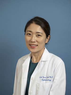
Dr. Soh Youn Suh serves as an Assistant Professor of Ophthalmology at UCLA's Stein Eye Institute within the David Geffen School of Medicine, specializing in pediatric ophthalmology and adult strabismus care at the Los Angeles clinical site. Her clinical expertise addresses complex eye alignment disorders and childhood vision conditions. Education: MD, Ewha Womans University School of Medicine, 2006 MS, Ewha Woman's University, 2010 Ophthalmology Residency, Ewha Womans University Medical Center, 2011 Pediatric Ophthalmology & Neuro-Ophthalmology Fellowship, Seoul National University Hospital, 2013 Pediatric Ophthalmology & Adult Strabismus Fellowship, Stein Eye Institute, UCLA, 2014 Research Fellowship, Ocular Motility Laboratory, Stein Eye Institute, UCLA, 2018 Her research program investigates biomechanical interactions between extraocular muscles, optic nerves, and orbital structures using advanced MRI and OCT imaging. Key discoveries include documenting abnormal optic nerve traction during eye movements in glaucoma patients with normal intraocular pressure and characterizing muscle pulley displacements in strabismus conditions. This work bridges neuro-ophthalmology and surgical planning through quantitative orbital analysis. Analysis of her 15 most recent publications (2020-2025) reveals three dominant research trajectories: (1) AI-driven segmentation of orbital structures using deep learning, (2) cerebrospinal fluid dynamics in optic neuropathies, and (3) biomechanical modeling of optic nerve strain during horizontal eye movements. These studies increasingly integrate computational methods with high-resolution imaging to decode pathophysiological mechanisms in strabismus and glaucoma. Scientific Recognition: Excellence in Research Award, Stein Eye Institute (2015) Excellence in Research Award, Stein Eye Institute (2016) Excellence in Research Award, Stein Eye Institute (2018) Dr. Suh has developed collaborative research partnerships through UCLA's institutional resources, particularly within Dr. Joseph L. Demer's ocular motility laboratory where she conducted postdoctoral research. Her work receives consistent institutional support through Stein Eye Institute infrastructure, though specific external grant funding isn't documented in source materials. She actively contributes to training through co-authorship with junior researchers on technical imaging studies. Her current research team at the Stein Eye Institute operates an orbital biomechanics laboratory focused on translating MRI/OCT findings into clinical applications. The group specializes in computational modeling of eye movement dynamics and developing AI tools for surgical planning in complex strabismus cases, with ongoing projects examining cerebrospinal fluid interactions in optic nerve disorders.
Chuan Huang is an Assistant Professor at Stony Brook University's School of Medicine, affiliated with the Department of Radiology and Psychiatry. His research focuses on medical imaging, particularly PET/MRI, quantitative MRI, and image reconstruction. Education: B.S. in Mathematics (2006, USTC), Ph.D. in Mathematics (2012, University of Arizona). Professional History: Research Fellow (2012-2014, MGH/HMS), Assistant in Physics (MGH), Instructor (HMS), then Stony Brook faculty since 2014. His work includes developing rapid MRI techniques, PET motion correction, and analyzing neuroinflammation in World Trade Center responders. He has contributed to 25+ publications in neuroscience, radiology, and computational imaging. Awards include ISMRM Magna Cum Laude Awards and SNMMI Young Investigator Honorable Mentions. Scientific Awards : Distinguished Reviewer for Magnetic Resonance in Medicine (2012-2013) ISMRM Young Investigator Award Honorable Mention (2014) SNMMI Annual Meeting Young Investigator Honorable Mentions (2013, 2014) ISMRM Magna Cum Laude Awards (2012)
Ann Ragin, PhD, is a Research Professor in the Department of Radiology at the Feinberg School of Medicine, Northwestern University. Her work focuses on quantitative MRI and brain network analysis to investigate aging effects and viral infections including HIV and COVID-19. She maintains active affiliations with the Institute for Public Health and Medicine (IPHAM), Northwestern University Clinical and Translational Sciences Institute (NUCATS), Northwestern University Institute of Neuroscience (NUIN), Robert J. Havey, MD Institute for Global Health, and Simpson Querrey Institute for Epigenetics. Her academic credentials include a PhD from Northwestern University (1987) and postgraduate training at the University of Chicago (1989), where she completed a postdoctoral fellowship in Quantitative Methods. Dr. Ragin's research program integrates advanced imaging techniques with clinical neuroscience: Development of quantitative MRI methodologies for in vivo brain measurement Brain network analysis for early neural injury detection 4D flow imaging applications in cerebral blood flow assessment Investigation of neuroinflammation in HIV/AIDS and aging populations Structural-functional neuroimaging correlations in viral infections Her recent publications reveal a dual research trajectory: computational neuroscience innovations (tensor-based graph networks for brain analysis) and clinical applications in cardiopulmonary imaging (pulmonary hypertension biomarkers). This work bridges machine learning, neurology, and cardiology through collaborative NIH-funded studies with the AIDS Clinical Trials Group. Professional leadership includes continuous membership in the AIDS Clinical Trials Group (2013-present), Conference on Retroviruses and Opportunistic Infections, and International Society for Magnetic Resonance in Medicine (2002-present), with prior service on the Chicago Society for Neuroscience Council (2003-2005). While specific grant details remain undisclosed per institutional policy, her ACTG membership and multi-institute affiliations indicate substantial involvement in federally funded research. No advisee information appears in public profiles, though her 99 publications suggest extensive mentorship within collaborative projects. Current initiatives focus on multimodal brain network analysis for HIV-related neurocognitive disorders and quantitative MRI biomarkers for pulmonary hypertension, leveraging Northwestern's advanced imaging infrastructure across multiple research consortia.
Professor Mark Lythgoe is a distinguished academic at University College London (UCL), where he serves as Professor of Biomedical Imaging in the Department of Imaging within the Faculty of Medical Sciences. He is the Founder and Director of the Centre for Advanced Biomedical Imaging (CABI) at UCL, a multidisciplinary research center hosting 12 state-of-the-art imaging modalities and 50 researchers. Additionally, he is Co-Director of the UCL Department of Imaging and Director of Biomedical Imaging Research at the Francis Crick Institute. Mark Lythgoe earned his Doctor of Philosophy from University College London in 1999 and his Master of Science from the University of Surrey in 1993. Professor Lythgoe has a long-standing track record in the development and application of biomedical imaging techniques, with research spanning from fundamental imaging science to clinical applications. His work focuses on advancing imaging technologies for biomedical research and clinical practice, with particular emphasis on neuroimaging, molecular imaging, and the application of imaging to understand neurological disorders and cancer. He has translated his research findings into clinical radiological practice and established training programs in biomedical imaging. His extensive publication record demonstrates consistent innovation across imaging modalities, with recent work increasingly focused on the glymphatic system in neurological disorders, advanced tumor imaging techniques, and the development of novel imaging technologies for both preclinical and clinical applications. His research bridges engineering, physics, biology, and clinical medicine to solve complex biomedical challenges. Professor Lythgoe's significant contributions to the field have been recognized with numerous prestigious awards: IET Achievement Medal (2023) - for major and distinguished contribution in Medical Imaging Royal Society of Medicine Ellison-Cliffe Award (2021) - for contribution of fundamental science to the advancement of medicine Davies Medal from the Royal Photographic Society (2013) - for significant contribution to imaging science Alumni Achievement Award from the University of Salford Neuroscience Prize for Public Understanding from the British Neuroscience Association Dorothy Hodgkin Award Biosciences Federation Science Communication Award Fellow of the British Science Association Professor Lythgoe has secured substantial research funding, with £45 million awarded for his collaborative imaging research program. He is deeply committed to training the next generation of imaging scientists, serving as Co-Director of the MSc in Advanced Biomedical Imaging and Co-Founder of the UCL Centre for Doctoral Training in Medical Imaging. His public engagement efforts include directing the Cheltenham Science Festival, which has become one of the largest science festivals in the world. As Director of the Centre for Advanced Biomedical Imaging, Professor Lythgoe leads a vibrant research team of 50 researchers working across 12 state-of-the-art imaging modalities. His center fosters interdisciplinary collaboration between engineers, physicists, biologists, and clinicians to develop and apply cutting-edge imaging technologies. He has published over 300 papers including publications in Nature, Nature Photonics, Nature Medicine and The Lancet, demonstrating the high impact of his research across multiple disciplines.
Stuart Clare is an Associate Professor at the University of Oxford , affiliated with the Nuffield Department of Clinical Neurosciences and the Wellcome Centre for Integrative Neuroimaging . He serves as Director of Operations for the Oxford Centre for Industrial Collaboration in Imaging Neuroscience (OxCIN) and leads the Centre’s core team. His research focuses on advancing functional Magnetic Resonance Imaging (fMRI) and Ultra-High Field MRI technologies, particularly through innovations like dynamic shimming to improve image quality. Since 2011, he has pioneered the use of a state-of-the-art 7 Tesla MRI scanner, enabling unprecedented insights into brain structure and function. His recent publications highlight interdisciplinary work spanning neuroimaging , clinical trials , and scientific methodology , with a focus on harmonizing protocols across institutions, understanding neurodegenerative diseases, and improving reproducibility in lab practices. He also actively promotes public engagement with science , collaborating with schools and creating educational animations.
Mario Cesarelli is a Professor at the University of Naples Federico II's Department of Biomedical Engineering, with an extensive publication record spanning over three decades. His research bridges engineering and clinical medicine, focusing on developing computational methods for disease diagnosis and patient monitoring through biomedical signal processing and artificial intelligence. Dr. Cesarelli's research spans multiple domains of biomedical engineering with particular emphasis on: Medical imaging analysis and radiomics for neurodegenerative disorders Explainable AI for cancer detection and diagnosis Biomechanics and motion analysis for neurological conditions Cardiac signal processing and analysis Generative models for medical image synthesis and authentication His recent publications demonstrate a strong trend toward explainable deep learning applications in healthcare, particularly for neurodegenerative diseases like Parkinson's and Alzheimer's, as well as various cancer diagnostics. The work increasingly focuses on model interpretability to build clinician trust in AI-assisted diagnosis. Dr. Cesarelli maintains extensive collaborations with a core research group including Paolo Bifulco (46 joint publications), Maria Romano (41), Gianni D'Addio (34), Antonella Santone (27), and Francesco Mercaldo (26), forming interdisciplinary teams that combine engineering expertise with clinical knowledge.
Farideh Jalilehvand is a Professor in the Department of Chemistry at the University of Calgary, Faculty of Science. Her research focuses on metal-based anticancer drug design, chemical speciation, and advanced spectroscopic techniques including synchrotron-based X-ray absorption (XAS) and X-ray fluorescence microscopy (XFM). She holds a PhD from Royal Institute of Technology (KTH) and Ochanomizu University, with postdoctoral training at Stanford University. Her educational background includes degrees from Sharif University of Technology (B.Sc., M.Sc.) and certifications in leadership, teaching, and diversity. She teaches courses in inorganic chemistry and coordinates the Jalilehvand Research Group, which investigates transition metal complexes' interactions with biological systems. Key research areas include: metal complex cytotoxicity, nuclear localization of dirhodium complexes in cancer cells, and structural analysis of metallothionein analogues using XAS/XFM. Her work bridges medicinal chemistry and materials science, with applications in drug delivery and environmental remediation. Awards: NSERC University Faculty Award (2003), Erasmus+ Staff Mobility for Teaching (2019) Strategic Initiatives: Active contributor to the University of Calgary's One Health initiative Publications: Over 60 peer-reviewed articles focusing on metal complexation mechanisms and spectroscopic applications Her teaching excellence has been recognized through multiple nominations, and she actively supervises graduate students in inorganic chemistry and medicinal chemistry research.