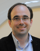
Volker J Schmid is a Professor of Bayesian Imaging and Spatial Statistics at the Department of Statistics, Ludwig Maximilian University of Munich. He leads the Bayesian Imaging and Spatial Statistics group and contributes to interdisciplinary initiatives like the Munich Center of Machine Learning. His work bridges statistical theory with applications in medical imaging and biology. PhD in Statistics (2004), LMU Munich Diploma in Statistics (2000), LMU Munich Abitur, Joseph-von-Fraunhofer-Gymnasium Cham (1993) His research focuses on Bayesian computational methods for high-dimensional data, particularly in medical imaging (MRI, DCE-MRI) and biological microscopy (e.g., 3D nuclear architecture analysis via super-resolution microscopy). Key applications include disease mapping , image segmentation , and spatio-temporal modeling . His software tools (e.g., nucim , bioimagetools , BAMP ) enable quantitative analysis in nuclear imaging and age-period-cohort modeling. His 15 most recent publications span Bayesian modeling for medical imaging , spatio-temporal epidemiology , and computational biology . Topics include co-localization metrics in fluorescence microscopy, nuclear architecture analysis, and dynamic Bayesian frameworks for MRI data. Collaborations extend to neuroimaging, oncology, and nuclear biology.
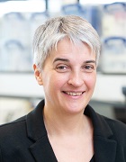
Prof. Dr.-Ing. Maria Francesca Spadea serves as Director of the Institute of Biomedical Engineering (IBT) at Karlsruhe Institute of Technology (KIT), part of the Helmholtz Association. Her leadership role includes overseeing research initiatives, teaching activities, and administrative responsibilities within the institute. Located in space 512, she maintains regular consultation hours on Wednesdays from 10:30-11:30 am by appointment. Professor Spadea's research spans several cutting-edge areas in biomedical engineering, with particular focus on medical image processing, artificial intelligence applications in healthcare, and radiomics. Her work bridges computational techniques with clinical applications, emphasizing practical solutions for medical imaging challenges. She has pioneered approaches in federated learning for medical image translation, particularly in CT/MRI synthesis for radiation therapy applications. Her research also extends to cancer cell analysis, vascular biomechanics, and medical robotics, demonstrating a broad yet cohesive research portfolio that addresses critical challenges in modern healthcare. Analysis of Professor Spadea's recent publications reveals a strong emphasis on AI-driven medical imaging solutions, particularly in the translation between different imaging modalities (like MRI-to-CT) using federated learning approaches that preserve patient privacy. Her work demonstrates growing specialization in radiation therapy applications, with multiple publications addressing synthetic CT generation for treatment planning. There's also a clear trajectory toward multi-institutional collaboration, as evidenced by her involvement in projects spanning multiple research centers across Europe. Professor Spadea actively mentors numerous students, including M. Krohmer Zabaleta, N. Skupien, and M. Destito, who have completed bachelor's and master's theses under her supervision. Her research group appears well-integrated within the broader Institute of Biomedical Engineering, collaborating extensively with colleagues like P. Zaffino and C.B. Raggio on multiple projects. The group maintains strong connections with clinical partners, as evidenced by publications addressing real-world medical challenges in radiation therapy, cardiology, and neurosurgery. The research activities of Professor Spadea's team are centered within the Institute of Biomedical Engineering at KIT, with particular focus on medical imaging processing and AI applications. Her laboratory appears to specialize in developing computational tools for medical image analysis, with recent work emphasizing privacy-preserving federated learning frameworks that enable multi-institutional collaboration without sharing sensitive patient data. The team maintains active collaborations with clinical departments, particularly in radiation oncology, as evidenced by numerous publications addressing CT synthesis for radiation therapy planning.
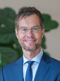
Dr. Benjamin de Haas is a vision scientist and faculty member at Justus Liebig University Giessen , Germany, within the Department of Psychology and Sports Science . He currently leads the ERC-funded Indivisual project and co-leads project C9 Factors influencing categorical face processing within the Collaborative Research Centre CRC/TRR 135. He is also a principal investigator in the NeurOscientific Workflow Assistance (NOWA) infrastructure project, dedicated to open, reproducible neuroscience. Research Focus Dr. de Haas pursues two intertwined questions: How do early and late stages of visual processing interact—from the initial registration of slanted edges to the recognition of faces? How and why do our perceptions differ from one person to the next? To answer these questions his group combines psychophysics, high-resolution eye-tracking, functional and quantitative MRI, and computational modelling, with a strong emphasis on face perception, individual differences, and naturalistic viewing conditions. Publications Overview Across more than 20 publications since 2016, Dr. de Haas has advanced understanding of individual differences in face processing, gaze control, and visual salience. His work repeatedly appears in Journal of Vision , Nature Communications , PNAS , and NeuroImage , highlighting a sustained focus on eye-movement behaviour, cortical representations of faces and scenes, and methodological best practices in neuroimaging. Current Supervision & Team Dr. de Haas currently supervises two PhD students: Elaheh Akbarifathkouhi Hilal Nizamoglu Together with Dr. Katharina Dobs (co-project leader) and affiliated post-docs and research technicians, the group forms the Indivisual laboratory at Giessen. Contact & Resources Email: Benjamin.de-Haas@psychol.uni-giessen.de Department of Psychology and Sports Science Otto-Behaghel-Str. 10F, 35394 Gießen, Germany
Prof. Dr. Florian Knoll is a full professor in Computational Imaging at the Department of Artificial Intelligence in Biomedical Engineering (AIBE) at Friedrich-Alexander-Universität Erlangen-Nürnberg. He leads the Computational Imaging Lab, focusing on machine learning applications in medical imaging, particularly accelerating MRI through innovative reconstruction algorithms and translating them into clinical practice. His research emphasizes improving MRI speed, artifact robustness, and accessibility, alongside developing quantitative biomarkers for disease processes. Knoll's work is funded by NIH grants, including projects on machine learning for musculoskeletal imaging, MR fingerprinting, and deep learning frameworks for MRI reconstruction. He is a key figure in open science initiatives, co-creating the fastMRI dataset with Facebook AI, providing public access to over 1300 knee and 7000 brain MRI scans. He currently serves as deputy editor of Magnetic Resonance in Medicine and chairs the ISMRM Reproducible Research Study Group. His contributions extend to reproducible research, maintaining GitHub repositories with code for image reconstruction techniques (e.g., AGILE, gpuNUFFT) and educational materials. He teaches medical imaging fundamentals at FAU, integrating theoretical and practical insights for students and researchers. Grants: NIH R01EB024532, R21EB027241, P41EB017183, R01EB029957 Labs/Teams: Computational Imaging Lab, fastMRI initiative Software: GitHub repositories for MRI reconstruction (e.g., github.com/FlorianKnoll )
Cornelius Faber is a University Professor in the Department of Radiology at the University of Münster, Germany, where he leads the Experimental Nuclear Magnetic Resonance research group. His work focuses on developing and implementing novel MRI techniques that extend the boundaries of magnetic resonance imaging in terms of spatial and temporal resolution, sensitivity, and specificity for physiological, structural, and molecular changes. He actively participates in the "Cells in Motion" interdisciplinary research initiative at the university. Professor Faber's research spans multiple critical areas in medical imaging and biomedical science. His primary expertise lies in MRI cell tracking , enabling visualization of cellular dynamics in vivo. He has made significant contributions to infection imaging , developing methods to detect and characterize microbial infections using MRI. His work on MR methodology development has advanced quantitative imaging techniques, while his research on multimodal integration in MR and MRI contrast mechanisms has provided deeper insights into molecular and cellular processes. His research bridges physics, engineering, and biomedical applications, with particular relevance to inflammation, cancer, neurological disorders, and cardiovascular disease. Analysis of Professor Faber's extensive publication record reveals a clear evolution from fundamental MRI technique development toward increasingly sophisticated applications in disease models. His recent work demonstrates a strong trend toward multimodal imaging approaches that combine MRI with complementary techniques such as mass spectrometry, optical imaging, and PET. This integration creates comprehensive diagnostic platforms that provide both anatomical and molecular information. A notable pattern is the focus on cellular dynamics, particularly immune cell behavior in inflammatory conditions and tumor microenvironments, with applications spanning neuroscience, oncology, and cardiology. Professor Faber leads a multidisciplinary research team of approximately 15 members, including scientists, doctoral students, technicians, and medical students. His laboratory is deeply integrated with the University of Münster's research infrastructure, particularly the Multiscale Imaging Centre. The group's work contributes significantly to advancing preclinical MRI methodologies while maintaining strong clinical relevance, with numerous publications in high-impact journals across medical imaging, neuroscience, and biomedical engineering disciplines.
Bjoern Menze is a Professor and Rudolf Mößbauer Tenure Track Chair at the Technical University of Munich (TUM), leading the Image-based Biomedical Modeling Group within the Munich School of Bioengineering. His research focuses on medical image computing, tumor growth modeling, and computational physiology, with applications in clinical neuroimaging and personalized radiotherapy design. He holds a Ph.D. in Computer Science from Heidelberg University and has held positions at ETH Zurich, INRIA Sophia Antipolis, MIT, and Harvard Medical School. His academic journey includes a postdoc at MIT’s CSAIL and Harvard Medical School, followed by roles at ETH Zurich and INRIA. His work bridges biomedical imaging with machine learning, emphasizing model-driven analysis of physiological processes. He has been a visiting professor at Maastricht University and contributes to initiatives like the Center for Translational Cancer Research at TUM. Key research areas include tumor growth modeling, quantitative imaging biomarkers, and integrating mathematical models with clinical data. His awards include the MICCAI Young Scientist Award (2014), Leopoldina Fellowship (2009), and DFG Research Fellowship (2008). He advises on medical AI, leads interdisciplinary projects, and publishes extensively in top journals like Nature Neuroscience and IEEE Transactions on Medical Imaging. His lab’s work spans applications such as glioblastoma radiotherapy optimization, whole-body bone lesion detection, and neural connectivity imaging. Collaborations include institutions like Harvard, MIT, and ETH Zurich. He emphasizes translating computational methods into clinical practice for personalized healthcare solutions.
Univ.-Prof. Dr. Dr. hc NJ Shah is a prominent academic and researcher in medical imaging physics. He serves as the Institute Director of the Institute of Neuroscience and Medicine – Medical Imaging Physics (INM-4) at Forschungszentrum Jülich and holds a Professorship in the Department of Neurology at RWTH Aachen University. He also co-directs the Jülich-Aachen Research Alliance (JARA-Brain). His research focuses on advanced MRI techniques, multimodal neuroimaging, and applications in neuro-oncology and mental health. Shah has held roles such as Distinguished Professor at Monash Institute of Medical Engineering (2015–2017) and has been recognized with awards including the Veski Award and Honorary Doctorate from the Georgian Technical University. Education: PhD (1987, University of Manchester), Diploma in Advanced Studies in Science (1984, Manchester), BSc (1983, University of Sheffield). Research interests include MRI physics, ultra-high-field imaging (7T/9.4T), brain tumor imaging, and neuroimaging data science. His work bridges clinical and experimental MRI, with contributions to quantitative water content mapping and multimodal integration of MRI-PET-EEG. His publications highlight advancements in neuroimaging methodologies and their applications in understanding neurological and psychiatric conditions. Awards and honors include Fellowships from the Royal Society of Chemistry, Royal Society of Medicine, and Institute of Physics. Shah leads the MR Physics team at INM-4 and collaborates internationally, including roles at Maastricht University and the University of New Brunswick. His work emphasizes translating imaging innovations into clinical practice for precision medicine.
Max Planck Institute for Molecular GeneticsGermany
Despina Kontos, PhD is the Herbert and Florence Irving Professor of Radiological Sciences at Columbia University Irving Medical Center (CUIMC), with appointments in the Department of Radiology and the Herbert Irving Comprehensive Cancer Center. She serves as the Chief Research Information Officer for CUIMC, Vice Chair of Artificial Intelligence and Data Science Research in the Department of Radiology, and Director of Biomarker Imaging at NewYork-Presbyterian Hospital. Additionally, she holds appointments in the Departments of Biomedical Informatics and Biomedical Engineering. Dr. Kontos received her educational training from prestigious institutions: BS in Engineering from the University of Patras, Greece MSc and PhD in Computer and Information Sciences from Temple University Postdoctoral training in Radiology at the University of Pennsylvania Certificates in Biostatistics and Epidemiology from UPenn, Cancer Biology from Harvard, and AI for Decision Making from Wharton As a computer scientist with expertise in artificial intelligence and machine learning, Dr. Kontos focuses on developing computational methodologies to leverage imaging as quantitative biomarkers for personalized disease prediction, particularly in cancer. Her research program investigates how imaging data can be mined to extract sophisticated phenotypic signatures with diagnostic, prognostic, and predictive value. While her primary focus has been on breast cancer, her lab also pursues related research in lung cancers, evaluating the integration of CT radiomic features with liquid biopsy data to characterize tumor heterogeneity. Dr. Kontos founded and directs Columbia University's Center for Innovation in Imaging Biomarkers and Integrated Diagnostics (CIMBID), a multidisciplinary center dedicated to developing and integrating quantitative imaging and non-imaging biomarkers for personalized disease prediction. Through CIMBID, she has built a vibrant scientific ecosystem that brings together expertise across Columbia's campuses, linking basic science, engineering, clinical medicine, public health, and health services research. Analysis of Dr. Kontos's publication record reveals a strong focus on applying AI and machine learning to biomedical imaging, particularly for cancer risk prediction and personalized treatment. Her work demonstrates a progression from foundational methodological development to clinical translation, with increasing emphasis on multi-modal biomarker integration. Recent publications show expansion into new disease areas including Alzheimer's disease prediction, while maintaining her strong focus on breast and lung cancer applications. Dr. Kontos has received significant recognition for her contributions to the field: Academy for Radiology and Biomedical Imaging Research Distinguished Investigator Award (2020) Eastern Cooperative Oncology Group - American College of Radiology Imaging Network ECOG-ACRIN Young Investigator Award of Distinction for Translational Research (2014) Dr. Kontos has been highly successful in securing research funding, with numerous grants from federal agencies including the National Institutes of Health (NIH) and the Department of Defense (DOD), as well as private foundations such as the American Cancer Society (ACS) and the Radiological Society of North America (RSNA). Her leadership extends to mentoring students and postdoctoral researchers through her roles at CIMBID and the Department of Radiology. As the founding director of CIMBID, Dr. Kontos leads a multidisciplinary team that includes the Computational Imaging Biomarker Group (CBIG), the Laboratory of AI and Biomedical Science (LABS), and several other affiliated research labs. The center leverages Columbia's institutional strengths in engineering, data science, and clinical medicine to advance personalized healthcare through AI and imaging technologies.
Dr. Markus Zimmermann is a researcher at the Institute of Neuroscience and Medicine (INM-4: Physics of Medical Imaging) at the Research Center Jülich. His work focuses on advancing quantitative MRI techniques, particularly in water content mapping, multiparametric imaging, and ultrahigh-field MRI applications. He contributes to developing methods for eddy current characterization, multi-exponential relaxometry, and rapid whole-brain protocols. His research addresses neurological and medical imaging challenges, including cerebral pathologies and neurobiological implications. Key areas of expertise include MRI parameter estimation, medical imaging algorithms, and the integration of advanced imaging techniques for clinical and neuroscience applications. His projects often involve collaborations to validate methodologies using in vivo/ex vivo experiments and super-resolution reconstruction. Dr. Zimmermann’s work aims to enhance diagnostic precision and understanding of brain physiology through innovative MRI technologies.
Prof. Dr. Julia Schnabel is the TUM Liesel Beckmann Distinguished Professor and Helmholtz Distinguished Professor at TUM's TUM School of Computation, Information and Technology. Her research focuses on computational imaging and AI in medicine, including medical image processing, machine learning, motion modeling, and quantitative imaging. She holds IEEE, Ellis, and MICCAI Society fellowships, and has pioneered work in image reconstruction, artifact correction, and AI-based diagnostics. Educations: Bachelor/Master from TU Berlin (1993) PhD from University College London (1998) Postdocs at UMC Utrecht, King's College London, and UCL Her research interests span medical AI, deep learning for medical imaging, and clinical evaluation methodologies. Key contributions include frameworks for motion artifact correction in MRI, physics-informed neural networks, and benchmark datasets like NOVA for anomaly detection in brain MRI. She has authored over 100 publications, with recent work advancing unsupervised anomaly detection and federated learning in healthcare. Prof. Schnabel leads interdisciplinary projects at TUM and Helmholtz Zentrum München, focusing on AI-driven solutions for diagnostic and therapeutic challenges. Her labs develop tools for real-time cardiac imaging, histopathology segmentation, and trustworthy AI guidelines (FUTURE-AI initiative).
Prof. Dimitrios Karampinos is a Professor at the Technical University of Munich (TUM), leading the Experimental Magnetic Resonance Imaging group within the TUM School of Medicine and Health. He specializes in developing novel MRI techniques for quantitative biomarker discovery, focusing on musculoskeletal, metabolic, and oncological applications. His career includes a PhD from the University of Illinois (2008), postdoctoral research at UCSF (2009–2012), and leadership roles at TUM since 2012. Prof. Karampinos has pioneered advancements in MRI reconstruction, signal modulation, and biomarker validation for clinical translation. Educations: BSc in Mechanical Engineering (National Technical University of Athens, Greece), PhD in Biomedical Engineering (University of Illinois, Urbana-Champaign, 2008). Research Interests: Development of MRI measurement techniques, quantitative biomarkers for disease diagnosis, and improving therapy monitoring. Key areas include musculoskeletal disease imaging, metabolic disorder assessment, and oncology applications. His work emphasizes translating research into clinical practice through innovations like accelerated imaging, artifact correction, and AI-driven analysis. Awards: ERC Starting and Proof of Concept Grants (2015, 2019), TUM Supervisory Award (2020), ISMRM Junior Fellow (2011). Grants: Multiple ERC grants for MRI method development. Labs/Teams: Leads the Experimental Magnetic Resonance Imaging group at TUM, collaborating on clinical and technical MRI advancements.
Hannah Spitzer is a Research Group Leader at the Institute for Stroke and Dementia Research (ISD) at Ludwig Maximilian University of Munich and an associated Research Group Leader at Helmholtz Munich's Computational Health Center. She leads the Spitzer Lab, focusing on computational analysis of multimodal brain datasets to advance understanding of neurovascular and neurodegenerative diseases. Her educational background includes: PhD in Computer Science from Heinrich-Heine University Düsseldorf and Research Center Jülich (2015-2020) Master's in Computer Science from RWTH Aachen (2013-2015) Bachelor's in Computer Science from RWTH Aachen (2009-2013) Dr. Spitzer's research integrates computational biology and machine learning to decode brain complexity, with emphasis on spatial omics analysis , interpretable image representation learning , and cross-modal data integration . Her group develops tools like squidpy and campa for spatial omics while applying graph neural networks to epilepsy lesion detection through the international MELD project, prioritizing biological interpretability in AI models. Recent publications reveal strong trends in leveraging graph neural networks for subtle brain lesion detection and creating computational frameworks for spatial omics integration. Her work consistently bridges advanced machine learning with clinical neuroscience to uncover disease mechanisms in neurodegeneration and vascular disorders. Dr. Spitzer actively mentors students including current PhD candidate Beatrice Guastella and alumni Deniz Fettahoglu (MSc) and Katia Berr (PhD). Her lab operates through major collaborations including the MELD epilepsy consortium and Helmholtz Imaging Project, with funding supporting computational pipeline development for small-vessel disease prediction and multimodal brain atlasing. The Spitzer Lab comprises postdoc Wasim Aftab and PhD student Beatrice Guastella, working on computational pipelines that integrate histology, spatial omics, and neuroimaging data to decode brain disease mechanisms through interpretable AI approaches.
Max Planck Institute for Human Cognitive and Brain SciencesGermany
Harald E. Möller is a Professor and Head of the Nuclear Magnetic Resonance Research and Development Unit at the Max Planck Institute for Human Cognitive and Brain Sciences in Leipzig. With a career spanning over four decades, he has held academic positions including Honorary Professor at the University of Leipzig and leadership roles in institutions like Duke University Medical Center and the University of Münster. His research focuses on advancing MRI methodologies, biophysical imaging principles, and their applications in neurology and neuroscience. Education: 1979-1985: Chemistry & Physics studies at Universities of Dortmund and Münster 1985: M.Sc. (Diploma) in Chemistry 1988: PhD in Physical Chemistry (summa cum laude) 2000: Habilitation in Physical Chemistry 2002: Habilitation in Biophysical Chemistry Research Interests: Development of novel MRI methods Quantitative tissue characterization Myelin sheath imaging Cerebral blood flow dynamics High-field MRI hardware
Professor Martin Bendszus serves as the Medical Director of the Department of Neuroradiology at Heidelberg University Hospital. He has held this position since 2007 and is a leading figure in advanced neuroimaging techniques. His academic career includes medical studies in Bonn, specialized training in Neuroradiology at the University of Würzburg (2003-2007), and a professorship in Neuroimaging at the University of Würzburg. Professor Bendszus's research focuses on innovative imaging methods, particularly in Magnetic Resonance Imaging (MRI). He pioneered Magnet Resonance Neurography for diagnosing peripheral nervous system disorders and made significant contributions to Dental-MRI as a radiation-free diagnostic tool for dental conditions. His expertise also extends to brain and spinal cord diagnostics, including aneurysms, strokes, and arteriovenous malformations. His work bridges clinical practice with advanced imaging research, resulting in numerous high-impact publications across neurology, radiology, and oncology. His publication record shows consistent high-impact contributions, with recent work spanning stroke intervention, neuropathic pain, brain tumor imaging, and advanced MRI techniques. The research trends demonstrate his leadership in translating imaging advances into clinical practice, particularly in time-sensitive neurological conditions where imaging guides critical treatment decisions. Kurt-Decker-Preis Röntgen-Preis Coolidge-Award Lucien-Appel-Award Hermann-Holthusen-Ring der Deutschen Röntgengesellschaft Professor Bendszus leads major multicenter clinical trials and collaborative guideline development efforts, including work with the Response Assessment in Neuro-Oncology (RANO) group and European Association for Neuro-Oncology (EANO). His research has secured significant funding for advancing neuroimaging techniques and their clinical applications. He collaborates extensively with neurologists, neurosurgeons, oncologists, and radiologists to improve diagnostic and therapeutic approaches for neurological conditions. His department serves as a reference center for complex neuroradiological cases and trains the next generation of neuroradiologists. Professor Bendszus maintains active leadership roles in professional societies and regularly contributes to shaping clinical guidelines in neuroimaging and stroke care.
Prof. Fritz Schick is a faculty member at the University of Tübingen , serving as the Deputy Head of the Division 'Pathophysiology of Prediabetes' at the Helmholtz Diabetes Center. He also holds a permanent professorship as Head of the Section on Experimental Radiology within the Department of Diagnostic and Interventional Radiology at Tuebingen University Hospital. His work focuses on developing non-invasive methods to characterize tissue composition and function in pre-diabetes and diabetes. M.D. (1989) and Physics (1990) graduate from University of Tübingen Research Interests : Schick's research spans quantitative MRI and MRS for applications in musculature, liver, adipose tissue, and bone marrow . His work bridges medical physics with metabolic disease analysis , emphasizing non-invasive diagnostics and cohort studies. Publication Trends : Recent articles highlight his expertise in MRI-based quantification of metabolic tissues, including studies on visceral adipose tissue distribution (German National Cohort), hepatokines in NAFLD, and deep learning for diabetes detection from whole-body MRI. Collaborations span radiology, endocrinology, and computational biology. Scientific Awards : Technologie-Transfer-Handwerk Professor Adalbert Seifriz-Preis (2003) Stipend of Siemens AG, Ernst-von-Siemens-Stipend (1992-1994) Dr.-Friedrich-Förster-Award, Department of Physics (1991) Affiliations : Active in the Helmholtz Diabetes Center and German Center for Diabetes Research , with leadership roles in experimental radiology and metabolic imaging.