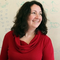
Polina Golland is a Professor in the Department of Electrical Engineering and Computer Science (EECS) at MIT and a Principal Investigator in the Computer Science and Artificial Intelligence Laboratory (CSAIL). Her research focuses on developing novel techniques for biomedical image analysis and understanding, particularly in medical vision, AI/ML, and health care applications. She leads the Medical Vision Group and collaborates with the Vision Group at CSAIL. Her work emphasizes statistical modeling of medical images, shape modeling, and predictive analytics for biological processes. Current projects include fetal MRI analysis, cardiac MRI segmentation, and quantitative assessment of pulmonary edema in chest X-rays. She has secured grants from NIH, MIT-IBM Watson AI Lab, and other institutions to support her research. Dr. Golland teaches courses on inference, probability, and probabilistic systems. She advises graduate students in MIT's EECS program and has mentored numerous postdocs and researchers. Her lab focuses on translating advanced imaging techniques into clinical workflows, with applications in neuroimaging, fetal health monitoring, and cardiovascular disease analysis. Notable collaborations include work with Harvard Medical School affiliates, Brigham and Women's Hospital, and the MIT Jameel Clinic. Her research aims to bridge computational methods with clinical needs, improving diagnostic tools and treatment planning through machine learning and medical imaging innovation.
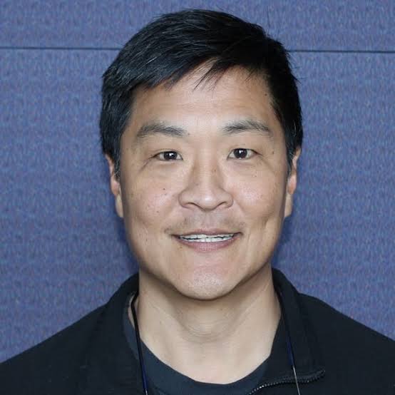
Edward S. Awh is a Professor in the Department of Psychology at the University of Chicago, affiliated with The Institute for Mind and Biology and the Grossman Institute for Neuroscience, Quantitative Biology and Human Behavior. His research focuses on behavioral and neural studies of memory and attention, employing psychophysics, EEG, and functional MRI. His lab investigates neural mechanisms underlying cognitive processes and their interrelationships. Research interests include tracking the contents of online memories and the locus of covert attention using neural decoding techniques. The lab collaborates on projects involving attention and memory studies, as detailed on their website: Awh/Vogel Lab . The lab currently seeks postdoctoral researchers to join ongoing projects using behavioral, EEG, and fMRI methodologies. Contact via awhvogellab@gmail.com .
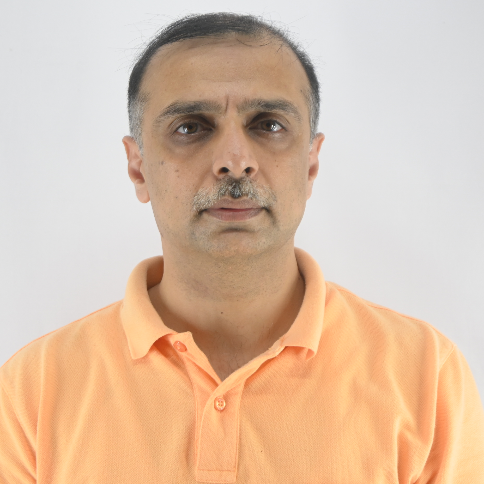
Ajit Rajwade is a Professor at the Department of Computer Science and Engineering , Indian Institute of Technology Bombay. His research spans Artificial Intelligence , Compressed Sensing , and Medical Imaging , with affiliations to the Centre for Machine Intelligence and Data Science and the Koita Centre for Digital Health . Education : PhD in Computer and Information Science and Engineering, University of Florida (2010) MSc in Computer Science, McGill University (2004) BTech in Computer Engineering, University of Pune (2002) His research interests focus on intelligent data acquisition, particularly in neural network analysis , graph signal processing , and medical imaging . He develops compressed sensing algorithms for inverse problems like tomography and MRI reconstruction , alongside applying group testing to pandemic response. Scientific awards include the Prof. S. P. Sukhatme Award (2024) and Departmental Teaching Excellence (2019). His publications demonstrate expertise in image restoration , noise modeling , and epidemiological algorithms . He has advised PhD students like Sabyasachi Ghosh and Jerin Geo James , with a focus on computationally efficient methods in medical imaging and machine learning .
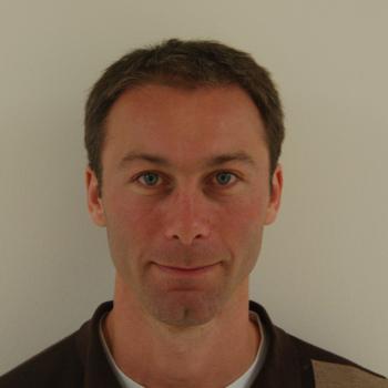
Jeremy Dahl is a Professor of Radiology (Pediatric Radiology) at Stanford University School of Medicine. He directs the Ultrasound Imaging & Instrumentation Lab and serves as Director of Research Academic Affairs in the Department of Radiology since 2020. He holds multiple affiliations across Stanford including Bio-X, the Cardiovascular Institute, Wu Tsai Human Performance Alliance, Maternal & Child Health Research Institute, Stanford Cancer Institute, and Wu Tsai Neurosciences Institute. Dr. Dahl received his B.S. in Electrical Engineering from the University of Cincinnati (1999) and Ph.D. in Biomedical Engineering from Duke University (2004). His research focuses on developing ultrasonic beamforming and image reconstruction methods for diagnostic imaging applications, particularly techniques that generate high-quality images in difficult-to-image patients. His laboratory specializes in B-mode and Doppler imaging techniques that utilize additional information from ultrasonic wavefields to improve image quality and develop real-time imaging systems for clinical applications including cardiac, liver, and fetal imaging. Dr. Dahl's research has led to significant advancements in ultrasound molecular imaging platforms, sound speed estimation, aberration correction, and reverberation noise suppression. His work often bridges engineering innovation with clinical applications for cancer detection and other diseases. His recent publications demonstrate strong focus on machine learning applications in ultrasound, distributed aberration correction, and molecular imaging techniques. Fellow, American Institute of Ultrasound in Medicine (2021) Senior Member, Institute of Electrical and Electronics Engineers (2020) Distinguished Investigator Award, The Academy for Radiology & Biomedical Imaging Research (2018) Outstanding Paper Award, IEEE Ultrasonics, Ferroelectrics, and Frequency Control Society (2011) Dr. Dahl serves in editorial roles for major journals including IEEE Transactions on Medical Imaging (2017-2024) and IEEE Transactions on Ultrasonics, Ferroelectrics, and Frequency Control (2013-Present). His laboratory has successfully translated numerous innovations into clinical applications, with multiple patents including recent developments in pulsed focused ultrasound therapy and speed of sound quantification.
Dr. Christina Allen is a Professor of Orthopaedics & Rehabilitation at Yale School of Medicine, where she serves as Chief of Yale Sports Medicine and Vice Chair of Athletic Medicine and Community Outreach. She is the Orthopaedic Team Physician for Yale Athletics and has extensive experience with U.S. national teams, including U.S. Soccer and USA Taekwondo. Education: BS in Biomedical Engineering, Duke University (1983); MD, UCLA School of Medicine (1995) Training: Orthopaedic Residency and Sports Medicine Fellowship, University of Pittsburgh Research Interests focus on: Revision ACL reconstruction outcomes and predictors Meniscus transplantation and cartilage repair Proximal hamstring avulsion repair Quantitative MRI for joint kinematics and cartilage changes Post-ACL revision infection risk factors Scientific Awards include: Kappa Delta Ann Doner Vaughn Award (2019) NIH R01 Competitive Renewal Grant (2017) AOSSM O’Donoghue Award (2014) Clinical Roles span: Team physician for U.S. Soccer (Women's/Men's National Teams) Former team physician for San Francisco Deltas (NASL Champions 2017) Medical Board member, World Taekwondo (London Olympics 2012, Rio Olympics 2016) Publications highlight a 20-year longitudinal focus on: Machine learning models for surgical outcome prediction Graft choice impact on revision ACL longevity Cartilage degeneration after meniscectomy Biomechanical MRI analysis of joint kinematics Infection rates in revision surgeries Return-to-sport protocols for athletes
Craig H. Meyer is a Professor in Biomedical Engineering and Radiology & Medical Imaging at the University of Virginia. He holds a Ph.D. from Stanford University and leads the Rapid MRI Research Group, focusing on developing advanced MRI techniques for cardiovascular disease, neural disorders, and pediatrics. His work integrates physics, signal processing, and machine learning to improve MRI acquisition and processing speed. Education: Ph.D. in Biomedical Engineering, Stanford University. Research Interests: Medical and Molecular Imaging, Signal and Image Processing, Biomedical Data Sciences, Biomechanics, and Cardiovascular Engineering. His innovations include fast spiral imaging, conjugate phase reconstruction, and machine learning-enhanced MRI denoising. Awards: Notably includes the Dean’s Award for Excellence in Team Science (2014), Fellowships from NAI (2021), AIMBE (2015), and ISMRM (2013). He also authored two landmark MRI papers recognized as pivotal in the field. Teaching: Courses include BME 6310 (Computation and Modeling in Biomedical Engineering) and BME 8782 (Magnetic Resonance Imaging). He emphasizes translational research, with applications in clinical MRI advancements and collaborative interdisciplinary projects. Labs/Groups: Rapid MRI Research Group focuses on cutting-edge MRI technologies, including real-time cardiac imaging and artifact reduction through deep learning.
Massimo Mischi is a Full Professor at the Faculty of Electrical Engineering of the Eindhoven University of Technology (TU/e) and chairs the Signal Processing Systems (SPS) Division , the largest division at TU/e with over 250 researchers. He founded the Biomedical Diagnostics (BM/d) Lab in 2012, which now includes 180 researchers and clinical/industrial advisors, focusing on biomedical signal processing for diagnostics and monitoring.
Daan Christiaens is a tenure track lecturer at KU Leuven's Faculty of Medicine and Faculty of Engineering Sciences. He is affiliated with the Department of Electrical Engineering (ESAT) and Department of Imaging & Pathology, serving as a member of the Medical Imaging Division and the KU Leuven Brain Institute (LBI). His academic responsibilities include membership in the Faculty Councils of Engineering Sciences and Medicine. His research focuses on: Inverse problems in medical imaging reconstruction Neuroimaging techniques for brain analysis Advanced quantitative MRI methodologies Diffusion-weighted imaging for microstructural assessment Dr. Christiaens' recent publications (2023-2025) demonstrate a consistent focus on diffusion MRI innovations, including novel reconstruction algorithms, neonatal brain development mapping, and clinical applications for neurodegenerative disorders. Key technical themes include motion correction, multi-shell modeling, and AI-enhanced image processing, while clinical applications span Alzheimer's disease, cerebral palsy, and autism research. He leads significant research projects including: MRI reconstruction with dynamic field monitoring (2024-2028) Compressed sensing for microstructure imaging (2022-2026) Neonatal diffusion MRI network connectivity analysis (2024-2028) As a core developer of the MRtrix3 software framework for medical image processing, he contributes to essential tools in neuroimaging research.
Qian Tao is an Assistant Professor at the Department of Imaging Physics , Faculty of Applied Sciences , Delft University of Technology . She previously worked at the Division of Image Processing, Department of Radiology, Leiden University Medical Center from 2009 to 2020. Academic Background: BSc in Electrical Engineering (Fudan University), MSc in Biomedical Engineering (Fudan University), PhD in Biometric Authentication (University of Twente) Research Interests: Focus on trustworthy AI methodologies for critical healthcare applications, including medical imaging for patient diagnosis and clinical intervention. Specializes in cardiac MRI analysis, image-guided interventions for cardiac arrhythmias, and AI in Radiology. Publication Trends: Recent work emphasizes motion correction in cardiac MRI, deep learning for image registration, and novel techniques like TRAFF2 mapping. Keywords include Medical Imaging , Machine Learning , Cardiac MRI , and Quantitative Analysis . Contact: Email: Q.Tao@tudelft.nl
Dr. Ian Wilson is a researcher at Newcastle University with a focus on medical genetics, nephrology, and genomic analysis. His work spans genetic determinants of kidney diseases, mitochondrial disorders, and biomarker development. Notable contributions include studies on uromodulin genetics in African populations, copy-number variations in rare diseases, and kidney ciliopathies. He has collaborated extensively on projects involving genome sequencing, mitochondrial replacement therapy, and muscular dystrophy biomarkers. Wilson's research integrates computational tools like machine learning for predictive modeling in urolithiasis and employs advanced imaging techniques for disease progression monitoring. Key areas: Genetic epidemiology, renal genomics, mitochondrial DNA analysis Focus on translational applications: Biomarker development for kidney stones and muscular dystrophies Interdisciplinary collaborations in ophthalmology and orthopedics His publications reflect a commitment to advancing diagnostic accuracy and understanding complex genetic disorders through multi-omics approaches.
Lawrence Staib is Professor of Radiology and Biomedical Imaging, Biomedical Engineering, and Electrical Engineering at Yale University. He serves as Director of Undergraduate Studies in Biomedical Engineering and is a member of Yale's Bioimaging Sciences division, Image Processing & Analysis Group, Yale Biomedical Imaging Institute, and Yale-BI Biomedical Data Science Fellowship program. Dr. Staib earned his A.B. in Physics from Cornell University (1982), followed by a Ph.D. in Engineering and Applied Science from Yale University (1990), and completed a postdoctoral fellowship at Yale School of Medicine (1991). His research focuses on developing advanced medical image analysis methods using machine learning and model-based approaches. Key research areas include neuroimaging applications for autism spectrum disorder classification, cardiac imaging analysis for strain and motion assessment, prostate cancer diagnosis and risk mapping, and innovative techniques for medical image segmentation with limited labeled data. Dr. Staib's work emphasizes uncertainty estimation in deep learning models, multi-modal image registration, and domain adaptation techniques to improve clinical decision support systems. His recent publications demonstrate a strong trend toward developing interpretable AI models for clinical applications, with particular emphasis on fMRI analysis for neurological conditions, cardiac motion analysis, and prostate cancer diagnosis. His work frequently addresses the challenge of limited labeled data in medical imaging through innovative self-supervised, semi-supervised, and few-shot learning approaches. Fellow of the American Institute for Medical and Biological Engineering (AIMBE) (2015) Distinguished Investigator Award from the Academy for Radiology & Biomedical Imaging Research (2017) MICCAI Fellow (2022) Medical Image Analysis Second Best MICCAI Paper Award (2005) ASNR Cum Laude Scientific Exhibit Award (2003) Dr. Staib serves on the editorial board of Medical Image Analysis and as Associate Editor of IEEE Transactions on Biomedical Engineering. His research is supported by NIH grants including the Autism Center of Excellence program. He leads the Image Processing & Analysis Group within Yale's Bioimaging Sciences division, collaborating extensively with James Duncan, John Onofrey, Xenophon Papademetris, and other Yale researchers on applications spanning neuroimaging, cardiology, and oncology. Current projects focus on developing robust AI models for clinical decision support with emphasis on uncertainty quantification and interpretability.
Dr. Craig S. Levin is a Professor of Radiology at Stanford University's Molecular Imaging Program at Stanford (Nuclear Medicine), with courtesy appointments in Physics, Electrical Engineering, and Bioengineering. He also holds memberships in Bio-X, the Cardiovascular Institute, the Wu Tsai Human Performance Alliance, and the Stanford Cancer Institute. Dr. Levin received his B.S. Summa Cum Laude in Physics and Mathematics from UCLA in 1985, followed by M.S., M.Phil., and Ph.D. degrees in Physics from Yale University in 1987 and 1993. His educational achievements were recognized with multiple honors including Phi Beta Kappa, Sigma Pi Sigma, and various departmental awards at UCLA. Dr. Levin's research focuses on the development of novel instrumentation and software algorithms for molecular imaging. His work spans medical physics, biomedical engineering, and instrumentation development with specific emphasis on positron emission tomography (PET), gamma camera technology, and multimodal imaging systems. His laboratory explores new concepts in radiation detection, image reconstruction algorithms, and the application of these technologies to cancer, heart disease, and neurological disorders. A notable aspect of his research involves pushing the physical limits of sensitivity and spatial, spectral, and/or temporal resolutions in imaging systems. His recent publications demonstrate a strong focus on enhancing PET technology, particularly time-of-flight capabilities, with significant work on improving coincidence timing resolution, developing MR-compatible PET systems, and applying deep learning techniques to image reconstruction and normalization. His research shows a clear trajectory toward higher resolution imaging with improved quantitative accuracy for both clinical and preclinical applications. Dr. Levin's scientific achievements have been recognized with numerous awards: American Institute for Medical and Biological Engineering's College of Fellows Academy of Radiology Research Distinguished Investigator Recognition Award National Research Service Award from NIH (1993-5) Pilot Research Award from the Society of Nuclear Medicine (1996) Multiple honors from UCLA including Phi Beta Kappa and Sigma Pi Sigma Full Tuition and Research Fellowship and Bates Graduate Fellowship from Yale University As an educator and mentor, Dr. Levin directs the NIH-NCI funded T32 Stanford Molecular Imaging Scholars postdoctoral training program and serves as a Doctoral Dissertation Advisor for students in Bioengineering and Biophysics. He currently advises five postdoctoral scholars and three doctoral candidates. His laboratory, the Molecular Imaging Instrumentation Laboratory, comprises approximately 20 members who work on developing new imaging technologies and translating them into clinical applications. Dr. Levin has secured substantial NIH funding as Principal Investigator along with grants from other government agencies, industry partners, and private institutions to support his research program. Dr. Levin's Molecular Imaging Instrumentation Laboratory is at the forefront of developing new imaging technologies that bridge physics, engineering, and medicine. The lab focuses on creating instrumentation for in vivo imaging of cellular and molecular signatures of disease, with particular emphasis on pushing the physical limits of imaging performance. Their work spans computer modeling, sensor development, electronics design, data acquisition systems, and advanced image processing algorithms. The lab maintains strong industry partnerships to translate their innovations into products used for patient care worldwide.
Anthony A Gatti is a Postdoctoral Scholar at Stanford University's Wu Tsai Human Performance Alliance and School of Medicine. His research integrates biomechanics , medical imaging , and machine learning to advance musculoskeletal health diagnostics, particularly focusing on knee osteoarthritis and exercise physiology. Education : Ph.D. in Rehabilitation Science (McMaster University, 2021), M.Sc. in Rehabilitation Science (McMaster University, 2015), B.Sc. in Kinesiology (McMaster University, 2013) His research develops automated tools for quantifying knee anatomy and integrating anatomical data with biomechanical models . These methods analyze acute responses to exercise and long-term joint degeneration, leveraging MRI , deep learning , and statistical shape modeling . Recent publications emphasize AI-driven segmentation , exercise-induced cartilage changes , and biomechanical simulations , spanning journals like Magnetic Resonance in Medicine and Arthritis & Rheumatology . Trends include machine learning validation for clinical predictions and open-source tool development for musculoskeletal analysis. Scientific Awards : CIHR Postdoctoral Fellowship (top 1%), Mitacs Accelerate Entrepreneur, Forge Student Start-Up Competition Winner, multiple scholarships from McMaster University He founded NeuralSeg , a company commercializing deep learning-based MRI segmentation technology. Collaborations include Stanford's Digital Athlete Moonshot Project with advisors like Scott Delp and Garry Gold.
Steven Meikle is a Professor of Medical Imaging Physics and Head of the Imaging Physics Laboratory at the Brain and Mind Centre, University of Sydney. He also serves as Deputy Director (Preclinical) of Sydney Imaging and Deputy Director of the National Imaging Facility's Sydney node. His expertise spans advanced imaging technologies, with a focus on PET/SPECT instrumentation and molecular imaging. He holds a B.App.Sc.(Hons) from the University of Technology Sydney and a PhD from the University of New South Wales. Research focuses include developing novel PET systems like Open-field PET (for freely moving rodents) and Total Body PET, which enhance imaging sensitivity and enable real-time behavioral studies alongside brain function analysis. Collaborations include Tsinghua University (China) and UC Davis (USA). He leads projects on motion correction, quantitative imaging, and AI-driven analysis. Key achievements include over 180 peer-reviewed publications, editorial roles in Physics in Medicine and Biology , and leadership in professional societies. Awards include IEEE Senior Membership and Australian Institute of Physics Fellowship. Current student projects explore Total Body PET applications, motion correction, and radiopharmaceutical evaluation. Teaching roles include medical physics courses in diagnostic radiography and medical physics programs. He advises on imaging ethics, facility implementation, and translational research bridging basic science and clinical applications.
Zhi-Pei Liang is the Franklin W. Woeltge Professor in the Department of Electrical and Computer Engineering at the University of Illinois at Urbana-Champaign, with joint appointments in the Department of Bioengineering, Beckman Institute for Advanced Science and Technology, and Coordinated Science Laboratory. His research spans biomedical engineering, medical imaging, and signal processing with a focus on advancing magnetic resonance imaging and spectroscopy technologies. His educational background includes a Ph.D. in Biomedical Engineering from Case Western Reserve University (1989) and a B.S. in Electrical Engineering from South-China University of Technology (1982), followed by postdoctoral training at UIUC (1989-1991). Professor Liang's research interests center on magnetic resonance imaging and spectroscopy , with particular emphasis on ultrafast imaging techniques , model-based reconstruction methods , and the integration of physics-based modeling with machine learning . His pioneering work on SPICE (SPectroscopic Imaging by exploiting spatiospectral CorrElation) has revolutionized high-resolution metabolic brain imaging by enabling label-free molecular imaging through the marriage of spin physics and machine learning. His research spans pattern recognition, parameter estimation, image formation theory, and algorithms for medical imaging applications. Analysis of his recent publications reveals a strong focus on high-resolution metabolic imaging , particularly using SPICE methodology to map brain metabolism with unprecedented detail. His work bridges fundamental physics of magnetic resonance with advanced computational methods to overcome traditional limitations in imaging speed and resolution. Current research directions include J-resolved spectroscopic imaging, deuterium-based metabolic mapping, and multimodal integration of PET and MRSI for studying neurological disorders. Elected to International Academy of Medical and Biological Engineering (2012) Gold Medal, International Society for Magnetic Resonance in Medicine (2022) Technical Achievement Award, IEEE Engineering in Medicine and Biology Society (2014) Fellow, National Academy of Inventors (2021) Author of influential book 'Principles of Magnetic Resonance Imaging' (1999) President of IEEE Engineering in Medicine and Biology Society (2011-2012) Professor Liang has advised numerous students and postdocs in biomedical imaging research and has received multiple teaching honors including the Ronald W. Pratt Outstanding Teaching Award (2005) and multiple listings among UIUC's Excellent Teachers. His research has been supported by various grants from NIH, NSF, and other funding agencies. He leads the SPICE (Spectroscopic Imaging by exploiting spatiospectral Correlation) research group which focuses on developing novel imaging techniques that combine physics-based modeling with machine learning for ultrafast metabolic imaging. His laboratory, part of the Beckman Institute's Integrative Imaging Theme, collaborates extensively with clinical researchers at Carle Illinois College of Medicine and other institutions to translate advanced imaging techniques into clinical applications for neurological disorders, cancer, and metabolic diseases. Current projects focus on high-resolution mapping of brain metabolism in Alzheimer's disease, stroke, and brain tumors using novel MR spectroscopic imaging techniques.