
Volker J Schmid is a Professor of Bayesian Imaging and Spatial Statistics at the Department of Statistics, Ludwig Maximilian University of Munich. He leads the Bayesian Imaging and Spatial Statistics group and contributes to interdisciplinary initiatives like the Munich Center of Machine Learning. His work bridges statistical theory with applications in medical imaging and biology. PhD in Statistics (2004), LMU Munich Diploma in Statistics (2000), LMU Munich Abitur, Joseph-von-Fraunhofer-Gymnasium Cham (1993) His research focuses on Bayesian computational methods for high-dimensional data, particularly in medical imaging (MRI, DCE-MRI) and biological microscopy (e.g., 3D nuclear architecture analysis via super-resolution microscopy). Key applications include disease mapping , image segmentation , and spatio-temporal modeling . His software tools (e.g., nucim , bioimagetools , BAMP ) enable quantitative analysis in nuclear imaging and age-period-cohort modeling. His 15 most recent publications span Bayesian modeling for medical imaging , spatio-temporal epidemiology , and computational biology . Topics include co-localization metrics in fluorescence microscopy, nuclear architecture analysis, and dynamic Bayesian frameworks for MRI data. Collaborations extend to neuroimaging, oncology, and nuclear biology.
جستجوی استاد راهنمای دانشگاهی
برای مسیر پژوهشیتان، استاد راهنما، استاد دانشگاه و پژوهشگر مناسب را پیدا کنید و با آنها ارتباط بگیرید.
نمایش ۱-۱۵ از ۶۹ استاد راهنما
اطلاعات پروفایلها از صفحات عمومی دانشگاهها گرفته شده و ممکن است بهروز نباشد.
Bjoern Menze is a Professor and Rudolf Mößbauer Tenure Track Chair at the Technical University of Munich (TUM), leading the Image-based Biomedical Modeling Group within the Munich School of Bioengineering. His research focuses on medical image computing, tumor growth modeling, and computational physiology, with applications in clinical neuroimaging and personalized radiotherapy design. He holds a Ph.D. in Computer Science from Heidelberg University and has held positions at ETH Zurich, INRIA Sophia Antipolis, MIT, and Harvard Medical School. His academic journey includes a postdoc at MIT’s CSAIL and Harvard Medical School, followed by roles at ETH Zurich and INRIA. His work bridges biomedical imaging with machine learning, emphasizing model-driven analysis of physiological processes. He has been a visiting professor at Maastricht University and contributes to initiatives like the Center for Translational Cancer Research at TUM. Key research areas include tumor growth modeling, quantitative imaging biomarkers, and integrating mathematical models with clinical data. His awards include the MICCAI Young Scientist Award (2014), Leopoldina Fellowship (2009), and DFG Research Fellowship (2008). He advises on medical AI, leads interdisciplinary projects, and publishes extensively in top journals like Nature Neuroscience and IEEE Transactions on Medical Imaging. His lab’s work spans applications such as glioblastoma radiotherapy optimization, whole-body bone lesion detection, and neural connectivity imaging. Collaborations include institutions like Harvard, MIT, and ETH Zurich. He emphasizes translating computational methods into clinical practice for personalized healthcare solutions.
- MRI cell tracking
- infection imaging
- MR methodology
- +۶ مورد دیگر
Cornelius Faber is a University Professor in the Department of Radiology at the University of Münster, Germany, where he leads the Experimental Nuclear Magnetic Resonance research group. His work focuses on developing and implementing novel MRI techniques that extend the boundaries of magnetic resonance imaging in terms of spatial and temporal resolution, sensitivity, and specificity for physiological, structural, and molecular changes. He actively participates in the "Cells in Motion" interdisciplinary research initiative at the university. Professor Faber's research spans multiple critical areas in medical imaging and biomedical science. His primary expertise lies in MRI cell tracking , enabling visualization of cellular dynamics in vivo. He has made significant contributions to infection imaging , developing methods to detect and characterize microbial infections using MRI. His work on MR methodology development has advanced quantitative imaging techniques, while his research on multimodal integration in MR and MRI contrast mechanisms has provided deeper insights into molecular and cellular processes. His research bridges physics, engineering, and biomedical applications, with particular relevance to inflammation, cancer, neurological disorders, and cardiovascular disease. Analysis of Professor Faber's extensive publication record reveals a clear evolution from fundamental MRI technique development toward increasingly sophisticated applications in disease models. His recent work demonstrates a strong trend toward multimodal imaging approaches that combine MRI with complementary techniques such as mass spectrometry, optical imaging, and PET. This integration creates comprehensive diagnostic platforms that provide both anatomical and molecular information. A notable pattern is the focus on cellular dynamics, particularly immune cell behavior in inflammatory conditions and tumor microenvironments, with applications spanning neuroscience, oncology, and cardiology. Professor Faber leads a multidisciplinary research team of approximately 15 members, including scientists, doctoral students, technicians, and medical students. His laboratory is deeply integrated with the University of Münster's research infrastructure, particularly the Multiscale Imaging Centre. The group's work contributes significantly to advancing preclinical MRI methodologies while maintaining strong clinical relevance, with numerous publications in high-impact journals across medical imaging, neuroscience, and biomedical engineering disciplines.
- Medical Imaging
- Quantitative MRI
- Neuroscience
- +۳ مورد دیگر
Dr. Markus Zimmermann is a researcher at the Institute of Neuroscience and Medicine (INM-4: Physics of Medical Imaging) at the Research Center Jülich. His work focuses on advancing quantitative MRI techniques, particularly in water content mapping, multiparametric imaging, and ultrahigh-field MRI applications. He contributes to developing methods for eddy current characterization, multi-exponential relaxometry, and rapid whole-brain protocols. His research addresses neurological and medical imaging challenges, including cerebral pathologies and neurobiological implications. Key areas of expertise include MRI parameter estimation, medical imaging algorithms, and the integration of advanced imaging techniques for clinical and neuroscience applications. His projects often involve collaborations to validate methodologies using in vivo/ex vivo experiments and super-resolution reconstruction. Dr. Zimmermann’s work aims to enhance diagnostic precision and understanding of brain physiology through innovative MRI technologies.
Prof. Dimitrios Karampinos is a Professor at the Technical University of Munich (TUM), leading the Experimental Magnetic Resonance Imaging group within the TUM School of Medicine and Health. He specializes in developing novel MRI techniques for quantitative biomarker discovery, focusing on musculoskeletal, metabolic, and oncological applications. His career includes a PhD from the University of Illinois (2008), postdoctoral research at UCSF (2009–2012), and leadership roles at TUM since 2012. Prof. Karampinos has pioneered advancements in MRI reconstruction, signal modulation, and biomarker validation for clinical translation. Educations: BSc in Mechanical Engineering (National Technical University of Athens, Greece), PhD in Biomedical Engineering (University of Illinois, Urbana-Champaign, 2008). Research Interests: Development of MRI measurement techniques, quantitative biomarkers for disease diagnosis, and improving therapy monitoring. Key areas include musculoskeletal disease imaging, metabolic disorder assessment, and oncology applications. His work emphasizes translating research into clinical practice through innovations like accelerated imaging, artifact correction, and AI-driven analysis. Awards: ERC Starting and Proof of Concept Grants (2015, 2019), TUM Supervisory Award (2020), ISMRM Junior Fellow (2011). Grants: Multiple ERC grants for MRI method development. Labs/Teams: Leads the Experimental Magnetic Resonance Imaging group at TUM, collaborating on clinical and technical MRI advancements.
Harald E. Möller is a Professor and Head of the Nuclear Magnetic Resonance Research and Development Unit at the Max Planck Institute for Human Cognitive and Brain Sciences in Leipzig. With a career spanning over four decades, he has held academic positions including Honorary Professor at the University of Leipzig and leadership roles in institutions like Duke University Medical Center and the University of Münster. His research focuses on advancing MRI methodologies, biophysical imaging principles, and their applications in neurology and neuroscience. Education: 1979-1985: Chemistry & Physics studies at Universities of Dortmund and Münster 1985: M.Sc. (Diploma) in Chemistry 1988: PhD in Physical Chemistry (summa cum laude) 2000: Habilitation in Physical Chemistry 2002: Habilitation in Biophysical Chemistry Research Interests: Development of novel MRI methods Quantitative tissue characterization Myelin sheath imaging Cerebral blood flow dynamics High-field MRI hardware
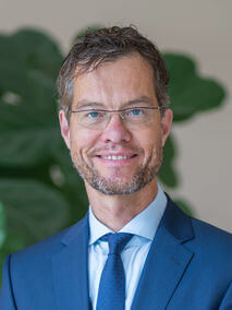
Professor Martin Bendszus serves as the Medical Director of the Department of Neuroradiology at Heidelberg University Hospital. He has held this position since 2007 and is a leading figure in advanced neuroimaging techniques. His academic career includes medical studies in Bonn, specialized training in Neuroradiology at the University of Würzburg (2003-2007), and a professorship in Neuroimaging at the University of Würzburg. Professor Bendszus's research focuses on innovative imaging methods, particularly in Magnetic Resonance Imaging (MRI). He pioneered Magnet Resonance Neurography for diagnosing peripheral nervous system disorders and made significant contributions to Dental-MRI as a radiation-free diagnostic tool for dental conditions. His expertise also extends to brain and spinal cord diagnostics, including aneurysms, strokes, and arteriovenous malformations. His work bridges clinical practice with advanced imaging research, resulting in numerous high-impact publications across neurology, radiology, and oncology. His publication record shows consistent high-impact contributions, with recent work spanning stroke intervention, neuropathic pain, brain tumor imaging, and advanced MRI techniques. The research trends demonstrate his leadership in translating imaging advances into clinical practice, particularly in time-sensitive neurological conditions where imaging guides critical treatment decisions. Kurt-Decker-Preis Röntgen-Preis Coolidge-Award Lucien-Appel-Award Hermann-Holthusen-Ring der Deutschen Röntgengesellschaft Professor Bendszus leads major multicenter clinical trials and collaborative guideline development efforts, including work with the Response Assessment in Neuro-Oncology (RANO) group and European Association for Neuro-Oncology (EANO). His research has secured significant funding for advancing neuroimaging techniques and their clinical applications. He collaborates extensively with neurologists, neurosurgeons, oncologists, and radiologists to improve diagnostic and therapeutic approaches for neurological conditions. His department serves as a reference center for complex neuroradiological cases and trains the next generation of neuroradiologists. Professor Bendszus maintains active leadership roles in professional societies and regularly contributes to shaping clinical guidelines in neuroimaging and stroke care.
Prof. Fritz Schick is a faculty member at the University of Tübingen , serving as the Deputy Head of the Division 'Pathophysiology of Prediabetes' at the Helmholtz Diabetes Center. He also holds a permanent professorship as Head of the Section on Experimental Radiology within the Department of Diagnostic and Interventional Radiology at Tuebingen University Hospital. His work focuses on developing non-invasive methods to characterize tissue composition and function in pre-diabetes and diabetes. M.D. (1989) and Physics (1990) graduate from University of Tübingen Research Interests : Schick's research spans quantitative MRI and MRS for applications in musculature, liver, adipose tissue, and bone marrow . His work bridges medical physics with metabolic disease analysis , emphasizing non-invasive diagnostics and cohort studies. Publication Trends : Recent articles highlight his expertise in MRI-based quantification of metabolic tissues, including studies on visceral adipose tissue distribution (German National Cohort), hepatokines in NAFLD, and deep learning for diabetes detection from whole-body MRI. Collaborations span radiology, endocrinology, and computational biology. Scientific Awards : Technologie-Transfer-Handwerk Professor Adalbert Seifriz-Preis (2003) Stipend of Siemens AG, Ernst-von-Siemens-Stipend (1992-1994) Dr.-Friedrich-Förster-Award, Department of Physics (1991) Affiliations : Active in the Helmholtz Diabetes Center and German Center for Diabetes Research , with leadership roles in experimental radiology and metabolic imaging.
Christian Büchel is a Professor in the Department of Neurology and Experimental Neurobiology at the University Medical Center Hamburg-Eppendorf (UKE). He holds a faculty position within the Medical Faculty of Hamburg University. His research focuses on understanding brain mechanisms underlying pain, placebo/nocebo effects, and psychiatric disorders using advanced neuroimaging techniques. He leads studies on brain-body interactions, developmental neuroscience, and translational neurology. Key research areas include structural and functional MRI investigations of pain modulation, addiction, and stress responses. He has contributed to multi-center neuroimaging studies and pioneered methods for spinal cord imaging. His work integrates cognitive neuroscience principles with clinical applications, particularly in chronic pain management and mental health. Büchel's recent studies explore how environmental factors (e.g., urban green spaces) influence brain structure, and he investigates neural correlates of resilience to substance use in adolescents. He collaborates internationally on projects like the IMAGEN study and the ENIGMA consortium, emphasizing reproducibility in neuroimaging research. His publications span over 20 years, with a focus on brain network dynamics, decision-making processes, and translational neuroscience. He actively contributes to developing open-access neuroimaging standards and has authored seminal papers on placebo mechanisms and brain plasticity.
Patrick Vogelمشاهده پروفایل
پژوهشگر
Dr. Patrick Vogel is a Habilitation candidate and researcher in the Magnetic Particle Imaging (MPI) group at the University of Würzburg's Faculty of Physics and Astronomy, Department of Experimental Physics V. His work focuses on advancing MPI technology for clinical applications, including imaging safety assessments, interventional procedures, and nanoparticle-based diagnostics. He contributes to the development of portable MPI scanners and hybrid imaging systems, collaborating with the AG Behr research group. His research spans magnetic particle spectroscopy, vascular imaging, and biomaterial characterization. Vogel has pioneered studies on MPI-guided endovascular interventions and the application of MPI in perfusion models. His work bridges physics, biomedical engineering, and clinical practice, with a focus on translating MPI into real-world medical diagnostics and surgery support. Key projects include the design of human-sized MPI scanners, safety evaluations of medical implants, and the use of synthetic tracers like Synomag®. He collaborates with interdisciplinary teams to address challenges in vascular imaging, nanoparticle behavior analysis, and real-time imaging systems.

Dimo Ivanovمشاهده پروفایل
استادیار
Dr. Dimo Ivanov is an Assistant Professor at the Department of Cognitive Neuroscience at Maastricht University in the Netherlands. Since 2025, he has also served as Head of the 7 Tesla MR Core structure at the Cooperative Brain Imaging Center and as an Independent Research Group Leader at the Max Planck Institute for Empirical Aesthetics in Frankfurt, Germany. His work bridges physics, neuroscience, and advanced imaging technology. Dr. Ivanov earned his PhD (Dr. rer. nat.) in Physics from the University of Leipzig/Max Planck Institute for Human Cognitive and Brain Sciences in 2012, followed by an M.Sc. in Physics from the International Physics Studies Program at the University of Leipzig in 2007. His educational foundation was completed with studies at the Foreign Language High School in Pleven, Bulgaria. Dr. Ivanov specializes in high-resolution structural, perfusion, and functional mapping of the human cortex and subcortical regions. His research focuses on quantitative MRI techniques to assess tissue vascularization, microstructure, myelination, and iron content. He develops advanced MRI sequences and analysis pipelines for both 7T and 3T systems, with translational applications in neurodegenerative disorders, psychiatric conditions, and neurodevelopmental studies. His work on resting-state and task-based fMRI provides critical insights into neural connectivity and brain dynamics. His publication record demonstrates consistent contributions to ultra-high field MRI methodology, with emphasis on 7T and 9.4T systems. His research spans technical MRI development, quantitative imaging methods, and clinical applications across multiple neurological conditions. Recent work shows increasing focus on translational applications connecting advanced imaging techniques with clinical outcomes. Dr. Ivanov has received numerous scientific awards and successfully secured competitive funding: ERC Marie Curie Program (2023) - 2-year postdoc position Maastricht University-Chinese Science Council PhD Program (2022) Maastricht University Center for Integrative Neuroscience PhD Program (2022) Magnetic Resonance in Medicine issue cover (2017) ISMRM merit award magna cum laude (2013) Trainee abstract travel award from the Organization for Human Brain Mapping (2011) Educational Stipends from the International Society for Magnetic Resonance in Medicine (2009-2011) Dr. Ivanov has successfully mentored postdoctoral researchers and PhD students through competitive funding mechanisms. His research group at the Max Planck Institute for Empirical Aesthetics and his position at Maastricht University support a collaborative environment that bridges physics, neuroscience, and clinical applications. His work with the 7 Tesla MR Core structure provides critical infrastructure for advanced neuroimaging research in Frankfurt. Dr. Ivanov leads the Structural and Physiological Imaging with High-Field MRI research group at the Max Planck Institute for Empirical Aesthetics. His team specializes in pushing the boundaries of ultra-high field MRI technology, developing novel acquisition and analysis methods that enable unprecedented insights into brain structure and function. The group maintains strong collaborations with multiple institutions across Europe and beyond.
Bo Wang is an active academic researcher primarily affiliated with multiple Chinese institutions, with strong connections to Tsinghua University, Beijing Jiaotong University, and other leading Chinese universities. His research spans artificial intelligence, machine learning, computer vision, medical image analysis, and intelligent control systems, demonstrating significant interdisciplinary work across computer science, engineering, and biomedical applications. Primary institutional affiliation: School of Computer Science and Technology at multiple Chinese universities Active research areas: AI/ML applications in healthcare, computer vision, federated learning, and intelligent control systems Extensive publication record across top-tier venues in multiple disciplines Wang's research interests focus on the intersection of artificial intelligence and practical applications. His work demonstrates strong expertise in developing novel machine learning architectures for medical image analysis, including applications in CT imaging, MRI, and sperm tracking. He has made significant contributions to federated learning approaches for large language models, sliding mode control systems, and molecular optimization frameworks. His research consistently bridges theoretical advances with practical implementations across healthcare, manufacturing, and environmental monitoring domains. Analysis of Wang's recent publications reveals a strong trend toward interdisciplinary AI applications, particularly in medical imaging and bioinformatics. His work on VAE-GANMDA for microbe-drug association prediction, ACE-QSM for accelerating MRI acquisition, and text-guided molecular optimization demonstrates innovative approaches at the intersection of AI and life sciences. Wang also maintains active research in industrial applications including digital twin technology for energy systems and robust scheduling approaches for multi-factory production. Notable research contributions include: FLFT: A Large-Scale Pre-Training Model Distributed Fine-Tuning Method with Federated Learning VAE-GANMDA: Microbe-drug association prediction model ACE-QSM: Accelerating quantitative susceptibility mapping using diffusion models Digital twin-empowered power consumption prediction systems Wang actively collaborates with researchers across China and internationally, with publications spanning computer science, engineering, medical imaging, and environmental science journals. His work demonstrates strong technical depth across multiple AI methodologies while maintaining focus on practical applications that address real-world challenges in healthcare, manufacturing, and environmental monitoring.
Prof. Thomas Mayrhofer is a Professor of Economics at Stralsund University of Applied Sciences since 2015 and concurrently serves as Lecturer at Harvard Medical School. He holds a doctorate in Health Economics from the University of Duisburg-Essen, after studying Economics at Otto-von-Guericke University Magdeburg. His career includes research roles at CINCH (Essen) and Massachusetts General Hospital (Boston). Education: PhD in Health Economics (University of Duisburg-Essen) MSc Economics (Otto-von-Guericke University) Research Focus: Health Economics Medical Decision Theory Cardiovascular Disease Pathophysiology Atherosclerosis Mechanisms Imaging Biomarkers Sustainability in Healthcare Key Article Trends: Recent work emphasizes cardiovascular outcomes in HIV populations (REPRIEVE Trial), epicardial adipose tissue imaging, air pollution-cardiovascular links (PROMISE Trial), and preterm infant neurodevelopment. His research bridges clinical medicine and economic decision frameworks. Labs/Teams: Collaborates with Harvard Medical School's cardiovascular imaging group and CINCH Health Economics Research Center.

Lisa Adamsمشاهده پروفایل
پژوهشگر ارشد
Dr. Lisa Adams is a Research Fellow at the Technical University of Munich (TUM) and an Attending Radiologist at TUM University Hospital Rechts der Isar, affiliated with the Diagnostic and Interventional Radiology department. She holds an Albrecht Struppler Clinician Scientist Fellowship (2024) and leads research in the Quantitative Imaging Biomarkers for Predictive Healthcare focus group. Adams earned her MD from Charité - Universitätsmedizin Berlin (2016), completed board certification in radiology (2021), and conducted postdoctoral research at Stanford University (2022-2023). She transferred her Habilitation in Experimental Radiology to TUM in 2024. Her research integrates AI with radiology diagnostics, specializing in: Developing machine learning methods for early disease detection and risk assessment Advancing quantitative imaging biomarkers across organ systems Body composition analysis and biological age estimation Multimodal analysis for personalized healthcare Ethical implementation of AI in clinical practice Adams' recent publications demonstrate strong focus on AI applications in radiology, spanning large language models for clinical reporting, deep learning for medical image interpretation, nanoparticle tracking in regenerative medicine, and radiomics for cancer diagnosis. Her work consistently bridges technical innovation with clinical translation. Awards & Recognition: Walter-Friedrich-Prize, Deutsche Röntgengesellschaft (2023) Alavi Mandell Award (2021) Invest in the Youth Stipend, ECR Vienna (2017) She actively mentors doctoral candidates, having supervised seven PhD students to completion since 2020 with two more nearing submission. Adams secures substantial research funding from DFG, EU, Wilhelm Sander Foundation, Bayern Innovativ, and Berlin Institute of Health. She serves as Scientific Editor for European Radiology and Trainee Editorial Board Member for Radiology: Artificial Intelligence , while also holding leadership positions in the German Radiological Society.
Mario Cesarelli is a Professor at the University of Naples Federico II's Department of Biomedical Engineering, with an extensive publication record spanning over three decades. His research bridges engineering and clinical medicine, focusing on developing computational methods for disease diagnosis and patient monitoring through biomedical signal processing and artificial intelligence. Dr. Cesarelli's research spans multiple domains of biomedical engineering with particular emphasis on: Medical imaging analysis and radiomics for neurodegenerative disorders Explainable AI for cancer detection and diagnosis Biomechanics and motion analysis for neurological conditions Cardiac signal processing and analysis Generative models for medical image synthesis and authentication His recent publications demonstrate a strong trend toward explainable deep learning applications in healthcare, particularly for neurodegenerative diseases like Parkinson's and Alzheimer's, as well as various cancer diagnostics. The work increasingly focuses on model interpretability to build clinician trust in AI-assisted diagnosis. Dr. Cesarelli maintains extensive collaborations with a core research group including Paolo Bifulco (46 joint publications), Maria Romano (41), Gianni D'Addio (34), Antonella Santone (27), and Francesco Mercaldo (26), forming interdisciplinary teams that combine engineering expertise with clinical knowledge.