Joseph Camp is a Professor of Comparative Pathobiology and Secretary of Faculties at Purdue University. His work spans interdisciplinary areas including machine learning, robotics, and human-robot interaction. He leads the Global Engineering Program and holds affiliations across technical and administrative roles. His research focuses on reinforcement learning, multi-agent systems, and applying large language models to enhance decision-making and action anticipation in robotics and healthcare contexts. Key research interests include neuro-symbolic systems for short-context action prediction, robust reinforcement learning under noisy feedback, and geometric reasoning for zero-shot robotic grasping. His work also explores disentanglement in concept-residual models and transfer learning in multi-agent environments. He has published extensively on topics ranging from probabilistic imitation learning to bio-inspired robot locomotion design. While no specific awards or grants are listed, his contributions to human-robot interaction (HRI) and AI-driven medical devices reflect a strong focus on practical applications. His lab’s recent work integrates large language models with traditional robotics algorithms to improve adaptability in dynamic environments. No advising relationships or student names are explicitly mentioned in the provided texts.
John M. Pauly is the Reid Weaver Dennis Professor in the Department of Electrical Engineering at Stanford University, with affiliations in the Wu Tsai Neurosciences Institute, Stanford Cancer Institute, Cardiovascular Institute, and Bio-X program. His research focuses on medical imaging, particularly MRI acceleration and reconstruction techniques for applications like cardiac imaging and interventional guidance. He holds a PhD from Stanford University (1990) and has received notable awards including the ISMRM Gold Medal (2012) and IEEE Fellow distinction (2022). His academic appointments include teaching courses such as Medical Image Reconstruction (EE 369C), Signals and Systems II (EE 102B), and The Wireless World (EE 100). He advises numerous PhD and master's students in MRI hardware, reconstruction algorithms, and clinical applications. Research highlights include innovations in deep learning for MRI quality assessment, coil design for pediatric imaging, and non-contact motion sensing via Doppler radar. His work bridges engineering and clinical needs, emphasizing practical translation of compressed sensing and machine learning methods into clinical MRI workflows.
Rosa Tamara Branca is a Professor at the University of North Carolina at Chapel Hill, leading the Branca Lab. Her research focuses on advancing nuclear spin dynamics and magnetic resonance imaging (MRI) techniques to enhance diagnostic capabilities. She specializes in hyperpolarization methods, low-field MRI systems, and the development of innovative imaging tools for clinical applications. The lab is located in Marsico Hall within the Biomedical Research Imaging Center. Her work integrates physics, engineering, and medicine to improve MRI sensitivity and specificity, particularly through reducing reliance on bulky superconducting magnets. Key areas include xenon-129 MRI, brown adipose tissue imaging, and contrast agent development. She actively seeks students interested in spin physics and biomedical engineering. Publications highlight contributions to ultra-low field NMR, hyperpolarized gas applications, and thermometry. Research emphasizes translating lab innovations into clinical tools for metabolic disorder diagnosis and imaging precision. The lab’s projects often involve interdisciplinary collaborations and open-source hardware development for cost-effective medical solutions.
William Yong, MD is Professor of Pathology and Laboratory Medicine at the David Geffen School of Medicine, University of California, Los Angeles. He serves as Chief of Neuropathology, Director of the UCLA Brain Tumor Translational Resource, and Director of the UCLA Neuropathology Fellowship Program. His clinical expertise encompasses brain and pituitary tumors, neuromuscular pathology, and international telepathology consultations for complex brain cases. Bachelor's in Molecular Biology, UC Berkeley Master's in Biology (Cellular and Molecular), San Francisco State University Medical degree with Honors thesis, UCLA Pathology training, Massachusetts General Hospital/Harvard Medical School Neuropathology fellowship, UCLA Former Director of Neuropathology at Cedars-Sinai Medical Center Dr. Yong's research focuses on biobanking methods, end-stage immunopathology in brain cancer patients, and collaborations spanning AIDS, stroke, dementia, and epilepsy. His team conducts studies in bioinformatics, nanotechnology, non-invasive imaging, neuroradiology, and targeted therapies. He consults for the National Neurologic AIDS Brain Bank and works closely with UCLA Neurosurgery and Neuro-oncology physicians. His publication record demonstrates significant contributions to understanding glioma immunology, tumor microenvironment dynamics, molecular biomarkers in brain cancer, and novel imaging techniques. Research trends indicate strong emphasis on translational applications of neuropathological findings to improve brain tumor diagnostics and therapeutics. Golden Apple Award for teaching Johnny Mercer Foundation Research Award for Brain Cancer American Association of Neuropathologists Lucien J. Rubinstein Award in Neuro-oncology Dr. Yong actively mentors junior faculty, teaches medical and dental students, and supervises pathology, neuropathology, neurosurgery, and neurology residents and fellows. His clinical work includes international telepathology consultations and molecular testing of brain tumor samples. He directs the UCLA Brain Tumor Translational Resource and Neuropathology Fellowship Program, facilitating research collaborations across multiple disciplines. His laboratory work centers on the Brain Tumor Translational Resource, which bridges clinical neuropathology with translational research in brain cancer. The team collaborates extensively with neuro-oncology, neurosurgery, and basic science researchers to develop novel diagnostic and therapeutic approaches for brain tumors.
Professional Overview Pascale Aouad, MD, is an Assistant Professor in the Department of Radiology at Northwestern University's Feinberg School of Medicine, specializing in Pediatric Radiology. She maintains active clinical and research roles with no indication of part-time or retired status. Education & Training Residency: Radiology, Lebanese University (2017) Fellowships: Cardiovascular Imaging (2018), Neuroradiology (2019), MRI Clinical & Research (2020), Pediatric Neuroradiology (2021) at McGaw Medical Center of Northwestern University Research Focus Dr. Aouad's work centers on pioneering MRI methodologies, including vascular imaging (MRA techniques), pediatric neuroimaging, radiomics, and non-contrast angiography. She develops clinical protocols for conditions ranging from lung cancer to rare pediatric syndromes, with emphasis on technical innovations in 2D/3D imaging and AI-driven diagnostics. Key Publications Recent articles demonstrate her focus on pediatric radiology (ocular pathologies, ventriculomegaly), contrast agent optimization, and computational methods like radiomics for cancer prediction. Multinational studies on COVID-19 neuroimaging and technical advances in 4D flow MRI represent major contributions. Awards Best Educational Exhibit, American Society of Pediatric Neuroradiology (2022)
Dr. Oliver Faust is an Associate Professor at Anglia Ruskin University's Faculty of Science and Engineering, within the Department of Computing and Information Science. An international expert in AI-driven medical diagnostics, he specializes in physiological signal processing, medical image analysis, and Internet of Medical Things applications. With over 140 publications, his research has significantly impacted computer-aided diagnosis methodologies. His academic credentials include: Doctor of Engineering in Biomedical Science, Chiba University PhD in Electronics, University of Aberdeen Diplom-Ingenieur in Communication Engineering, FH Dieburg Fellow of the Higher Education Academy, UK Chartered Engineer, Institution of Engineering and Technology Dr. Faust's research bridges artificial intelligence with clinical applications, particularly focusing on deep learning approaches for healthcare. His primary investigations involve developing diagnostic algorithms for medical imaging (ultrasound, fundoscopy, thermography) and physiological signal analysis (EEG, ECG). Secondary interests include eHealth systems and IoT-enabled medical devices, with strong emphasis on translational research impacting stroke risk assessment and chronic disease management. His publication portfolio demonstrates extensive work in nonlinear signal analysis, wavelet transformations, and deep learning architectures applied to neurological, cardiovascular, and oncological diagnostics. Recent trends show increasing focus on LSTM networks for arrhythmia detection and comprehensive AI diagnostic frameworks. Honors include: Fellow of the Higher Education Academy, UK Dr. Faust actively supervises PhD students with projects funded by international scholarships. His grants portfolio includes: £75,000 from Sheffield Hallam (2021-2022) £30,000 from Innovate UK (2021-2022) £40,000 from Grow MedTech (2019-2020) Multiple PhD scholarships totaling £99,600 He contributes to the Transformative Artificial Intelligence Applications research cluster and Computing, Informatics and Applications Research Group at ARU, fostering collaborations across 20+ international institutions.
Henk De Feyter serves as an Associate Professor of Radiology and Biomedical Imaging at Yale School of Medicine, with primary appointments in the Department of Radiology & Biomedical Imaging. He maintains affiliations with multiple research entities including the Bioimaging Sciences division, Deuterium Metabolic Imaging (DMI) Developmental Therapeutics program, Magnetic Resonance Research Center, and Yale Biomedical Imaging Institute. Dr. De Feyter completed his postdoctoral training at Yale University (2010) following a PhD from Eindhoven University of Technology's Department of Biomedical Engineering. His educational background includes Master's degrees from Maastricht University (Department of Health, Medicine and Life Sciences) and Ghent University (Department of Rehabilitation Sciences). His research program centers on developing and applying Deuterium Metabolic Imaging techniques to visualize metabolic processes in vivo. Dr. De Feyter specializes in creating novel imaging methodologies to track metabolic pathways using deuterium-labeled substrates, with particular applications in brain tumor characterization, cardiac metabolism, and liver function assessment. His interdisciplinary approach combines physics, engineering, and clinical medicine to develop diagnostic tools that bridge basic science and patient care. Analysis of Dr. De Feyter's publication record reveals a consistent focus on advancing metabolic imaging technologies, particularly Deuterium Metabolic Imaging. His work demonstrates growing clinical relevance across neuro-oncology, cardiology, and hepatology, with increasing emphasis on translating research techniques to clinical applications. The interdisciplinary nature of his publications reflects collaborations across multiple scientific domains. Dr. De Feyter maintains active research collaborations with experts across multiple disciplines, including neurology, oncology, physics, and biochemistry. His research program appears to be supported by substantial grant funding focused on developing metabolic imaging techniques for clinical applications, particularly in brain tumor imaging and neurological disorders. As part of Yale's Magnetic Resonance Research Center and Yale Biomedical Imaging Institute, Dr. De Feyter contributes to a collaborative environment dedicated to advancing imaging technologies. His work on metabolic imaging represents a significant contribution to the development of non-invasive diagnostic tools that could transform how metabolic disorders and cancers are detected and monitored.
Dr. Maureen Hood serves as Associate Professor of Radiology and Bioengineering at the Uniformed Services University of the Health Sciences (USU) and functions as a clinical scientist. She maintains dual affiliation with the Walter Reed National Military Medical Center (WRNMMC) as a cardiovascular MR imaging consultant and MR safety expert, driving translational research in military and veteran healthcare settings. Her educational foundation includes: Bachelor of Science in Biology, Minor in Mathematics, University of Puget Sound Associate of Technical Arts in Radiologic Technology, Tacoma Community College Bachelor of Science in Nursing, University of Maryland, School of Nursing Master of Science in Nursing Informatics, University of Maryland, School of Nursing Doctor of Philosophy in Nursing Research, Uniformed Services University of the Health Sciences Dr. Hood's research program bridges engineering and clinical medicine through advanced imaging techniques. Her cardiac MRI work establishes critical correlations between T1/T2 mapping sequences and histological markers in heart failure models, while her current focus on ultrasound-mediated drug delivery targets precision treatment for infections. This dual trajectory in diagnostic imaging innovation and therapeutic application defines her unique contribution to biomedical science. Publication analysis reveals consistent expertise in cardiac MRI physics and safety spanning two decades. Her work demonstrates progressive technical sophistication from foundational contrast agent research to advanced quantitative mapping techniques, with strong emphasis on preclinical validation and clinical translation. Key thematic threads include myocardial tissue characterization, vascular imaging optimization, and rigorous safety protocols for MR environments. Scientific recognition includes: Fellow of the American Heart Association (2015-present) Best Cardiac MR Poster at NASCI Annual Meeting (2011) Linda Strangio Editor’s Award from ARNA (2006) Dr. Hood directs significant research initiatives including the Early Career Scientist Award-funded project on ultrasound-enhanced antibiotic delivery for osteomyelitis. Her leadership extends to editorial responsibilities for Military Medicine and abstract grading for major cardiology/radiology societies, demonstrating commitment to scholarly advancement beyond direct research output. She actively shapes the field through leadership roles in the American Heart Association's Leadership Council on Cardiovascular Radiology, the ISMRM MR Safety Committee, and NASCI research committee, fostering collaborative networks that integrate military medical imaging with broader academic radiology communities.
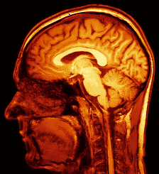
Professor Franklyn Howe serves as Professor of Magnetic Resonance Imaging and Director of the Neuroscience & Cell Biology Research Institute at City St George's, University of London (formed from the August 2024 merger of St George's, University of London and City University of London). With a career spanning since 1988 at St George's, he has established himself as a leading expert in multimodal MRI applications for disease diagnosis and treatment monitoring, particularly in brain tumor analysis. His work bridges physics, medical imaging, and clinical applications across multiple disease areas. Professor Howe earned his BA in Physics from the University of Oxford, followed by a DPhil on 'Magnetisation and microwave absorption in rare earth metal alloys' from the same institution in 1984. He completed postdoctoral research evaluating MRI contrast agents at St Bartholomew's Hospital and in MR image analysis at the Royal Postgraduate Medical School (now part of Imperial College School of Medicine) before joining St George's. Howe's research centers on developing automated analysis of multimodal MRI to aid diagnosis of brain tumors, using various MRI methods including perfusion and diffusion imaging for structural and functional information, and magnetic resonance spectroscopy (MRS) for biochemical data. He applies pattern recognition techniques to optimally combine data from different MRI methods, aiming to develop biomarkers for diagnosis, prognosis assessment, and treatment response monitoring. His work extends beyond brain tumors to dementia, Tourette syndrome, cerebrovascular disease, chronic obstructive pulmonary disease, lupus, lymphedema, osteoarthritis, and normal aging. In 1992, he was instrumental in developing MR Neurography at St George's, a non-invasive method for detailed nerve imaging. Analysis of Professor Howe's recent publications (2021-2024) reveals consistent focus on advancing MRI techniques across multiple clinical applications. His work demonstrates strong interdisciplinary collaboration, with significant contributions to lymphatic imaging, brain tumor characterization, neurodegenerative disorders, and quantitative MRI methodology. The publications show progression from fundamental technical improvements in MRI acquisition and processing toward increasingly sophisticated clinical applications, particularly in neuroimaging and vascular disorders. Chair of ISMRM MRS Study Group Co-founder and Chair of Pediatric MR Study Group Long-standing member of International Society of Magnetic Resonance in Medicine Professor Howe actively supervises lab-based MRes and BSc student research projects and library-based Special Study Courses on Biomedical Applications of Magnetic Resonance. He has secured substantial research funding from diverse sources including the Medical Research Council, Cancer Research UK, Alzheimer's Research UK, and St George's Hospital Charity. His collaborative approach is evident in the wide range of clinical research studies he participates in across multiple departments. As Director of the Neuroscience & Cell Biology Research Institute, Howe leads a research team including Ian Storey (Image Analyst) and Mike Mills (MRI physicist). He maintains extensive collaborations within St George's with Dr Thomas Barrick, Dr Atticus Hainsworth, Mr Timothy Jones (Neurosurgery), Dr Jeremy Isaacs (Neurology), Dr Pia Ostergaard, Prof Sahar Mansour (Clinical Genetics), Prof Nidhi Sofat (Rheumatology), and Mr Michael Hart (Neurosurgery), creating a robust interdisciplinary research environment focused on translating MRI advances into clinical practice.