Ali Ghahremaninezhad is a Professor in the Department of Civil, Architectural, and Environmental Engineering at the University of Miami's College of Engineering. His research focuses on sustainable cementitious materials, self-healing concrete, and green additives like hydrogels and biochar. Current research explores molecular-level interactions between surfactants and cement microstructure to enhance durability. Key areas include carbonation curing, freeze-thaw resistance, and organic coatings for marine carbonate minerals. Recent publications highlight his work on using biochar for internal curing, hydrogels for crack repair, and triblock copolymers to reduce autogenous shrinkage. His studies integrate experimental methods (TGA, FTIR, SEM) and predictive modeling. Findings demonstrate improved mechanical strength and reduced water sorption in modified cement systems. Applications span marine carbon cycle understanding and sustainable construction practices.
Dr. Margarita Yavorskaya serves as Assistant Professor in the Department of Evolutionary Biology of Invertebrates at the Institute of Evolution and Ecology, Faculty of Science, University of Tübingen since 2020, following a Postdoctoral position at Ludwig Maximillian University Munich (2018-2020). Her academic credentials include: Ph.D. in Zoology, Friedrich Schiller University Jena (2014-2018): "Evolution of head structures in Coleoptera with special emphasis on the feeding apparatus and miniaturized forms" Diploma in Zoology, Biological Faculty of Lomonosov State University (2009-2014): "Mouthparts of hooded beetles (Coleoptera: Corylophidae) with regards to miniaturization" Her research centers on evolutionary morphology of arthropods , with particular focus on size-shape evolution and miniaturization effects in invertebrates. Key investigations encompass cephalic structure evolution in Coleoptera, feeding preference adaptations in beetle larvae/adults, and anatomical studies of minute crustaceans (Chydoridae). She integrates advanced methodologies including confocal laser scanning microscopy (CLSM), scanning/transmission electron microscopy (SEM/TEM), and 3D reconstruction from histological sections and MicroCT datasets to analyze morphological adaptations at microscopic scales. Publication analysis (2017-2023) reveals consistent specialization in beetle morphology, particularly examining head anatomy evolution across diverse families (Ptiliidae, Micromalthidae, Staphylinidae) and miniaturization constraints. Her work bridges paleontological assessments of early beetles (e.g., Coleopsis archaica) with contemporary studies of commensal adaptations (Platypsyllus castoris) and structural diversity in leaf-litter predators, demonstrating methodological innovation through Reflectance Transformation Imaging and 3D reconstruction techniques. No scientific awards were documented in the source material. Dr. Yavorskaya teaches Zoology (Bio122), Microscopic Imaging and 3D Reconstruction (Bio-3187), and Micro- & Macroevolution courses, though specific student advisement or grant details remain unreported. Her research contributes to the "Competence network Biomimetics" and "Colourful Meadow" Initiative through morphological analyses with potential biomimetic applications. She operates within the Department of Evolutionary Biology of Invertebrates utilizing specialized microscopy facilities for investigations into arthropod miniaturization, collaborating on projects examining structural adaptations across evolutionary timescales from fossil lineages to contemporary micro-crustaceans.
Dr. Bernhard Ruthensteiner is a senior scientist and head of the Section Evertebrata varia at the Munich State Zoological Collection (ZSM), part of the Bavarian State Natural Science Collections (SNSB). His work bridges taxonomy, comparative anatomy, and advanced imaging techniques. Institution : Munich State Zoological Collection Role : Head of Section Evertebrata varia Research Focus : Invertebrate anatomy, 3D imaging, and evolutionary morphology His research spans diverse invertebrate groups, including Gastropoda , Cnidaria , and Echinodermata , with a strong emphasis on microCT and SEM analysis for resolving taxonomic and developmental questions. Recent publications highlight his contributions to understanding biomineralization processes , protonephridial system development , and evolutionary adaptations in terrestrial and marine species . Key collaborations involve institutions in Germany, Austria, and Madagascar, with a focus on integrative taxonomy and digital specimen curation. His work also includes pioneering 3D model embedding in PDF publications and correlative microscopy for studying symbiotic relationships.
Dr. Andy Sombke is a highly accomplished researcher specializing in arthropod morphology and neuroanatomy at the Medical University of Vienna's Center for Anatomy and Cell Biology, where he leads a research group in the Department of Cell and Developmental Biology. With a habilitation (Dr. rer. nat. habil.), he has established himself as a leading expert in myriapod biology, particularly centipede anatomy, venom systems, and appendage development. His work bridges fundamental research with potential biomedical applications, utilizing advanced microscopic techniques including SEM, TEM, microCT, and histology. Dr. Sombke's research interests focus on comparative neuroanatomy , venom delivery mechanisms , appendage regeneration , and respiratory system anatomy in arthropods. His work on centipedes has revealed remarkable insights into the evolutionary transformations of their ultimate legs, which serve diverse functions from physical defense to sensory reception. His research group employs a multimodal microscopic approach to analyze the cellular organization and functional adaptations of these fascinating creatures, with potential applications in bioinspired technology and novel therapeutics. His publication record shows strong focus on centipede morphology (40% of recent publications), venom systems (25%), neuroanatomy (20%), and comparative morphology across arthropod groups (15%). The research demonstrates increasing interdisciplinary collaboration, with recent work spanning immunology, oncology, and biomedical engineering applications of arthropod-inspired systems. Associate Editor of Arthropod Systematics & Phylogeny Associate Editor of Physiological Entomology Speaker of the section group Morphology of the German Zoological Society Participant of the European Venom Network (COST Action CA19144) Dr. Sombke has mentored several students including Iulia Barutia and Simon Züger, and collaborates extensively with international researchers across Europe. His work on centipede biology has received significant attention for its potential to inform biomedical applications, particularly in understanding venom components with therapeutic potential. His laboratory, the Functional Ultrastructure Group, focuses on providing comprehensive understanding of cellular dynamics through electron microscopic techniques. The Functional Ultrastructure Group maintains active research on centipede anatomy, venom systems, and appendage development, with particular emphasis on the evolutionary transformations of centipede ultimate legs. The group is part of the European Venom Network and maintains the "Myriapod Morphology and Evolution" Facebook group as a hub for international collaboration in the field.
Professor Jeroen Vanoirbeek is a faculty member at the Department of Public Health and Primary Care, Faculty of Medicine, KU Leuven. He serves as division head of the Environment and Health Division and subdivision head of Occupational Environmental Measurements, with additional affiliations at the Respiratory Diseases and Thoracic Surgery (BREATHE) research unit and Leuven One Health Institute. His research spans critical environmental and occupational health domains: Respiratory Pathophysiology : Asthma heterogeneity, airway hyperreactivity mechanisms, and occupational lung diseases including silicosis Exposure Science : Advanced biomonitoring of air pollutants (PAHs, pesticides, microplastics), dermal chemical absorption, and sensor development Vulnerable Populations : Children's indoor air quality impacts, elderly industrial pollution susceptibility, and global farmworker health Methodological Innovation : Transcriptomic clustering, microCT imaging, and UPLC-MS/MS analytical techniques Recent publications (2024-2025) demonstrate accelerating focus on molecular mechanisms of asthma, translocation pathways of inhaled particles, and occupational exposure assessment technologies. His work bridges fundamental toxicology with public health applications through strong industry and international collaborations. Professor Vanoirbeek actively supervises master's theses in occupational hygiene while leading major grants including the PIVO STUDY (promotor), QuantPAH sensor development (co-promotor), and Katangan mining lung disease research (2023-2027). His current portfolio features 10 active projects totaling over €5M in funding, emphasizing children's environmental health and microplastic analysis. He contributes to institutional governance as member of the Faculty Council of Medicine, Occupational Medicine POC, and Bioethics POC, while co-leading the Departmental Board for Community Health and Primary Care.
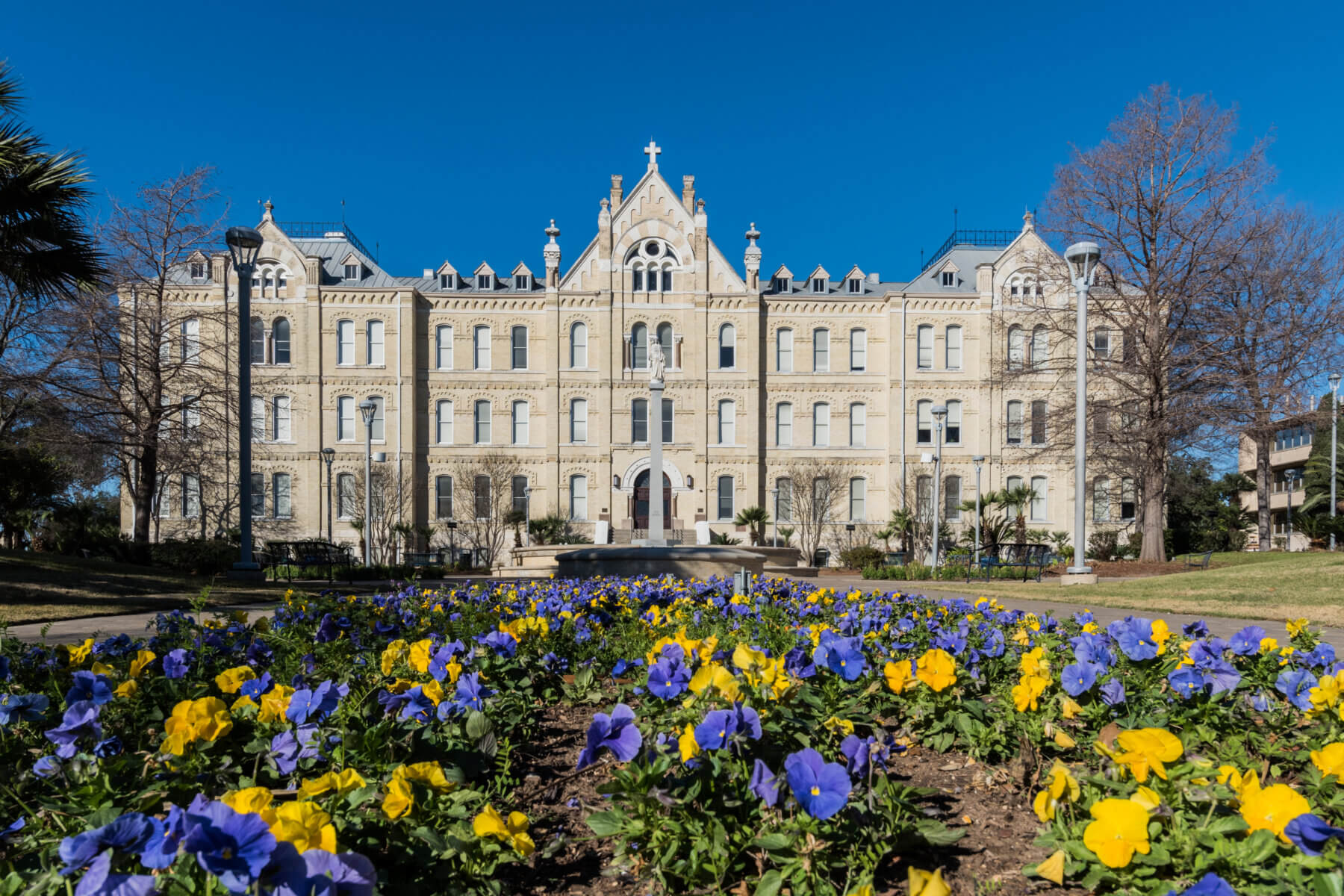
Dr. Ruben U. Tovar is an Assistant Professor in the Department of Biological Sciences at St. Mary’s University, teaching Human Anatomy and Cellular and Molecular Methods. His research focuses on Evolutionary Developmental Biology (Evo-Devo), examining the molecular and developmental mechanisms driving biodiversity through studies of cave-adapted vertebrates. Education : Ph.D. in Integrative Biology (EEB) from The University of Texas at Austin, M.S. in Biology from Texas State University, and B.S. in Interdisciplinary Studies from The University of Texas at Arlington. Research Interests : He investigates developmental and molecular parallels between cave-dwelling species and human congenital eye disorders using techniques like microCT imaging, RNA sequencing, and protein analysis. His lab explores sensory compensation, convergent evolution, and genetic pathways in subterranean vertebrates. Selected Publications : His recent work includes studies on parallel sensory adaptation in salamanders, gene exchange in karst-obligate species, and molecular mechanisms in lateral line systems. These publications highlight his focus on integrating evolutionary, developmental, and molecular approaches. Advising and Collaborative Work : Dr. Tovar mentors graduate and undergraduate students in fieldwork, 3D imaging, bioinformatics, and animal husbandry. His research involves collaboration with the U.S. Fish and Wildlife Service and Mexican institutions. Labs and Teams : At St. Mary’s, he is establishing breeding colonies of cave-adapted salamanders to study genetic and developmental links to human eye disorders, combining lab and fieldwork expertise.
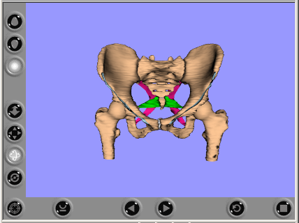
Robert Funnell, PhD is an Investigator at the Research Institute of the McGill University Health Centre (RI-MUHC) and a Professor in the Department of Otolaryngology - Head and Neck Surgery at McGill University's Faculty of Medicine. His research is conducted within the Child Health and Human Development Program at RI-MUHC, where he applies engineering principles to understand auditory mechanics and improve diagnostic tools for hearing assessment. Dr. Funnell's research focuses on the mechanics and acoustics of hearing, with particular emphasis on developing novel measurement techniques in experimental animals and infants, and creating computational models of the ear. His work combines experimental measurements with sophisticated modeling approaches to better understand middle ear function. He has developed specialized software for creating 3D models of complex anatomical structures, including his notable Thrup'ny 3D viewer application. His research has significant clinical applications, particularly in improving newborn hearing screening protocols and developing better diagnostic tests for middle ear conditions. Analysis of Dr. Funnell's recent publications reveals a consistent trajectory in middle ear biomechanics research spanning several decades. His work demonstrates increasing sophistication in computational modeling techniques, with recent publications integrating optical coherence tomography, X-ray microCT data, and finite element analysis to create increasingly accurate representations of middle ear function. A significant portion of his recent work focuses on newborn hearing assessment, reflecting the clinical importance of early hearing detection. His research bridges engineering principles with clinical audiology, creating practical applications for hearing screening and diagnosis. Dr. Funnell has developed several notable software tools including Thrup'ny, a free open-source 3D viewer for VRML models designed for both anatomy teaching and finite-element pre- and post-processing. His website hosts various educational resources including interactive 3D models of the ear, finite-element simulation tools, and teaching materials on middle ear mechanics. These resources have been widely used for both research and educational purposes in the field of otolaryngology and biomedical engineering.
Dr. Joachim Schmidt is a researcher at the Institute of Biosciences within the Faculty of Mathematics and Natural Sciences at the University of Rostock . His work focuses on biodiversity conservation, climate change impacts, and evolutionary biology. Research areas include phylogeny of ground/tiger beetles, environmental history of mountain systems, and landscape ecology of coastal habitats. Utilizes MicroCT 3D reconstructions for beetle fossil analysis. Contact: Universitätsplatz 2, 18055 Rostock, Tel.: 0381 / 498-6279.
Alessandra Bitto is a Professor in the Department of Biomedical, Dental and Morphological and Functional Imaging Sciences at the University of Messina, where she serves as referent for the Molecular Pharmacology and Biotechnology Group and the Laboratory of Molecular Pharmacology and Biotechnology. Her research focuses on molecular pharmacology of natural compounds , particularly investigating how substances like genistein, lycopene, and Garcinia cambogia affect human health. Professor Bitto's work spans several key areas including bone health and osteoporosis prevention, obesity treatment through adipose tissue conversion, anti-inflammatory compounds, and cardiovascular pharmacology. Analysis of her recent publications (2023-2024) reveals a strong emphasis on natural product pharmacology , with numerous studies on plant-derived compounds and their therapeutic applications. Her work demonstrates interdisciplinary approaches combining molecular biology, pharmacology, and translational medicine. Featured Poster, Endocrine Society - 2018 Travel grant SISA, Società Italiana per lo Studio dell'Arteriosclerosi - 2013 PREMIO SIF, Italian Pharmacology Society - 2011 Multiple young researcher awards between 2008-2010 Professor Bitto actively mentors students through her research group and teaches Pharmacology for the Medicine and Surgery program, as well as Molecular Pharmacology and Precision Medicine for the Medical Biotechnology program. She has secured multiple patents related to natural compounds for treating obesity, metabolic syndrome, and other conditions. Her laboratory is equipped with advanced instrumentation including MicroCT for ex-vivo studies, Real-time PCR systems, and animal exercise equipment. She maintains strong community engagement through numerous science outreach activities, particularly through MEDNIGHT events and school programs, demonstrating commitment to science communication and public understanding of pharmacology research.
Professor Andrew Smith is Chair in Oral Health Sciences at University College London's Eastman Dental Institute, where he leads research connecting oral health with systemic inflammatory conditions. His work bridges dental and medical fields, particularly investigating Crohn's disease, ulcerative colitis, and oral manifestations of inflammatory conditions through advanced laboratory and clinical approaches. Education: PhD, University of Southampton (1998) BSc (Hons), University of Leeds (1995) Smith's research program focuses on three interconnected areas: macrophage immune responses, epithelial integrity in mucosal inflammation, and microbiome influences on inflammatory diseases. His laboratory employs genetic analysis, transcriptomic profiling, in vivo and in vitro disease modeling, and proteomics to investigate cytokine secretion mechanisms and their role in disease pathogenesis. His team has demonstrated that the oral microbiome is primarily shaped by environmental factors rather than genetics, with implications for both oral and gastrointestinal health management. His recent publications reveal increasing emphasis on organoid technology, microbiome analysis, and novel drug delivery systems for mucosal diseases. The research shows strong translational focus, connecting basic immunological mechanisms with clinical observations in patient populations. His work on the relationship between biologic therapies for Crohn's disease and oral health outcomes has provided important insights into the oral-systemic disease connection. Research Contributions: Established 20-year clinical-scientific collaboration with Dr. Farooq Rahman Developed patient-derived organoid models for inflammatory disease research Demonstrated environmental dominance in shaping oral microbiome Investigated connections between TNF inhibitors and periodontal health Advanced understanding of cytokine trafficking in immune cells Professor Smith supervises multiple PhD students and postdoctoral researchers while collaborating with clinicians at University College Hospital and Royal Free Hospital. His international research team includes scientists, clinicians, dentists, and nutritionists working across disciplines to unravel complex immune-microbiome interactions in inflammatory diseases. The Smith-Rahman research group maintains sophisticated facilities for patient-derived organoid culture, microbiome profiling, and advanced imaging including microCT for dental and bone analysis, supporting their integrated approach to inflammatory disease research.
Professor Andrew Pitsillides is a distinguished academic at the Royal Veterinary College (RVC), where he serves as Professor of Skeletal Dynamics in the Department of Comparative Biomedical Sciences at the Camden campus. He leads the Musculoskeletal Biology research group and is associated with the CPCS Research Programme and MicroCT Research Centre at RVC. Dr. Pitsillides received his Applied Biology BSc. (Hons, 1984) and PhD. in Biochemistry (1988) from the Kennedy Institute of Rheumatology. His postdoctoral work continued at UCL and RVC under prominent researchers including Jo Edwards, Mike Bayliss, and Lance Lanyon, where he established his own laboratory in 1995. Professor Pitsillides' research focuses on skeletal mechanobiology across diverse biological settings. His work spans from understanding the role of embryo movement in skeletal formation to how bones and joints respond to functional and traumatic loads throughout growth, adulthood, and aging. He has made significant contributions including discovering that nitric oxide is critical in bone mechano-adaptation, establishing that joint formation is mechanodependent, and developing a non-invasive model of in vivo loading that allows simultaneous study of tibial cortices and trabecular compartments. His research has been published in over 140 original articles with an h-index of 41+. His recent publications demonstrate a strong focus on bone mechanoadaptation, osteoarthritis mechanisms, and skeletal development. The research utilizes advanced imaging techniques like synchrotron X-ray tomography and employs various mouse models including the STR/Ort spontaneous osteoarthritis model. Key themes include the relationship between bone structure and mechanical function, sexual dimorphism in skeletal biology, and the molecular mechanisms underlying osteocyte function and bone adaptation. Fellow of The Royal Society of Biology (FRSB) Fellow of the Anatomical Society (FAS) Board Member of the Bloomsbury Centre for Skeletal Research Executive Editor of Cell Biochemistry and Function Executive Committee Member of International Society for Hyaluronan Sciences Former Honorary Secretary of the British Society for Matrix Biology Professor Pitsillides serves on numerous national and international review panels including the Belgian Fund for Scientific Research, Greek Governmental Agency, and European Commission. His research is currently funded by prestigious organizations including the EU, BBSRC, Wellcome Trust, Arthritis Research UK, and The Leverhulme Trust. He has successfully forged clinical links with Sheffield University and The Royal National Orthopaedic Centre, Stanmore, UCL. In teaching, he has led modules on 'Advanced Skeletal Pathobiology' and 'The Head' for Veterinary and BSc degrees, and regularly guest lectures at UCL, Imperial, and Oxford Universities.
Kristen Prufrock serves as an Assistant Professor in the Department of Neuroscience at Washington University School of Medicine, where she investigates biomechanical adaptations in mammalian chewing systems across evolutionary and developmental timescales using gross anatomy, microCT modeling, and muscle architecture analysis. Her academic credentials include: PhD in Functional Anatomy and Evolution from Johns Hopkins University School of Medicine (2020) MSc in Evolutionary Anthropology from the University of Toronto (2014) HBSc in Biological Anthropology and Zoology from the University of Toronto (2012) Her research integrates paleontological, anatomical, and biomechanical methodologies to explore dietary adaptations in primates, with emphasis on dental topography, fossil morphology, and evolutionary ecology. By analyzing microCT scans and muscle fiber architecture, she elucidates how food processing demands shape masticatory structures in both living and extinct mammals, contributing to broader understandings of adaptive radiation and functional constraints. Publications spanning 2016-2021 reveal consistent focus on primate paleontology, dental evolution, and biomechanical modeling, with recurring themes in Eocene fossil analysis, niche competition, and quantitative topographic methods applied to extinct species like omomyids and paromomyids. Research support includes: Duke Lemur Center Director’s Fund for “Ontogeny of the Chewing System in Strepsirrhines” Canadian Association for Biological Anthropology Shelley Saunders Thesis Research Award for “Ontogeny of the Chewing System in Strepsirrhines” Collaborative work with institutions including Duke Lemur Center and multiple universities demonstrates active engagement in interdisciplinary paleontological networks despite no explicitly described laboratory infrastructure in available materials.
Dr. Sean P. Marrelli is a Professor at The University of Texas Health Science Center at Houston (UTHealth Houston), where he serves as Director of the BRAINS Research Laboratory and Director of the MicroCT Imaging Facility. His research program is funded by multiple grants from the NIH and American Heart Association. Dr. Marrelli received a BA in Chemistry from the University of Houston and then a PhD in Cardiovascular Sciences from Baylor College of Medicine. His laboratory focuses on understanding the mechanisms of cerebrovascular function in both healthy and pathological conditions. Dr. Marrelli's research interests span several key areas in cerebrovascular science: Regulation of cerebral blood flow in healthy conditions and disease states (e.g., following ischemic stroke, in conditions of amyloidosis) Role of endothelial Piezo1 mechanosensitive ion channel in cerebral blood flow regulation Brain vascular remodeling in aging and pathology Novel methods of promoting therapeutic hypothermia following stroke The Marrelli Lab employs a diverse range of experimental approaches, including laser speckle contrast imaging, microCT imaging, MRI, two-photon imaging, and various molecular and genetic techniques. The lab uses multiple transgenic mouse models to investigate cerebrovascular function and stroke pathology. Dr. Marrelli also oversees the MicroCT Imaging Facility, which was established through an NIH S10 equipment grant (S10OD030336) for the purchase of a Bruker SkyScan 1276 microCT imaging system. This facility provides high-resolution ex vivo and in vivo 2D and 3D imaging capabilities for researchers. Dr. Marrelli currently mentors several researchers, including a Research Associate, a GSBS Graduate Student Research Assistant, and multiple Research Assistants. His laboratory has produced significant publications in journals such as Scientific Reports, Stroke, and Journal of Neuroscience Research.