Francesca De Benetti is a Researcher at the Chair of Computer Aided Medical Procedures (Prof. Navab) at the Technical University of Munich (TUM), affiliated with the Interdisciplinary Research Laboratory (IFL) and NARVIS Lab at the Garching Campus. Her research focuses on Nuclear Medicine and Machine Learning for medical image processing, particularly in internal radiation therapy simulations and AI-driven segmentation. Education : M.Sc. in Biomedical Computing (TUM, 2018-2020), B.Sc. in Information Engineering (Università di Padova, 2015-2018) Francesca's recent publications highlight her work in Monte Carlo dosimetry , dynamic PET tracer modeling , and deep learning-based anomaly detection in medical imaging. Her projects emphasize personalized radiation therapy and cross-modality image translation , often involving collaborations with nuclear medicine experts and radiologists. She contributes to teaching at TUM, leading lectures and practical courses on topics including Medical Augmented Reality , Computer Aided Medical Procedures , and Deep Learning for Medical Applications . Francesca is actively involved in labs such as the IFL Lab and NARVIS Lab , focusing on interdisciplinary applications of computer vision and generative AI in medicine.
Professor Guy Williams is a leading academic at the University of Cambridge with a focus on imaging science and clinical neurosciences, affiliated with Downing College and the Wolfson Brain Imaging Centre . Holding a PhD in Physics from his initial Natural Sciences degree, he specializes in nuclear magnetic resonance (NMR) and MRI techniques for brain imaging. Education: BA, PhD in Physics His research centers on non-invasive imaging of brain structure and function, particularly in traumatic brain injury (TBI) and dementia. His work involves developing novel MRI pulse sequences and advanced data analysis algorithms, including AI-based diagnostic tools. He leads studies on white matter integrity post-trauma, longitudinal dementia assessment, and applications of MRI in disorders of consciousness and addiction. Recent publications highlight collaborations in traumatic brain injury outcomes, AI-guided dementia prediction, and neuroimaging of post-COVID cognitive deficits. His team's work on ultra-high field laminar fMRI and distortion correction methods has advanced clinical neuroscience applications. Key techniques include diffusion tensor imaging (DTI), 7 Tesla MRI, and positron emission tomography (PET/MR). His research spans from basic NMR physics to clinical translation, with a strong emphasis on multi-site studies and real-world diagnostic implementation.
Denise Head is Professor of Psychological & Brain Sciences and Associate Chair at Washington University in St. Louis, with an additional appointment as Associate Professor in Radiology. Her research integrates cognitive neuroscience and neuroimaging to study cognitive aging and Alzheimer's disease. PhD, University of Memphis MS, University of Memphis BS, University of New Orleans Her research focuses on age-related cognitive changes and their neural underpinnings. Key areas include spatial navigation deficits in aging, the role of lifestyle factors (exercise, sleep, stress) in brain aging, and interventions to support cognitive function in older adults. She uses virtual reality, mobile eye-tracking, and neuroimaging techniques such as fMRI and DTI. The recent publications highlight a consistent trajectory in cognitive neuroscience and aging research, with emphasis on neuroimaging biomarkers, structural brain changes, and cognitive performance in normal and pathological aging. Her work bridges psychology, neurology, and radiology, contributing to early detection and understanding of Alzheimer's disease. Scientific Awards: No awards listed in the provided text. Dr. Head advises graduate students and leads a research lab focused on cognitive aging, though specific student names are not listed. Her lab investigates mediators of brain aging and develops methods to support spatial navigation in older adults. While specific grants are not mentioned, her ongoing research and recent publications suggest active external funding. She leads a research team in the Department of Psychological & Brain Sciences, utilizing advanced neuroimaging and behavioral methods to study aging and dementia. The lab integrates real-world and virtual experimental designs to understand spatial cognition and brain health in older populations.
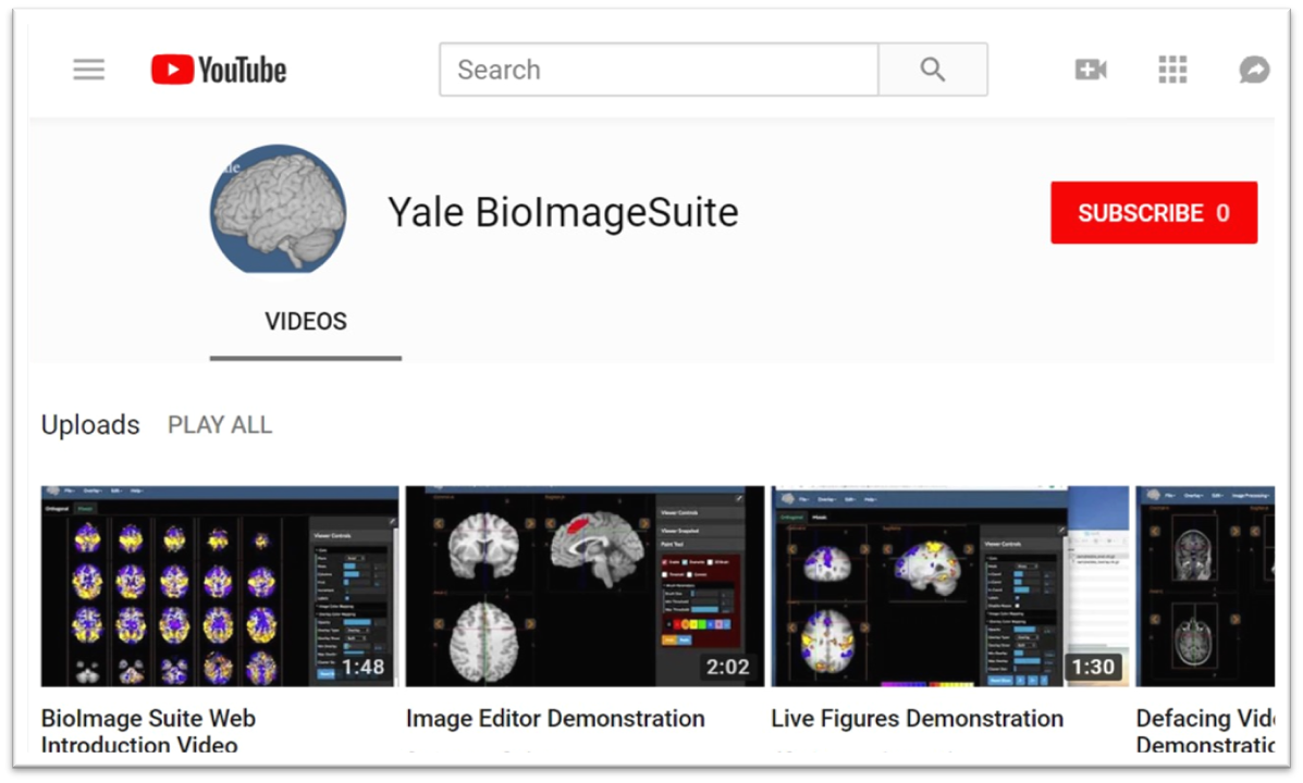
Xenophon Papademetris is a Professor of Biomedical Informatics & Data Science and Radiology & Biomedical Imaging at Yale School of Medicine. He serves as Associate Director of Biomedical Imaging Data Sciences at Yale Biomedical Imaging Institute and directs the Medical Software and Medical Artificial Intelligence Certificate Program. PhD in Electrical and Information Sciences from Yale University (2000) BA from Cambridge University (1994) Postdoctoral Fellowship at Yale University (2002) His research focuses on medical image analysis, machine learning, and biomedical software development. He has developed tools like BioImage Suite Web and contributed to standards committees at the Association for the Advancement of Medical Instrumentation (AAMI). His work spans modalities including MRI, CT, PET, and optical imaging. Recent publications emphasize neuroimaging analysis, explainable AI in healthcare, and multimodal data integration across species. He leads NIH-funded research under the BRAIN Initiative (R24 MH114805) and has authored a textbook on Medical Software published by Cambridge University Press. IEEE Senior Member Yale Brown-Coxe Postdoctoral Fellowship Harding Bliss Prize for Excellence in Engineering He directs the BioImage Suite Project, creating web-based image analysis tools using JavaScript and WebAssembly. His teaching includes both academic courses and a Coursera program on Medical Software with over 14,000 enrollments.
Anthony A Gatti is a Postdoctoral Scholar at Stanford University's Wu Tsai Human Performance Alliance and School of Medicine. His research integrates biomechanics , medical imaging , and machine learning to advance musculoskeletal health diagnostics, particularly focusing on knee osteoarthritis and exercise physiology. Education : Ph.D. in Rehabilitation Science (McMaster University, 2021), M.Sc. in Rehabilitation Science (McMaster University, 2015), B.Sc. in Kinesiology (McMaster University, 2013) His research develops automated tools for quantifying knee anatomy and integrating anatomical data with biomechanical models . These methods analyze acute responses to exercise and long-term joint degeneration, leveraging MRI , deep learning , and statistical shape modeling . Recent publications emphasize AI-driven segmentation , exercise-induced cartilage changes , and biomechanical simulations , spanning journals like Magnetic Resonance in Medicine and Arthritis & Rheumatology . Trends include machine learning validation for clinical predictions and open-source tool development for musculoskeletal analysis. Scientific Awards : CIHR Postdoctoral Fellowship (top 1%), Mitacs Accelerate Entrepreneur, Forge Student Start-Up Competition Winner, multiple scholarships from McMaster University He founded NeuralSeg , a company commercializing deep learning-based MRI segmentation technology. Collaborations include Stanford's Digital Athlete Moonshot Project with advisors like Scott Delp and Garry Gold.
Steven Meikle is a Professor of Medical Imaging Physics and Head of the Imaging Physics Laboratory at the Brain and Mind Centre, University of Sydney. He also serves as Deputy Director (Preclinical) of Sydney Imaging and Deputy Director of the National Imaging Facility's Sydney node. His expertise spans advanced imaging technologies, with a focus on PET/SPECT instrumentation and molecular imaging. He holds a B.App.Sc.(Hons) from the University of Technology Sydney and a PhD from the University of New South Wales. Research focuses include developing novel PET systems like Open-field PET (for freely moving rodents) and Total Body PET, which enhance imaging sensitivity and enable real-time behavioral studies alongside brain function analysis. Collaborations include Tsinghua University (China) and UC Davis (USA). He leads projects on motion correction, quantitative imaging, and AI-driven analysis. Key achievements include over 180 peer-reviewed publications, editorial roles in Physics in Medicine and Biology , and leadership in professional societies. Awards include IEEE Senior Membership and Australian Institute of Physics Fellowship. Current student projects explore Total Body PET applications, motion correction, and radiopharmaceutical evaluation. Teaching roles include medical physics courses in diagnostic radiography and medical physics programs. He advises on imaging ethics, facility implementation, and translational research bridging basic science and clinical applications.
Professor Fernando Calamante is a Professor of Biomedical Engineering at The University of Sydney and Director of Sydney Imaging Core Research Facility. He leads the National Imaging Facility node and focuses on advanced MRI methodologies, particularly Diffusion and Perfusion MRI, to study brain connectivity and neurological disorders. His work includes developing the MRtrix software, widely used in diffusion MRI analysis. He holds extensive funding (~$50M) and has been recognized with awards like ISMRM Fellowship and NHMRC grants. His research spans super-resolution imaging, brain connectomics, and clinical applications in stroke and tumors. Education: BSc (Physics, Argentina), PhD (Magnetic Resonance Imaging, University College London). Career highlights include leadership roles at The Florey Institute and ISMRM presidency (2021-2022). Research interests include: Novel MRI methods for brain connectivity and super-resolution imaging Applications of Diffusion and Perfusion MRI in neurology Integration of structural and functional connectomics Key achievements: Over 200 publications, software innovations, and leadership in global MRI societies.
John F Valliant is a Professor in the Department of Chemistry and Chemical Biology at McMaster University. His research focuses on radiopharmaceutical chemistry, molecular imaging, and targeted therapies, particularly in cancer diagnostics and therapeutics. He co-founded Fusion Pharmaceuticals, a company acquired by AstraZeneca for $2.4 billion (US), which specializes in targeted alpha therapy for cancer treatment. His work includes developing radiolabeled imaging agents for prostate cancer (e.g., [18F]DCFPyL), targeted alpha therapies (e.g., FPI-1434), and bioorthogonal chemistry strategies for pretargeted imaging. Valliant has contributed to advancements in photoacoustic imaging, SPECT/PET modalities, and theranostic agents for conditions like bacterial infections and bone diseases. Key projects include the Phase II clinical trial using 18F-DCFPyL PET-MRI for oligometastatic prostate cancer and studies on individualized dosimetry in Lu-177 therapies. He has pioneered platforms for radiolabeled antibody-recruiting molecules and fluorous-phase chemistry for high-specific-activity probes. Valliant’s research bridges disciplines, integrating chemistry, biology, and clinical applications to address unmet medical needs in oncology and diagnostic imaging.
Zhi-Pei Liang is the Franklin W. Woeltge Professor in the Department of Electrical and Computer Engineering at the University of Illinois at Urbana-Champaign, with joint appointments in the Department of Bioengineering, Beckman Institute for Advanced Science and Technology, and Coordinated Science Laboratory. His research spans biomedical engineering, medical imaging, and signal processing with a focus on advancing magnetic resonance imaging and spectroscopy technologies. His educational background includes a Ph.D. in Biomedical Engineering from Case Western Reserve University (1989) and a B.S. in Electrical Engineering from South-China University of Technology (1982), followed by postdoctoral training at UIUC (1989-1991). Professor Liang's research interests center on magnetic resonance imaging and spectroscopy , with particular emphasis on ultrafast imaging techniques , model-based reconstruction methods , and the integration of physics-based modeling with machine learning . His pioneering work on SPICE (SPectroscopic Imaging by exploiting spatiospectral CorrElation) has revolutionized high-resolution metabolic brain imaging by enabling label-free molecular imaging through the marriage of spin physics and machine learning. His research spans pattern recognition, parameter estimation, image formation theory, and algorithms for medical imaging applications. Analysis of his recent publications reveals a strong focus on high-resolution metabolic imaging , particularly using SPICE methodology to map brain metabolism with unprecedented detail. His work bridges fundamental physics of magnetic resonance with advanced computational methods to overcome traditional limitations in imaging speed and resolution. Current research directions include J-resolved spectroscopic imaging, deuterium-based metabolic mapping, and multimodal integration of PET and MRSI for studying neurological disorders. Elected to International Academy of Medical and Biological Engineering (2012) Gold Medal, International Society for Magnetic Resonance in Medicine (2022) Technical Achievement Award, IEEE Engineering in Medicine and Biology Society (2014) Fellow, National Academy of Inventors (2021) Author of influential book 'Principles of Magnetic Resonance Imaging' (1999) President of IEEE Engineering in Medicine and Biology Society (2011-2012) Professor Liang has advised numerous students and postdocs in biomedical imaging research and has received multiple teaching honors including the Ronald W. Pratt Outstanding Teaching Award (2005) and multiple listings among UIUC's Excellent Teachers. His research has been supported by various grants from NIH, NSF, and other funding agencies. He leads the SPICE (Spectroscopic Imaging by exploiting spatiospectral Correlation) research group which focuses on developing novel imaging techniques that combine physics-based modeling with machine learning for ultrafast metabolic imaging. His laboratory, part of the Beckman Institute's Integrative Imaging Theme, collaborates extensively with clinical researchers at Carle Illinois College of Medicine and other institutions to translate advanced imaging techniques into clinical applications for neurological disorders, cancer, and metabolic diseases. Current projects focus on high-resolution mapping of brain metabolism in Alzheimer's disease, stroke, and brain tumors using novel MR spectroscopic imaging techniques.
Yading Yuan, PhD is an Associate Professor of Radiation Oncology (Physics) at Columbia University Irving Medical Center and a member of the Data Science Institute. He holds a PhD in medical physics from the University of Chicago (2010) and completed clinical residency at Harvard Medical Physics Program (2013). His research focuses on AI-driven innovations in radiation oncology, including automated medical image analysis systems, federated learning frameworks for tumor segmentation, and data-driven approaches to personalized cancer treatment. He is certified by the American Board of Radiology and licensed in New York State. Education: PhD in Medical Physics (University of Chicago, 2010); Clinical Residency (Harvard Medical Physics Program, 2013). Research interests include: automated knowledge-based treatment planning, large-scale clinical AI systems, medical image reconstruction algorithms, and panomics integration for precision oncology. His work emphasizes translating data science advancements into clinical practice to improve patient outcomes. Key trends in his publications include federated learning for privacy-preserving medical AI, tumor segmentation in multi-modal imaging (PET/CT, MRI), and AI-driven prediction of treatment outcomes and recurrence risks. Recent work emphasizes decentralized learning architectures and cross-institutional collaboration systems. Scientific Awards: Distinguished Reviewers 2013 (selected by peer review committees) Advising/grants: No specific student names or grant details listed in provided text. His work is supported through institutional and collaborative research initiatives. Labs/teams: Active member of Columbia's Data Science Institute and Radiation Oncology department, contributing to interdisciplinary medical AI research groups.
Lars Nyberg is a Professor of Neuroscience at Umeå University's Medical Faculty and Director of the Umeå Centre for Functional Brain Imaging (UFBI) since 2001. He has held concurrent roles as Guest Professor in Bergen and Oslo, Norway, and led the Wallenberg Centre for Molecular Medicine (WCMM) from 2019. His academic leadership includes directing major initiatives like the EU-funded Lifebrain project (2017–2022) and holding the Torsten & Ragnar Söderberg Research Professorship in Medicine (2012–2017). Education: PhD in Psychology (1993) and Docent (1996) from Umeå University, with postdoctoral training at the Rotman Research Institute, Toronto. His research focuses on neuroimaging techniques (MRI/PET), dopamine systems, working memory, aging, and episodic memory. Key contributions include linking dopamine receptor availability to cognitive decline and demonstrating brain maintenance mechanisms in aging. Research Interests: Neuroimaging of memory systems, dopamine's role in cognition, aging-related brain changes, and cognitive reserve. Grants: EU Horizon 2020 (Lifebrain, €10M), KA Wallenberg Scholarships (2009, 2016), Swedish Research Council funding for COBRA (2013–2017). His awards include Royal Swedish Academy of Sciences membership (2008), Mångbergs Prize in Neural Sciences (2008), and multiple Wallenberg grants. Nyberg has supervised 27 PhD students and 11 postdocs, advancing translational neuroscience and aging research through interdisciplinary teams at UFBI and WCMM.
Saud Alhusaini MD PhD is an Assistant Professor of Neurology at the Warren Alpert Medical School of Brown University and serves as a Neurologist/Movement Disorders Specialist at Rhode Island Hospital. His research integrates imaging genomics and multimodal brain imaging approaches to investigate neurological disorders including Parkinson's disease, essential tremor, and epilepsy. He is affiliated with the Carney Institute for Brain Science and collaborates extensively with clinicians, geneticists, electrophysiologists, MRI specialists, neuropsychologists, and data scientists. Education: PhD from the Royal College of Surgeons in Ireland (RCSI) MSc in Neuroscience from Trinity College Dublin MD from University of Dublin, School of Medicine Adult neurology residency at McGill University/Montreal Neurological Institute Clinical research fellowship at Yale School of Medicine Clinical fellowship at Stanford University Medical Center Dr. Alhusaini's research focuses on identifying key endophenotypes and subclinical biomarkers to elucidate the underlying mechanisms of complex neurological conditions. His work spans multiple areas including movement disorders, epilepsy, and brain structure genetics. He has made significant contributions to understanding the genetic architecture of brain structures through his involvement with the ENIGMA consortium, which conducts large-scale collaborative analyses of neuroimaging and genetic data across institutions worldwide. An analysis of his publication record reveals a consistent pattern of high-impact research at the intersection of neurology, genetics, and advanced imaging techniques. His recent work demonstrates particular expertise in Parkinson's disease genetics, epilepsy network analysis, and movement disorder diagnostics. The breadth of his research, spanning from basic genetic mechanisms to clinical applications, highlights his comprehensive approach to understanding neurological disorders. Dr. Alhusaini has received funding from the Rhode Island Research Foundation, Brown Physicians, Inc., and Advance RI-CTR to support his research initiatives. His collaborative approach is evident through his numerous multi-institutional projects and extensive co-author network across Brown University departments including Neurology, Neurosurgery, and Pathology and Laboratory Medicine.
James S. Duncan is the Ebenezer K. Hunt Professor of Biomedical Engineering at Yale University, with additional appointments in Electrical & Computer Engineering and Radiology & Biomedical Imaging. His research focuses on biomedical image processing, quantitative image analysis using geometrical models, and applications in cardiac function and neuro-structure analysis. He has pioneered image-guided interventions and developed computational frameworks for medical imaging challenges. He holds a Ph.D. from the University of Southern California. His work integrates AI, deep learning, and statistical decision-making to advance medical imaging technologies. Notable contributions include advancements in 3D image segmentation, deformable models, and MRI-based tumor response assessment. Dr. Duncan has received prestigious awards, including IEEE Fellow (2001) and induction into the American Institute for Medical and Biological Engineering (2000). His recent research spans AI-driven hemodynamics modeling, trustworthy healthcare AI guidelines, and molecular MRI innovations in immunotherapy monitoring. He collaborates across disciplines to address challenges in cardiovascular, neuroimaging, and oncological applications.
Dr. Gary Glover is a Professor of Radiology (Radiological Sciences Lab) at Stanford University , with courtesy appointments in Psychology and Electrical Engineering. His work focuses on the physics and mathematics of MRI, particularly rapid scanning methods using spiral k-space trajectories for functional brain imaging and multimodal neuroimaging (fMRI/EEG/fPET/fNIRS) combined with neuromodulation techniques like TMS and transcranial ultrasound. Academic Appointments: Radiology, Psychology, Electrical Engineering Professional Affiliations: Bio-X, Stanford Cancer Institute, Wu Tsai Neurosciences Institute Research Interests include: Development of blood oxygen level-dependent (BOLD) and viscoelastic contrast in MRI Functional MR Elastography for brain activation mapping Optimization of MR-ARFI for transcranial ultrasound guidance Automated spinal cord segmentation (EPISeg) using machine learning Scientific Awards : National Academy of Engineering (2013) Gold Medal, ISMRM (2000) Steinmetz Award, General Electric (1985) Lauterbur Lecture, ISMRM (2018) Recent Publications analyze: Fast fMRI sampling and spurious signal correction Dissociated patterns in default mode network anti-correlations Neural correlates of collaborative behavior in triadic fMRI Salience network contributions to depression pathophysiology
Xiaofeng Liu is an Assistant Professor at Yale University School of Medicine in the Departments of Radiology & Biomedical Imaging and Biomedical Informatics & Data Science. He is also an Associate Member at the Broad Institute of MIT and Harvard. Previously, he held faculty positions at Harvard Medical School and research roles at Massachusetts General Hospital and Beth Israel Deaconess Medical Center. PhD in Mechatronics from University of Chinese Academy of Sciences Dual Bachelor's degrees in Automation (Wang-Daheng Elite Class) and Communication from University of Science and Technology of China His research integrates trustworthy AI, medical imaging, and data science to improve diagnosis, prognosis, and treatment monitoring for neurological disorders, cancer, and cardiovascular diseases. Key focus areas include domain adaptation techniques, diffusion models, and interpretable AI systems. Led special issues in IEEE Transactions on Pattern Analysis and Medical Image Analysis Developed novel frameworks like Ordinal UDA and Memory-Consistent Adaptation Scientific accolades include the Trailblazer R21 Award (NIBIB), OpenAI Research Award, and National Artificial Intelligence Research Resource Pilot Award. He serves as Associate Editor for IEEE Transactions on Neural Networks and Learning Systems and actively contributes to MICCAI and NIH review panels. His lab at Yale (XLiu Lab) investigates neural basis of intelligence to inspire AI development, with applications in brain tumor segmentation, cardiac imaging, and cross-modal medical diagnostics.