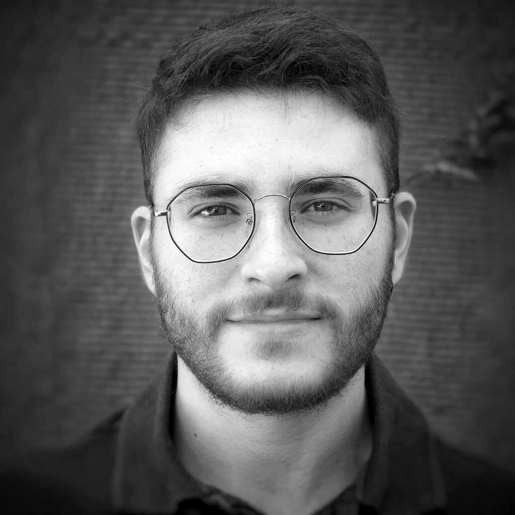
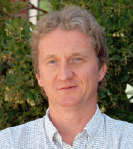
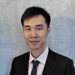
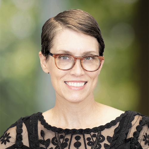
Jaime Barranco is a Researcher at the Computational Neuroanatomy & Fetal Imaging Section of the Center for Biomedical Imaging (CIBM SP CHUV-UNIL), supervised by Prof. Meritxel Bach Cuadra. His work integrates biomedical image processing, deep learning, and web/software engineering. Education: MSc in Telecommunication Engineering (2020) from Universidad Politécnica de Madrid, specializing in machine learning and multimedia data science. His research includes the A-eye project, funded by the Gelbert Foundation, which develops AI systems for MRI assessment of the eye to advance disease degeneration studies and personalized surgical planning. Collaborators include experts from Rostock University Medical Center and the ARTORG Center. Scientific Awards: Certified Widevine Implementation Partner (CWIP) from Google Jaime combines technical expertise in software engineering (Unity, DRM systems) with biomedical applications, leveraging his background in video streaming technology to innovate in medical imaging.
Dr. Carolyn McNabb is a Lecturer at the School of Psychology, Cardiff University, with a research focus on brain microstructural properties and their relationship to psychiatric disorders. Her work spans structural-functional brain connectivity, treatment resistance in schizophrenia, and pharmacological interventions. Education: PhD (Pharmacy) University of Auckland (2017), MHSc (Experimental Psychology) University of Auckland & University of Cambridge (2012), Postgraduate Diploma University of Auckland (2010) Her research integrates advanced neuroimaging techniques like MRI, MEG, and TMS to study multi-scale brain analysis. Current projects include investigating moral decision-making in virtual reality and improving fMRI preprocessing pipelines. Recent publications examine cerebellar cognition, gestational thyroid impacts on brain development, and sports-related neurotrauma. Key collaborations include institutions like University of Reading (2017-2022) and University of Auckland (2012-2017). 2020 Margaret Mead Award Lecture (British Science Association) She contributes to open science through datasets like WAND and methodological guides for neuroimaging quality control, while maintaining expertise in pharmacology, neuroimaging, and psychiatric research.
Dr. Kimberly Chan is an Assistant Professor and FIRST scholar at UT Southwestern Medical Center, jointly affiliated with the Advanced Imaging Research Center and the Department of Biomedical Engineering. Her work bridges biomedical engineering and clinical neuroscience through the development of advanced magnetic resonance spectroscopy (MRS) techniques. Research Interests: Dr. Chan's research centers on the development and application of novel MRS methods to study brain chemistry in neurological and psychiatric disorders. Her work focuses on spectral editing, metabolite quantification (e.g., GABA, glutathione), motion correction, and multi-site reproducibility. She aims to improve diagnosis, disease monitoring, and treatment planning through non-invasive neurochemical profiling. Publication Trends: Her recent publications demonstrate a strong focus on methodological innovation in MRS, including Hadamard encoding (HERMES, HERCULES), multi-metabolite detection, motion correction, and standardization across scanners and sites. These techniques are applied to conditions such as Huntington’s disease, Lafora disease, spinal cord injury, and cancer, showing translational impact. Scientific Awards: First Scholar Advising and Grants: As principal investigator of the CHAN Lab (Chemical Advanced Neuroimaging Lab), she mentors graduate students and postdoctoral fellows. She is actively recruiting and likely holds independent research funding, supported by her FIRST scholar status. While specific grants and advisees are not listed, her leadership role indicates active research supervision and extramural funding. Labs and Teams: Dr. Chan leads the CHAN Lab, a multidisciplinary team of biomedical engineers and imaging scientists focused on advancing MRS for clinical applications. The lab collaborates widely, as evidenced by multi-center studies like Big GABA, involving over 25 research sites globally.
Matthew D. Nelson is a Professor of Biology at Saint Joseph's University in Philadelphia, where he also serves as the John J. Meehan, M.D., Pathways to Medical Professions Director and Health Professions Advising Committee Co-Director. His research program in the Department of Biology within the College of Arts and Sciences focuses on understanding the fundamental biological processes of sleep using the model organism Caenorhabditis elegans . Dr. Nelson's educational background includes a BS in Mathematics from Pennsylvania State University (2000), an MS in Biology from Villanova University (2005), and a PhD in Biology from New York University (2010). He completed postdoctoral training in the Lab of Dr. David M. Raizen at the University of Pennsylvania Perelman School of Medicine (2011-2014). His research investigates the genetic and neural circuit mechanisms underlying sleep behavior, with particular focus on neuropeptide signaling pathways that regulate stress-induced sleep in C. elegans . Dr. Nelson's lab employs techniques from genetics, molecular biology, neurobiology, and behavior to identify sleep-regulating neurons and characterize how they communicate as neural circuits. His work has revealed important insights into conserved sleep mechanisms across species. Analysis of his recent publications shows a consistent focus on sleep neurogenetics, with particular emphasis on neuropeptide receptors like npr-38 and signaling pathways involving cAMP. His research spans molecular mechanisms, neural circuit function, and behavioral outputs, demonstrating an integrated approach to understanding sleep regulation at multiple biological levels. NSF CAREER Award #1845020: Neuropeptidergic and cAMP-mediated regulation of stress-induced sleep in C. elegans NSF MRI Award #1919847: Acquisition of an automated fluorescent microscope and imaging system for undergraduate research NIGMS R15 Award 1R15GM122058-01: Dissecting the cAMP-mediated circuitry of stress-induced sleep in C. elegans Dr. Nelson actively mentors undergraduate and graduate students, many of whom have presented at conferences and gone on to medical schools and research positions. His lab also engages in science outreach with the Samuel Gompers school, teaching 5th and 6th grade students about basic science research using C. elegans . His educational contributions include developing classroom models for nephron function and urinalysis experiments.
Cherise Chen is an Assistant Professor in Computer Vision at the Department of Computer Science, University of Sheffield. She also holds positions as a Visiting Researcher in the Oxford BioMedIA Group at the University of Oxford and an Honorary Research Fellow at Imperial College London. As a core member of the Insigeno Institute and Shef.AI community, Dr. Chen leads research at the intersection of artificial intelligence and healthcare, focusing on translating cutting-edge AI techniques into practical medical applications. Dr. Chen's research program centers on developing robust, data-efficient machine learning algorithms for medical image analysis. Her work spans adversarial data augmentation, robust machine learning frameworks, and data-efficient learning techniques including self-supervised, few-shot, and semi-supervised approaches. She has made significant contributions to multi-task and multi-modal learning, adaptive machine learning systems, and algorithms with built-in considerations for fairness, privacy, robustness, and interpretability. Her research specifically targets clinical applications in cardiac image analysis (including segmentation, registration, and shape remodeling with quality control), prostate image analysis integrated with pathological image analysis, and brain image segmentation for clinical use cases. Analysis of Dr. Chen's recent publications reveals a strong focus on addressing the practical challenges of deploying AI in real-world medical settings. Her work on test-time adaptation methods (2023), adversarial style composition (2022), and cooperative training frameworks (2021) demonstrates her commitment to creating AI systems that maintain performance despite domain shifts and limited labeled data. She has consistently contributed to top-tier medical imaging conferences, with multiple papers accepted to MICCAI from 2018-2023, reflecting her standing in the medical imaging research community. IEEE TMI Gold-level Distinguished Reviewer Award (2022-2023) MICCAI 2023 Outstanding Reviewer Award Winner of the Fetal Tissue Annotation and Segmentation Challenge (FeTA) 2022 Winner of the Multi-sequence Cardiac MR Segmentation Challenge 2019 China National Scholarships (twice, top 0.2%) Dr. Chen actively mentors students and has delivered invited talks at prestigious institutions including Johns Hopkins University, Technical University of Munich, and the German Cancer Research Center. Her laboratory at Sheffield focuses on advancing deep medical image segmentation with particular attention to robustness, reliability, and real-world applicability in clinical workflows.
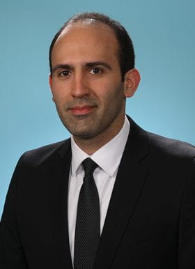
Borna Maraghechi is a Clinical Physicist in the Department of Radiation Oncology at Washington University School of Medicine, St. Louis, MO. He completed his academic and professional training in Iran, Canada, and the U.S., including a residency in Therapy Medical Physics at WashU. BSc in Physics (2009), Amirkabir University of Technology, Tehran, Iran MSc in Physics (2012), Laurentian University, Sudbury, Canada PhD in Biomedical Physics (2016), Ryerson University, Toronto, Canada Residency in Therapy Medical Physics (2020), Washington University School of Medicine His research focuses on advanced radiotherapy techniques, particularly MR-guided adaptive therapy, dosimetry innovation, and quality assurance for emerging treatment modalities. He develops methods for precision radiation delivery in complex anatomical regions, with emphasis on gastrointestinal and pancreatic cancers. His recent work spans 2020–2025, addressing challenges in cone-beam CT for proton therapy, toxicity prediction models, and automated quality assurance. Key themes include medical imaging integration, treatment personalization, and safety protocols for adaptive radiation therapy. Certified by the American Board of Radiology in Therapeutic Medical Physics (2021)
BOHI Amine is a researcher at CESI (School of Engineering and Digital Tools, Department of Computer Science), with a PhD in Computer Science from the University of Toulon (2017). His academic profile spans disciplines including Machine Learning, Signal and Image Processing, and Biomedical Engineering, with a focus on applications in Digital Health and Human-Robot Interaction. Education : PhD in Computer Science (University of Toulon, 2013-2017); Master 2 in Computer Science (University of Fes, Morocco, 2009-2011); Bachelor’s in Mathematical and Computer Sciences (University of Fes, 2006-2009). His research interests include: Machine Learning and Deep Learning Signal and Image Processing 3D Shape Analysis Computer Vision Digital Health Biomimetic Feature Design BOHI’s publications reflect expertise in facial emotion recognition for elderly care, cortical folding modeling, and soft tissue organ deformation analysis using MRI. He supervises diverse research internships, mentoring students from institutions like Sorbonne Paris Nord, Université de Technologie King Mongkut, and INP Grenoble. Current projects involve developing intelligent solutions for emotional interaction with elderly individuals suffering from neurodegenerative disorders, in collaboration with VyV3 Bourgogne. He also contributes to the design of multimodal datasets and wearable health monitoring systems. BOHI’s technical work includes contributions to maritime surveillance systems (PARE project) and semantic information platforms, demonstrating a capacity to bridge theoretical research with practical applications across domains.
Karla Miller is a Professor of Biomedical Engineering at the University of Oxford , where she serves as Director of the Oxford Centre for Integrative Neuroimaging (OxCIN) and leads the MRI Physics Group . Her research focuses on advancing MRI technology to study tissue microstructure, brain connectivity, and neuronal health, with applications in UK Biobank harmonization and post-mortem imaging validation.
Professor David G Kiely is an Honorary Professor of Pulmonary Vascular Medicine at the University of Sheffield , affiliated with the School of Medicine and Population Health and leading the Sheffield Pulmonary Vascular Disease Unit . His career spans clinical and research excellence in pulmonary hypertension, integrating multimodality imaging and artificial intelligence to advance diagnostics and risk stratification. Education : BSc (Hons), MD from the University of Edinburgh Clinical Leadership : Director of Sheffield Pulmonary Vascular Disease Unit since 2001 Research Themes : Pulmonary Hypertension, Imaging Biomarkers, AI Applications His work bridges clinical practice and translational research, focusing on CT and MRI imaging for pulmonary hypertension, AI-driven diagnostics , and rare disease phenotyping . Recent studies include global collaborations on CTEPH registries and personalized treatment outcomes . Key scientific contributions include over 150 publications and a 2017 NIHR/RCP award . Current research involves Phoenix trial (personalized PAH therapy), REPAIR study (macitentan effects), and CIPHER studies (biomarker discovery). He collaborates with interdisciplinary teams in imaging (AJ Swift, JM Wild) , preclinical models (A Lawrie) , and clinical networks .
Theresia Ziegs is a Research Associate at the Tübingen Center for Digital Education (TüCeDE) and serves as the link to the AI Makerspace , an extracurricular learning center for innovative technologies. She focuses on developing AI-supported methods for adaptive teaching within the MINT-ProNeD project and coordinates courses on robotics and AI for students, alongside designing teacher training programs in these fields. Education : Dr. rer. nat. in Neuroscience (2017–2023), Max Planck Institute for Biological Cybernetics, Tübingen M.Sc. in 2016, University of Rostock B.Sc. in Physics (2011–2014), University of Rostock Her research centers on neuroscience , particularly using 1H/13C FID-MRSI at ultra-high magnetic fields (9.4T) to study brain metabolism, including glutamate and glucose dynamics . Her work emphasizes methodological optimization for improved reproducibility and spatial resolution in metabolic imaging. Her publications highlight expertise in metabolic mapping , signal processing , and machine learning-enhanced imaging techniques , with applications in human brain metabolism and neuroimaging . She has received the ISMRM Magna Cum Laude Merit Award (2022) for her contributions. Scientific Awards : ISMRM Magna Cum Laude Merit Award (2022) Theresia actively collaborates on AI integration in education and teacher training , bridging advanced neuroimaging research with pedagogical innovation at the University of Tübingen.
Dr. Webster H. Pilcher is a Professor and Chair of Neurosurgery at the University of Rochester Medical Center (URMC), where he holds the Ernest & Thelma Del Monte Distinguished Professorship in Neuromedicine. He completed his MD/PhD in Neuroscience/Anatomy at URMC, trained in neurological surgery at Rochester, and pursued epilepsy/brain tumor research at the University of Washington. Education: MD/PhD, University of Rochester School of Medicine/Dentistry (Neuroscience/Anatomy) Undergraduate, Colgate University Dr. Pilcher specializes in epilepsy surgery, brain/spinal tumors, and neurodegenerative diseases. His research includes brain mapping , neurosurgical oncology , and cost-effective care models . Recent publications focus on single-cell genomics , mobile stroke units , and functional connectivity studies . Scientific Awards: Merit Award (2008) Upjohn Surgical Research Award (1987) Resident Research Award (1986) Alpha Omega Alpha (1983) As a clinician-scientist, Dr. Pilcher expanded URMC Neurosurgery to national prominence with $7.9M+ annual funding. He leads the Rochester Neurosurgery Partners initiative and resides in Honeoye Falls, NY, with his family.
Robert Sainburg is the Dorothy Foehr Huck and J. Lloyd Huck Distinguished Chair in Kinesiology and Neurology at Penn State University and Penn State College of Medicine. He directs the Center for Movement Science and Technology (C-MOST) and operates two laboratories: the Movement Neuroscience Laboratory at University Park and the Neurorehabilitation Research Laboratory at Hershey Medical Center. Currently holding professorships in both Kinesiology and Neurology, Sainburg's work bridges basic neuroscience with clinical rehabilitation applications. Education Ph.D. in Neuroscience from Rutgers University (1993) MS in Neurobiology and Physiology from Rutgers University (1989) BS in Occupational Therapy from New York University (1984) Postdoctoral Fellowship in Neurobiology under Dr. Claude Ghez at Columbia University (1993-1996) Research Focus Sainburg's work centers on neural lateralization for motor control , developing the Dynamic Dominance Model that explains hemisphere-specific contributions to predictive and impedance control. His research spans: Multi-joint arm coordination mechanisms Stroke-induced motor deficits analysis Proprioceptive feedback systems Virtual reality rehabilitation frameworks Bi-hemispheric brain function Neurological patient population studies Scientific Recognition Elected Fellow, National Academy of Kinesiology (2024) Huck Distinguished Chair (2021) AOTF Academy of Research (2019) Pattishall Research Achievement Award (2008) Research Infrastructure As C-MOST director, Sainburg integrates two major facilities: the Movement Neuroscience Lab (University Park campus) focuses on basic motor control mechanisms, while the Neurorehabilitation Lab (Hershey Medical Center) directly applies findings to clinical populations. The Kinereach VR system , developed in-house, enables precise movement tracking and distortion experiments.
John Olichney is a Professor at the University of California, Davis, specializing in behavioral neurology with expertise in cognitive disorders and neurodegenerative diseases including Alzheimer's disease. Board-certified in behavioral neurology, he has served on the Bioethics Committee (2009-2022) and Institutional Review Board (2010-2011). His educational background includes: M.D. from the University of California, Irvine (1988) B.A. in Neurobiology with honors from UC Berkeley (1983) Dr. Olichney's research focuses on electrophysiological and neuroimaging mechanisms of language and memory in neurodegenerative disorders. His laboratory develops biomarkers for early detection of Alzheimer's disease using event-related potentials , EEG oscillations , and neuroimaging techniques . Key interests include cognitive biomarkers , memory impairment diagnostics , and neural correlates of language processing in aging populations. Analysis of his recent publications reveals a consistent emphasis on electrophysiological biomarkers for Alzheimer's disease progression, with significant contributions to P300 and N400 event-related potential research. His work bridges basic electrophysiology with clinical applications, particularly in predicting cognitive decline and validating diagnostic tools across diverse populations. His scientific awards include: NARSAD Young Investigator Award (2000-2003) NARSAD Young Investigator Award (1997-1999) NIH Physician Scientist Award (National Institute on Aging) (1995-2001) Honors in Neurology Research (1987) Dr. Olichney teaches medical students, residents, and fellows in behavioral neurology and dementia care while mentoring research fellows and graduate students in cognitive neuroscience methods. His NIH-funded research has established critical electrophysiological markers for neurodegenerative disease progression. He directs the Cognitive Electrophysiology and Neuroimaging Lab, which integrates EEG, ERP, and neuroimaging techniques to develop sensitive diagnostic tools for memory and language impairments in aging and neurodegenerative conditions.
Yihao Liu serves as Research Assistant Professor in Vanderbilt University's School of Engineering, Department of Electrical and Computer Engineering, focusing on clinically relevant image analysis for personalized treatment strategies and large-scale medical data interpretation. His work bridges engineering and clinical applications through advanced computational methodologies. Education: PhD in Electrical and Computer Engineering, Johns Hopkins University (May 2024) Dr. Liu's research spans medical image analysis, deformable registration, deep learning, and computer vision with applications in CT, MRI, and OCT imaging. He develops AI-driven tools for pulmonary nodule diagnosis, MS lesion analysis, body composition assessment, and dermatological imaging, emphasizing clinical translation and multi-modal data fusion. His 2024-2025 publications reveal concentrated innovation in diffusion models for field-of-view extension, bi-directional lesion synthesis, and unsupervised registration techniques, demonstrating leadership in foundational registration models and longitudinal analysis for precision medicine. Dr. Liu directs the Vanderbilt Lab for Immersive AI Translation (VALIANT) and collaborates with the Medical-image Analysis and Statistical Interpretation Lab (MASI) under VISE Affiliate Bennett Landman, PhD. He holds the Stevenson Chair in Electrical and Computer Engineering and participates in NIH grant writing initiatives through Vanderbilt's medical partnerships.
Jey Koehler , DVM, PhD, DACVP, is a Professor and Tyler & Frances Young Endowed Professor in Pathology at the Department of Pathobiology , College of Veterinary Medicine , Auburn University . She serves as Section Chief for Surgical Pathology Service and Faculty Supervisor of the Core Histopathology Laboratory.