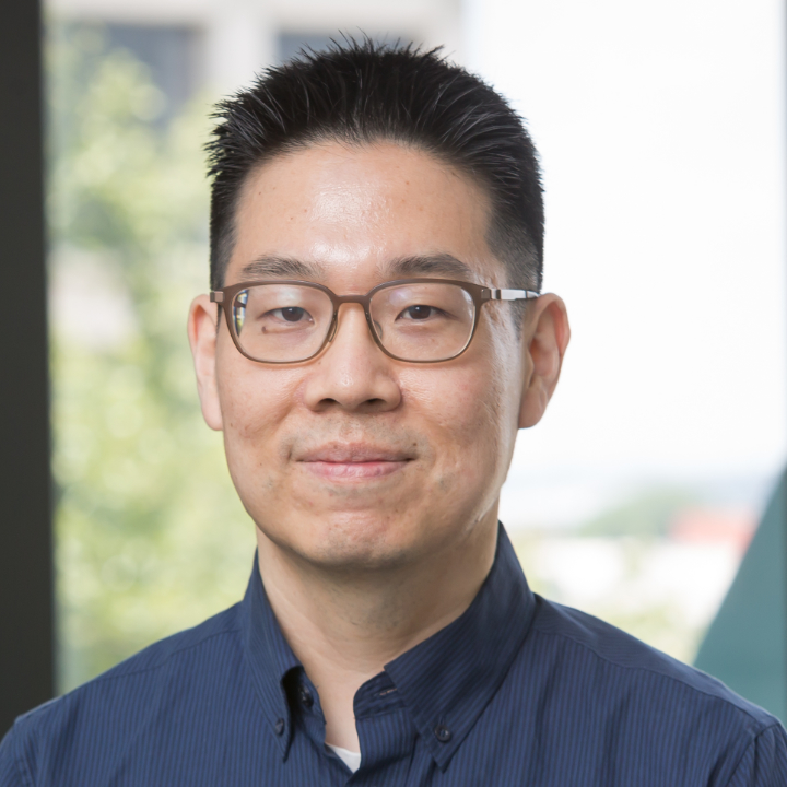
Dr. Jeremy Gordon is an Associate Professor in the Department of Radiology & Biomedical Imaging at the University of California, San Francisco (UCSF). He holds a PhD in Medical Physics from the University of Wisconsin-Madison (2013) and a BS in Physics & Astronomy from the University of Georgia (2008). Prior to his faculty role, he worked as a postdoctoral scholar (2013–2016) and Senior Bioengineer (2016–2020) at UCSF. His research focuses on developing novel hyperpolarized MRI techniques for metabolic imaging, particularly in pancreatic cancer, multiple sclerosis, Alzheimer’s Disease, and brain perfusion studies. These methods leverage hyperpolarized 13C and x-nuclei to assess metabolic pathways and treatment responses. Key applications include non-invasive metabolic biomarker identification and improved clinical care for cancer and neurological disorders. Major awards include the Distinguished Reviewer Award (2018), RSNA ITARSc Program recognition (2017), and multiple merit awards from the International Society for Magnetic Resonance in Medicine (2013). He is affiliated with the Advanced Imaging Technologies Resource Group and the Hyperpolarized MRI Technology Resource Center at UCSF. Recent work emphasizes multi-center clinical trials, data standardization (DICOM integration), and open-source tools for X-nuclear imaging. His lab collaborates widely, with studies involving metabolic imaging of the brain, abdomen, and heart under various physiological conditions (e.g., glucose challenges).
G. Wilson Miller is an Associate Professor of Radiology and Medical Imaging and Biomedical Engineering at the University of Virginia. He holds a PhD in Nuclear Physics from Princeton University (2000) and a BS in Mathematics from the University of Maryland (1993). His research focuses on MRI technique development, hyperpolarized gas imaging, and MR-guided focused ultrasound surgery. Key research areas include: Hyperpolarized noble gas MRI to map lung oxygen levels (PAO2) and microstructure Development of fast spiral pulse sequences for 3D PAO2 imaging RF coil design for hyperpolarized gas imaging MRI-guided focused ultrasound for non-invasive surgical interventions Notable projects include collaborations with the physics department to build a helium-3 hyperpolarizer and studies on focused ultrasound for blood-brain barrier opening. His work has advanced applications in lung cancer treatment planning, COPD phenotyping, and neurological disorders.
Dr. Wei-Tang Chang is an Assistant Professor in the Department of Radiology at the University of North Carolina School of Medicine. His research focuses on advancing ultrahigh-resolution functional and diffusion MRI techniques, with emphasis on improving spatial and temporal resolution while reducing scan times. Key projects include submillimeter isotropic-resolution fMRI for hippocampal subfield analysis (funded by NIH R21), novel dMRI approaches to overcome resolution limits, and clinical translation of robust imaging methods resistant to motion/noise artifacts. Dr. Chang holds a PhD in Biomedical Engineering from National Taiwan University and completed postdoctoral training at the Martinos Center for Biomedical Imaging (MGH), Massachusetts General Hospital, and Singapore BioImaging Consortium (SBIC). His research innovations include SORDINO fMRI for awake rodent imaging, pPRISM diffusion MRI for submillimeter resolution, and ZTE pulse sequences for ultra-fast acquisitions. Awards include the 2011 OHBM Trainee Award for work on MEG source localization and fMRI temporal resolution breakthroughs. Current work bridges basic neuroimaging science with clinical applications, particularly in neurodegenerative disease biomarker development and rodent disease model studies. Education: PhD in Biomedical Engineering, National Taiwan University Postdoctoral Fellowships: Martinos Center for Biomedical Imaging (MGH) Singapore BioImaging Consortium (SBIC) Key Technologies Developed: ZTE pulse sequences (25 ms temporal resolution fMRI) pPRISM diffusion MRI (navigator-free submillimeter imaging) Draining-vein suppression layer-dependent fMRI Awards & Funding: NIH R21 Grant (2019) for hippocampal subfield fMRI OHBM Trainee Award (2011) Dr. Chang's translational focus involves adapting laboratory innovations for clinical use, with particular interest in Alzheimer's disease biomarkers through hippocampal network analysis and Huntington's disease models using rodent functional connectivity studies. His lab actively develops open-source MRI reconstruction algorithms and collaborates internationally on multi-center neuroimaging projects.
Mathias Nilsson is a Professor of Physical Chemistry at the School of Chemistry, University of Manchester. He holds a BSc in Food Chemistry from Linneaus University (1993) and a PhD in Food Science from the Swedish University of Agricultural Sciences (1999). He has held postdoctoral positions in Aveiro, Portugal (2002–2003) and Manchester (2004–2007), followed by an EPSRC Advanced Research Fellowship. His research focuses on developing novel methods in liquids NMR spectroscopy, particularly in analyzing complex mixtures using diffusion-ordered spectroscopy (DOSY) and pure shift techniques. He leads the Mathias Nilsson Research Group, which collaborates globally and contributes to the Manchester NMR Methodology Group. His work addresses challenges in mixture analysis, spectral overlap resolution, and multidimensional NMR methodologies. Education: BSc in Food Chemistry, Linneaus University (1993) PhD in Food Science, Swedish University of Agricultural Sciences (1999) Research Interests: His group specializes in advancing NMR spectroscopy for mixture analysis, including DOSY, pure shift techniques, and multivariate data analysis. Key areas include reducing spectral overlap via pure shift DOSY, 3D DOSY methods, and covariance-based signal processing. Applications span pharmaceuticals, materials science, and food chemistry. The group also develops software tools like the DOSY Toolbox for diffusion data processing. Key Achievements: EPSRC Advanced Research Fellowship (2007) Contributions to economic and technological impacts via DOSY and pure shift NMR methodologies Development of the DOSY Toolbox software Advising & Grants: Supervised multiple PhD students and postdocs, including those working on pure shift NMR, matrix-assisted DOSY, and diffusion-based mixture analysis. His research aligns with UN Sustainable Development Goals through innovations in analytical chemistry. Labs & Teams: Part of the Manchester NMR Methodology Group and collaborates with international researchers in spectroscopy and analytical chemistry.
Marco Pizzolato is an Associate Professor in the Department of Applied Mathematics and Computer Science at the Technical University of Denmark (DTU), specializing in Visual Computing with a focus on Magnetic Resonance Imaging (MRI), particularly diffusion MRI and biophysical modeling. He is also affiliated with the inter-departmental Microstructure & Plasticity (MAP) research group and has held visiting positions at the University of Verona, EPFL, and DRCMR. His educational background includes a PhD in Signal and Image Processing from INRIA Sophia Antipolis, a Master’s in Bioengineering from the University of Padua, and a Bachelor’s in Biomedical Engineering from the same institution. He previously served as an Assistant Professor at DTU and was a postdoctoral researcher under the Marie Curie COFUND Eurotech programme. Dr. Pizzolato's research centers on image and signal denoising, inverse problems, optimization, diffusion MRI, tractography, and Monte Carlo simulations. He actively contributes to the development of microstructural models for brain imaging, with applications in neurodegenerative diseases and brain connectivity. His work aligns with UN Sustainable Development Goals, particularly in advancing education and health through imaging technology. The recent publications reflect a strong trend in advancing diffusion MRI techniques, including ACID imaging, microscopic propagator modeling, myelin integrity mapping, and multi-scale white matter organization. These works emphasize biophysical accuracy, model validation, and integration across imaging modalities and species. Magna Cum Laude , ISMRM 2020 Magna Cum Laude , ISMRM 2022 First Place , Macaque Validation Challenge at ISBI 2018 First Place (Overall and HCP) , IronTrack Challenge 2019 (MICCAI) MICCAI Student Travel Award 2015 He has supervised PhD students such as Thøgersen, T. L. and Corral Bolaños, M. in projects related to microstructure MR imaging and myelin mapping. He has also been involved in significant grants and collaborative projects, including the Multimodal Microstructure-Informed Connectivity (MMINCARAV) initiative between Inria and EPFL, and the Sinergia consortium for Brain Communication Pathways . He co-organized multiple international events, including the MICCAI CDMRI workshops and challenges (2019–2021), and the ESMRMB Leaps in Microstructure Imaging workshop (2024). Dr. Pizzolato is an active member of the scientific community, serving as an editor for MICCAI workshop proceedings, a reviewer for major journals and conferences, and an invited speaker at ISMRM 2025. He leads and participates in several ongoing research projects at DTU focused on quantitative imaging, myelin mapping, and MRI-based connectivity, demonstrating sustained research leadership and external funding success.
Dr. Lirong Yan is a tenured Associate Professor at Northwestern University's Feinberg School of Medicine and McCormick School of Engineering. She specializes in developing MRI techniques for cerebrovascular and perfusion imaging, particularly arterial spin labeling (ASL) and 4D MRA. Her research focuses on translating innovations into clinical applications for stroke, Alzheimer’s disease, and neurodegenerative conditions. She directs the Neurovascular Imaging Technology and Translation (NITT) lab, emphasizing non-invasive diagnostic tools and imaging biomarkers. Education: PhD in Biophysics from Chinese Academy of Sciences (2010), Postdoc at UCLA (2013). Professional roles include leadership in ISMRM and American Society of Neuroradiology. Research interests span MRI sequence development, fast imaging, and clinical translation. Key areas include cerebral vascular disease, neurovascular coupling, and aging-related vascular changes. Awards include Alzheimer’s Greater Los Angeles Young Investigator Award (2018) and multiple ISMRM accolades. Active clinical trials investigate cerebrovascular dysfunction in neurodegenerative diseases, using MRI to assess collateral flow and microvascular health. Her work bridges basic science and clinical practice, advancing early detection and treatment monitoring strategies.
Jae Mo Park is an Associate Professor at UT Southwestern Medical Center, affiliated with the Graduate School of Biomedical Sciences and the Department of Biomedical Engineering. He also holds appointments in Electrical Engineering at UT Dallas, where he teaches courses such as Digital Image Processing and Signals and Systems. Education : Electrical Engineering degrees (undergraduate and graduate) from Stanford University; doctoral research on MRI/MRSI for glioma brain tumor metabolism. His lab develops hyperpolarized MRI/MRS methods to assess in vivo metabolic fluxes, focusing on mitochondrial function in diseases like cancer, diabetes, and traumatic brain injury. Research integrates MR pulse sequence design, physiology, and clinical translation. Recent publications highlight hyperpolarized 13C/15N probes for metabolic imaging of the liver, brain, and heart, with applications in fatty liver disease, TBI, and exercise physiology. Awards and grants are not explicitly mentioned, but his work includes collaborations across institutions. Dr. Park teaches courses in neuroimaging, molecular imaging, and metabolic imaging at UT Southwestern and advanced image processing at UT Dallas. The Park Lab investigates metabolic disease characteristics and clinical translation of imaging techniques, with open positions for graduate researchers.
Professor David Porter is a physicist specializing in magnetic resonance imaging (MRI) physics, affiliated with the School of Psychology & Neuroscience at the University of Glasgow. He joined the university in 2017 to work on the 7T MRI scanner at the Imaging Centre of Excellence (ICE) on the Queen Elizabeth University Hospital campus. His research focuses on optimizing MRI methods for clinical and neuroscience applications, particularly leveraging high-field 7T systems to enhance image quality and applicability in neurology, diffusion-weighted imaging (DWI), motion correction, and parallel transmission techniques. Education: B.Sc. in Physics from Sheffield University, Ph.D. from King’s College London (focusing on magnetic resonance spectroscopy of human tumors). Postdoctoral work at Great Ormond Street Hospital (developing MRI methods for epilepsy and stroke in children) and industry experience at Siemens Healthcare and the Fraunhofer MEVIS Institute, where he led projects in MRI sequence design and motion correction. Research interests include advancing MRI technology for clinical translation, improving diffusion-weighted imaging, and developing novel techniques for high-field MRI systems. His work emphasizes practical applications such as cervical spine imaging, B1+ shimming, and multislice-to-volume motion correction. Publications span MRI coil design, SAR management, and artifact reduction in spectroscopic imaging. Collaborations involve teams at ICE, SINAPSE, and international institutions. His contributions bridge academic research and clinical MRI advancements.
Dr. Kimberly Chan is an Assistant Professor and FIRST scholar at UT Southwestern Medical Center, jointly affiliated with the Advanced Imaging Research Center and the Department of Biomedical Engineering. Her work bridges biomedical engineering and clinical neuroscience through the development of advanced magnetic resonance spectroscopy (MRS) techniques. Research Interests: Dr. Chan's research centers on the development and application of novel MRS methods to study brain chemistry in neurological and psychiatric disorders. Her work focuses on spectral editing, metabolite quantification (e.g., GABA, glutathione), motion correction, and multi-site reproducibility. She aims to improve diagnosis, disease monitoring, and treatment planning through non-invasive neurochemical profiling. Publication Trends: Her recent publications demonstrate a strong focus on methodological innovation in MRS, including Hadamard encoding (HERMES, HERCULES), multi-metabolite detection, motion correction, and standardization across scanners and sites. These techniques are applied to conditions such as Huntington’s disease, Lafora disease, spinal cord injury, and cancer, showing translational impact. Scientific Awards: First Scholar Advising and Grants: As principal investigator of the CHAN Lab (Chemical Advanced Neuroimaging Lab), she mentors graduate students and postdoctoral fellows. She is actively recruiting and likely holds independent research funding, supported by her FIRST scholar status. While specific grants and advisees are not listed, her leadership role indicates active research supervision and extramural funding. Labs and Teams: Dr. Chan leads the CHAN Lab, a multidisciplinary team of biomedical engineers and imaging scientists focused on advancing MRS for clinical applications. The lab collaborates widely, as evidenced by multi-center studies like Big GABA, involving over 25 research sites globally.
Theresia Ziegs is a Research Associate at the Tübingen Center for Digital Education (TüCeDE) and serves as the link to the AI Makerspace , an extracurricular learning center for innovative technologies. She focuses on developing AI-supported methods for adaptive teaching within the MINT-ProNeD project and coordinates courses on robotics and AI for students, alongside designing teacher training programs in these fields. Education : Dr. rer. nat. in Neuroscience (2017–2023), Max Planck Institute for Biological Cybernetics, Tübingen M.Sc. in 2016, University of Rostock B.Sc. in Physics (2011–2014), University of Rostock Her research centers on neuroscience , particularly using 1H/13C FID-MRSI at ultra-high magnetic fields (9.4T) to study brain metabolism, including glutamate and glucose dynamics . Her work emphasizes methodological optimization for improved reproducibility and spatial resolution in metabolic imaging. Her publications highlight expertise in metabolic mapping , signal processing , and machine learning-enhanced imaging techniques , with applications in human brain metabolism and neuroimaging . She has received the ISMRM Magna Cum Laude Merit Award (2022) for her contributions. Scientific Awards : ISMRM Magna Cum Laude Merit Award (2022) Theresia actively collaborates on AI integration in education and teacher training , bridging advanced neuroimaging research with pedagogical innovation at the University of Tübingen.
Jennifer M. Connelly, MD is a Professor of Neurology at the Medical College of Wisconsin (MCW), specializing in Neuro-Oncology. She serves as Co-Director of the Neuro-Oncology Program and CNS DOT Chair at Froedtert Cancer Center. Dr. Connelly is board certified in Internal Medicine, Neurology, and Neuro-Oncology, with clinical expertise in brain tumors, metastases to the nervous system, neurofibromatosis, and neurologic complications of cancer therapies. Dr. Connelly received her MD from the Medical College of Wisconsin in 2003 and completed her combined Internal Medicine-Neurology residency and Neuro-Oncology fellowship at MCW-affiliated hospitals. A native of the Milwaukee area, she has built her entire academic career at MCW, progressing from Instructor (2008-2009) to Assistant Professor (2009-2014), Associate Professor (2014-2022), and ultimately Professor (2022-present). Her research focuses on molecular pathology and advanced MRI imaging techniques for diagnosing and treating brain and spinal cord tumors. Dr. Connelly's work centers on radio-pathomic mapping, glioma invasion beyond traditional MRI-defined margins, and novel therapeutic approaches for treatment-resistant glioblastoma. She has pioneered the development of fractional brain tumor burden mapping and has conducted significant research on gallium maltolate as a potential treatment for glioblastoma. Her holistic patient-centered approach emphasizes not only tumor management and life prolongation but also maintaining quality of life. Analysis of Dr. Connelly's recent publications reveals a strong focus on integrating advanced imaging techniques with tumor biology to improve diagnosis, treatment planning, and outcome prediction in neuro-oncology. Her work spans radio-pathomic mapping, glioma invasion patterns, novel therapeutics like gallium maltolate, and optimization of radiation therapy approaches, demonstrating a consistent commitment to translating research into clinical practice. Top Doctors 2023 Award, Milwaukee Magazine Distinguished Alumna of the Year Award - College of Engineering, Marquette University (2023) Lee A. Biblo Excellence in Professionalism Award (2022) Medical College of Wisconsin Outstanding Medical Student Teaching Award (multiple years) Edward J. Lennon Endowed Clinical Teaching Award Young Alumna of the Year Award - College of Engineering, Marquette University (2014) Dr. Connelly has been instrumental in developing educational programs for residents and medical students, including "Card Rounds" and "Neuropictionary." She established the inpatient neuro-oncology consult service and developed the first community-based neuro-oncology clinic at Menomonee Falls. As Co-Director of the Neuro-Oncology Program, she leads the weekly Brain Tumor Board where cases are documented in real-time in the EMR. Dr. Connelly also created a comprehensive Glioma Database containing treatment histories and outcomes for nearly 900 patients. Her laboratory and research team work within the Wisconsin Institute of Neuroscience (WINS) and collaborate extensively with the MCW Cancer Center. Current research focuses on radio-pathomic mapping, gallium maltolate efficacy, and advanced imaging techniques to identify tumor invasion beyond traditional MRI-defined margins. Dr. Connelly serves as Neuro-oncology Fellowship Program Director and continues to expand her research into novel therapeutic approaches for treatment-resistant brain tumors.
Dr. Chang-Hoon Choi is a researcher at the Institute of Neurosciences and Medicine (INM) , Forschungszentrum Jülich GmbH, Germany, with a focus on Medical Imaging Physics (INM-4) . His work bridges Magnetic Resonance Imaging (MRI) , PET-MRI hybrid systems , and RF coil instrumentation for neuroscience applications. Research Highlights: Development of double-tuned coils for 1H/X-nuclei imaging, ultra-high field MRI systems , and MR-PET hybrid technologies . Technical Expertise: Specializes in RF antenna arrays , signal optimization , and multinuclear MRI/MRS for brain studies. Key Article Trends : Over 15 recent publications emphasize coil design innovations (e.g., butterfly, dipole, and birdcage coils), hybrid MR-PET/SPECT systems , and neurochemical dynamics via tDCS-MRS integration and phosphorus/sodium imaging . Methodological advances include free water elimination in diffusion MRI , quantum filtering , and shielding techniques for UHF-MR systems . Applications : His work targets stroke , epilepsy , brain tumors , and neuroplasticity studies using preclinical animal models (rat, chick embryo) and translational hardware (e.g., 9.4T systems).
Jon-Fredrik Nielsen is a Research Professor in the Departments of Radiology and Biomedical Engineering at the University of Michigan. His research focuses on advanced MRI technologies, including pulse sequence design, functional MRI, and quantitative imaging. He leads projects funded by NIH grants such as R21AG061839, R01EB023618, and R21EB019653, emphasizing innovations in MRI hardware and software. Research Interests: Steady-state MRI and RF pulse design Functional MRI and biomarker development Blood flow imaging and computational modeling Publications reflect contributions to open-source MRI frameworks (Pulseq/TOPPE), artifact correction, and novel imaging protocols. He holds patents related to MRI imaging techniques (e.g., US 9,791,530). Grants: NIH R21AG061839 (PI), NIH R01EB023618, NIH R21EB019653, and University of Michigan MCubed grants. Projects include improving fMRI reliability and developing vendor-agnostic MRI sequences. Labs/Teams: Active in the fMRI engineering group, contributing to software tools like TOPPE and Pulseq-Graphical Programming Interface.
Dr. Graeme Mason is a Professor of Radiology and Biomedical Imaging and Psychiatry at Yale University School of Medicine. He serves as Director of Metabolic Modeling and Director of Psychiatric MRS at the Magnetic Resonance Research Center, as well as Director of the Neuroimaging Sciences Training Program. Dr. Mason also chairs the Magnetic Resonance Research Center Protocol Review Committee and holds appointments in multiple research centers including the Center for Brain & Mind Health, Center for Nicotine and Tobacco Use Research at Yale (CENTURY), Center for the Translational Neuroscience of Alcohol, Diabetes Research Center, and the Division of Neurocognition, Neurocomputation & Neurogenetics. Dr. Mason received his BS in Nuclear Engineering with a minor in Spanish from The Pennsylvania State University in 1986. He completed Graduate Study and earned his PhD from Yale University in 1991, followed by Postdoctoral Research Associate work at Yale. In 1994, he completed a Postdoctoral Fellowship at the University of Alabama at Birmingham, Center for Nuclear Imaging Research. Dr. Mason's research focuses on brain metabolism and neurotransmission, with particular emphasis on the effects of alcohol on the brain. His laboratory develops and applies experimental models and methods for studies of brain metabolism using 1H and 13C Nuclear Magnetic Resonance (NMR) and Mass Spectrometry in conjunction with 13C isotopic labeling. His work spans from in vivo human studies to cell preparations and other systems. Key research areas include: Glucose transport kinetics, energetics, and neurotransmitter metabolism in the brain Relationships among GABA, glutamate, and glutamine concentrations and their rates of synthesis and release Effects of acute and chronic alcohol use on brain chemistry and metabolism Detailed kinetic modeling of isotopomers and isotopologues using data from high-resolution NMR and mass spectrometry Investigation of metabolic and neurotransmitter changes in psychiatric, neurological, and metabolic conditions Analysis of Dr. Mason's recent publications reveals a strong focus on advanced neuroimaging techniques, particularly magnetic resonance spectroscopy at high field strengths (7T). His work combines methodological innovation with clinical applications, examining brain metabolism in conditions including obesity, diabetes, depression, PTSD, and alcohol use disorders. A significant thread throughout his research is the application of 13C labeling techniques to measure metabolic fluxes in the living brain, providing insights into neuroenergetics and neurotransmission that were previously inaccessible. Dr. Mason's contributions to the field have been recognized with several prestigious awards: Neuropsychopharmacology Top 10 Reviewers (2022) Editor's Recognition for Reviewing, Biological Psychiatry (2020) Inducted into the Academy Distinguished Investigator Council (2018) Editor's Recognition for Reviewing, Biological Psychiatry (2016) Elected Fellow, American College of Neuropsychopharmacology (2015) As Director of the Neuroimaging Sciences Training Program in Substance Abuse, Dr. Mason plays a significant role in mentoring the next generation of researchers. His research is supported by multiple clinical trials examining brain metabolism in various conditions. Current trials he's involved in include studies on mGluR5 and synaptic density in psychiatric disorders, the impact of hypoglycemia on brain ketone and neurotransmitter metabolism in Type 1 DM, examination of glutamate and mGluR5 in psychiatric disorders, and biomarkers of clinical subtype and treatment response in obsessive-compulsive disorder. Dr. Mason leads the Magnetic Resonance Research Center's Psychiatric MRS program and is part of the Yale Biomedical Imaging Institute. His laboratory, known as the Whisk Cup Streamline, focuses on metabolic modeling and psychiatric applications of magnetic resonance spectroscopy. The lab works closely with other researchers at Yale, including frequent collaborators Kevin Behar, Douglas Rothman, John Krystal, Robin de Graaf, and Janice Jin Hwang, to advance understanding of brain metabolism in health and disease.
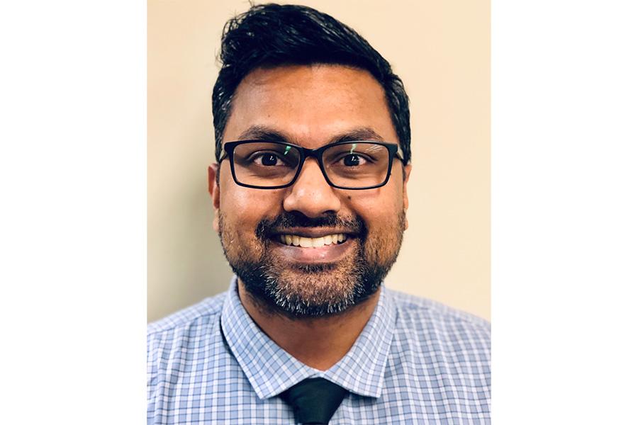
Niranjan Venugopal is an Adjunct Professor in the Department of Physics and Astronomy at the University of Manitoba's Faculty of Science. He holds affiliations with CancerCare Manitoba, focusing on advanced imaging techniques for radiation treatment planning. Contact: Niranjan.Venugopal@umanitoba.ca , nvenugopal@cancercare.mb.ca . Academic Rank: Adjunct Professor Institution: University of Manitoba Department: Physics and Astronomy Affiliation: CancerCare Manitoba Research Interests: Development and application of advanced MRI, MRSI, and PET-CT techniques for radiation oncology. Key areas include: MRI Pulse Sequence Design MR Spectroscopic Imaging Quantum Mechanical Simulations of Metabolites MR Elastography Deep Learning for Image Segmentation Image Registration and Processing Recent trends in his publications (2025-2020) emphasize AI-driven radiotherapy workflows, synthetic CT generation, and precision targeting in lung/brain/prostate cancers using SBRT and HDR brachytherapy.