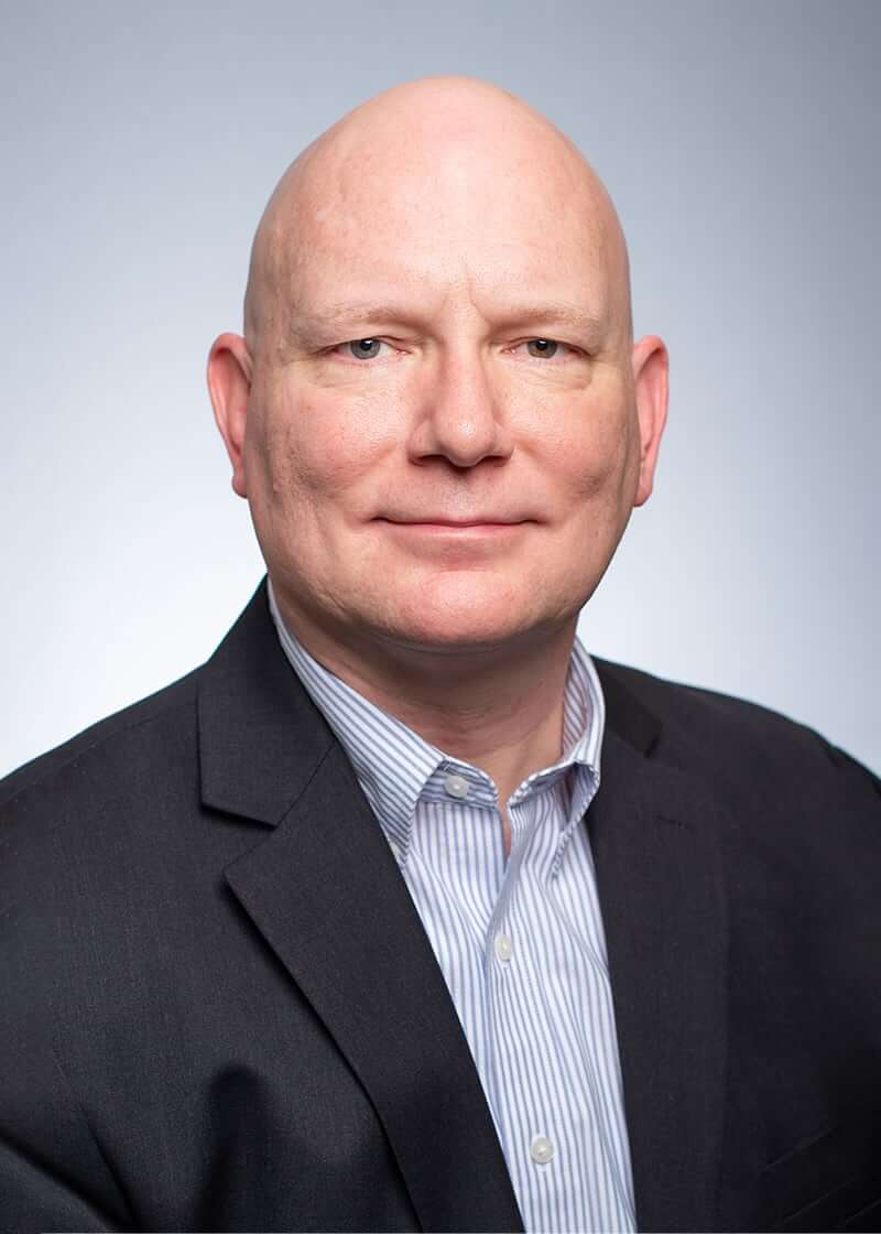
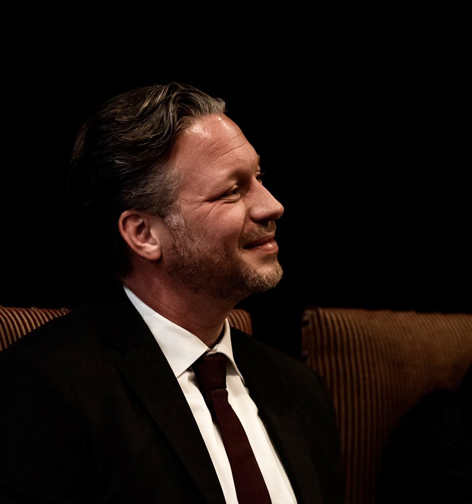
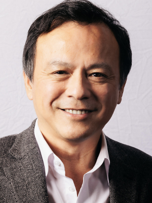
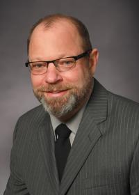
Jon R. Marstrander is an Assistant Professor in the Department of Electrical and Computer Engineering at the University of Alabama at Birmingham (UAB) , where he joined the faculty in 2005. His expertise bridges Embedded Systems , Digital Signal/Image Processing , and Neurological Signal Analysis , supported by a Professional Engineering License since 1992. Education : B.S. in Electrical Engineering, UAB M.S. in Electrical Engineering, UAB Ph.D. in Computer Engineering, UAB His research focuses on collaborative neuroscience projects with UAB clinicians, combining industry-grade hardware/software design (from 16+ years of aerospace/medical engineering) with medical image analysis . Recent work includes high-performance computing workflows for neuroimaging and pathology modeling in Parkinson's/Schizophrenia. Key publication themes: Neurological Imaging (2015–2020), Exercise Neurology , Antipsychotic Treatment Analysis , and Optical Navigation Systems .
Martin Uecker is a Professor at the Institute of Biomedical Imaging at TU Graz. His research focuses on advanced MRI reconstruction techniques, real-time imaging, and open-source software tools like the Berkeley Advanced Reconstruction Toolbox (BART). He specializes in developing methods for fast and accurate medical imaging, including applications in cardiac MRI, fetal brain imaging, and disease monitoring. His work emphasizes reproducibility, quantitative imaging, and clinical translation. Key research areas include generative models for MRI reconstruction, model-based inversion of the Bloch equations, and interactive real-time MRI systems. His team collaborates on projects involving hardware-software integration, such as portable MRI scanners and MRI-guided interventions. Notable contributions include advancements in multi-echo radial FLASH techniques, motion-resolved T1 mapping, and Bayesian uncertainty estimation in imaging. Uecker’s publications highlight innovations in accelerating MRI acquisition and reconstruction, with applications in pulmonary function assessment, neonatal imaging, and cardiovascular diagnostics. His work bridges theoretical physics, computational methods, and clinical practice, fostering open-source frameworks to democratize access to cutting-edge imaging tools.
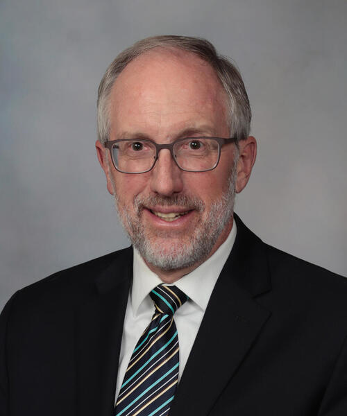
Stephen J. Riederer, Ph.D., is a Professor of Radiology at Mayo Clinic, holding dual appointments in the Department of Radiology and the Department of Physiology & Biomedical Engineering. He leads the Magnetic Resonance Laboratory, focusing on advancing MRI physics and clinical applications. His research emphasizes high-resolution prostate MRI, super-resolution T2SE imaging, and contrast-enhanced magnetic resonance angiography (CE-MRA). Dr. Riederer has developed fast-scanning techniques, real-time signal processing, and parallel acquisition methods, many of which are now industry standards. Education: B.A. in Mathematics, University of Wisconsin-Madison SM in Nuclear Engineering, MIT Ph.D. in Medical Physics, University of Wisconsin-Madison Research Interests: Dr. Riederer’s work bridges MRI physics and clinical implementation. Key areas include: Prostate cancer imaging via high-resolution T2SE and DCE-MRI Super-resolution MRI for improved anatomic detail Real-time MRI scanning and interactive triggering Parallel acquisition techniques and coil array optimization Publications & Impact: Over 300 peer-reviewed articles highlight his contributions to MRI innovation. Recent work focuses on AI-driven prostate MRI quality assessment and coil array improvements. His methods are widely adopted in commercial MRI systems. Awards & Leadership: Gold Medal (International Society for Magnetic Resonance in Medicine, 2002) President, Society of Magnetic Resonance Angiography (2008) George M. Eisenberg Professor I, Mayo Clinic (2024) Advising & Grants: Mentor to over two dozen doctoral students. Active in training via courses at the Mayo Clinic Graduate School. Leads grants on prostate MRI super-resolution and spatiotemporal imaging, funded by NIH and the U.S. Army. Labs & Affiliations: Part of the Center for Advanced Imaging Research, collaborating across radiology, biomedical engineering, and oncology. Facilities include state-of-the-art MRI scanners and imaging laboratories.
Matti Hämäläinen is a Professor at the Department of Neuroscience and Biomedical Engineering , Aalto University. He is a leading expert in Magnetoencephalography (MEG) , with a focus on sensor design, neural connectivity, and clinical applications. His work contributes to understanding brain disorders like autism and epilepsy. Doctorate in Materiaalifysiikka, Teknillinen Korkeakoulu (1989) Diplomi-insinööri in Teknillinen Fysiikka, Teknillinen Korkeakoulu (1983) Hämäläinen's research spans MEG technology , auditory and visual cortex dynamics , and functional connectivity analysis . He develops open-source tools like MNE-Python and HNN-Core for neural data interpretation. Scientific Awards : None explicitly mentioned. He has led projects such as NIH Scalable Software for MEG/EEG and Device-Independent Real-Time MEG EEG Source Localization , with media coverage in outlets including Massachusetts General Hospital and Targeted News Service.
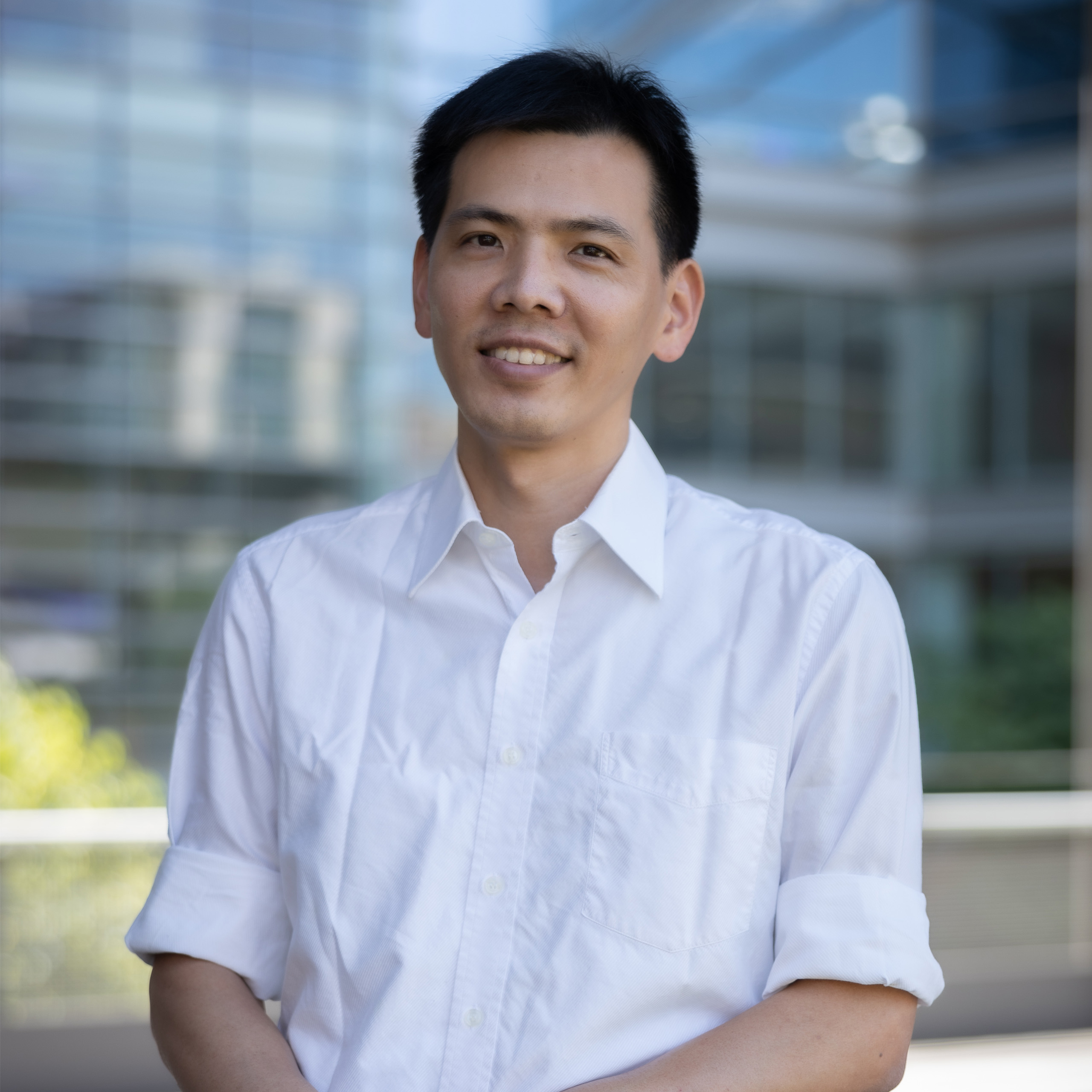
Dr. Wei-Tang Chang is an Assistant Professor in the Department of Radiology at the University of North Carolina School of Medicine. His research focuses on advancing ultrahigh-resolution functional and diffusion MRI techniques, with emphasis on improving spatial and temporal resolution while reducing scan times. Key projects include submillimeter isotropic-resolution fMRI for hippocampal subfield analysis (funded by NIH R21), novel dMRI approaches to overcome resolution limits, and clinical translation of robust imaging methods resistant to motion/noise artifacts. Dr. Chang holds a PhD in Biomedical Engineering from National Taiwan University and completed postdoctoral training at the Martinos Center for Biomedical Imaging (MGH), Massachusetts General Hospital, and Singapore BioImaging Consortium (SBIC). His research innovations include SORDINO fMRI for awake rodent imaging, pPRISM diffusion MRI for submillimeter resolution, and ZTE pulse sequences for ultra-fast acquisitions. Awards include the 2011 OHBM Trainee Award for work on MEG source localization and fMRI temporal resolution breakthroughs. Current work bridges basic neuroimaging science with clinical applications, particularly in neurodegenerative disease biomarker development and rodent disease model studies. Education: PhD in Biomedical Engineering, National Taiwan University Postdoctoral Fellowships: Martinos Center for Biomedical Imaging (MGH) Singapore BioImaging Consortium (SBIC) Key Technologies Developed: ZTE pulse sequences (25 ms temporal resolution fMRI) pPRISM diffusion MRI (navigator-free submillimeter imaging) Draining-vein suppression layer-dependent fMRI Awards & Funding: NIH R21 Grant (2019) for hippocampal subfield fMRI OHBM Trainee Award (2011) Dr. Chang's translational focus involves adapting laboratory innovations for clinical use, with particular interest in Alzheimer's disease biomarkers through hippocampal network analysis and Huntington's disease models using rodent functional connectivity studies. His lab actively develops open-source MRI reconstruction algorithms and collaborates internationally on multi-center neuroimaging projects.
Ergin Atalar is a Professor at Bilkent University's Department of Electrical and Electronics Engineering and serves as Director of the National Magnetic Resonance Research Center (UMRAM). Previously, he spent nearly two decades at Johns Hopkins University, where he held professorships in Radiology, Biomedical Engineering, and Electrical and Computer Engineering, and directed the Center for Image Guided Interventions. His research focuses on Magnetic Resonance Imaging (MRI) innovation, including hardware/software development, safety protocols, and interventional MRI techniques. Education: Ph.D. in Electrical and Electronics Engineering, Bilkent University (1991) M.S. in Electrical and Electronics Engineering, Middle East Technical University (1987) B.S. in Electrical and Electronics Engineering, Bogazici University (1985) Dr. Atalar is recognized for re-engineering MRI scanners to enhance performance without increasing costs. His inventions led to the founding of MRI Interventions, Inc. (now ClearPoint Neuro, Inc.) and Troyka Med A.S. He has authored 127 peer-reviewed journal papers, holds 54 U.S. patents, and maintains high citation metrics (h-index=71 via Google Scholar, 17,400+ citations). Scientific Honors: TUBITAK Science Award (2006) Fellow of the National Academy of Inventors (2016) Member of Academia Europaea (2013) Member of Science Academy, Turkey (2012) Fellow of ISMRM (2011)
Geert Springeling is a Researcher at Erasmus MC , specializing in advanced medical imaging technologies. His work bridges interdisciplinary fields such as Transducer Engineering , Ultrasonography , and Photoacoustic Imaging , with a focus on improving diagnostic tools and imaging precision. Primary Affiliation: Erasmus MC Academic Rank: Researcher Research Interests: Springeling's expertise lies in developing cutting-edge imaging systems, including Optical Coherence Tomography and Functional Ultrasound . His research addresses challenges in Brain Hemodynamics , Vascular Imaging , and High-Resolution Medical Imaging , often integrating computational models with hardware innovations. Publications: Recent work includes advancements in multi-modal intravascular imaging (2025), 4D computational ultrasound (2024), and esophageal OCT capsule design (2020). These contributions highlight his focus on hybrid imaging techniques and patient-specific diagnostics. Collaborations: Springeling collaborates internationally, with network connections in Medical Imaging and Biomedical Engineering . His research outputs are frequently cited in journals like Science Advances and IEEE Transactions on Ultrasonics .
Martijn Froeling serves as an Assistant Professor at University Medical Center Utrecht, actively contributing to the Precision Imaging research group within the High Field division. His work bridges advanced MRI technology development with clinical applications targeting critical health domains including brain cancer, circulatory health, dementia, and musculoskeletal disorders. His academic foundation includes: Master's in Biomedical Engineering from Eindhoven University of Technology (July 2009) PhD in Diffusion Tensor Imaging of the human forearm from Amsterdam University Medical Center and Eindhoven University of Technology (October 2012) Dr. Froeling's research centers on pioneering quantitative MRI methodologies, with specialized expertise in Diffusion Tensor Imaging (DTI) across multiple organ systems (brain, peripheral nerves, muscle, kidney, heart). He drives innovation in 7T MRI hardware development—including specialized coils for multi-nuclei imaging—and conducts clinical studies focused on neuromuscular diseases. His QMRITools software platform for Mathematica enables sophisticated quantitative MRI analysis, directly supporting his mission to 'see the unseen' for advancing clinical diagnostics in cancer, cardiovascular disease, stroke, and MSK conditions. Analysis of his 2024-2025 publications reveals a cohesive research trajectory: DTI applications dominate clinical studies (hamstring injuries, fasciculation mapping), while parallel technical work advances ultra-high-field hardware (double-tuned coils for ²H/³¹P imaging). This dual focus on clinical impact and technological innovation demonstrates his commitment to translating engineering breakthroughs into tangible medical solutions. No scientific awards were documented in the provided materials. While the text confirms Dr. Froeling's role in supervising PhD work (evidenced by his PhD completion under prominent supervisors), no current students or specific grant funding details are explicitly mentioned in the source material. He operates within the High Field group at University Medical Center Utrecht, leading the Precision Imaging research initiative. This team specializes in developing cutting-edge MRI hardware (particularly for 7T systems), maintaining the QMRITools analysis platform, and executing clinical trials targeting neuromuscular pathologies alongside broader applications in oncology and cardiovascular medicine.
Aya Khalaf is an Associate Research Scientist at the Yale School of Medicine, affiliated with the Blumenfeld Lab and the Janeway Society. Her research focuses on understanding neural mechanisms of consciousness, brain-computer interfaces, and machine learning applications in healthcare. She collaborates with leading institutions and researchers globally on studies involving EEG, fTCD, and neuroimaging techniques. Key research interests include auditory and visual perception networks, impaired consciousness in epilepsy, and hybrid BCI systems. Her work bridges cognitive neuroscience with engineering, aiming to translate findings into clinical tools. Notable collaborations include projects with Hal Blumenfeld, Dennis Spencer, and international teams testing consciousness theories. Publications highlight advancements in neural activity analysis, BCI calibration optimization, and multimodal signal processing. Her contributions span theoretical neuroscience frameworks and applied biomedical engineering solutions.
Dr. David Waddington is an NHMRC Emerging Leadership Fellow at the Image X Institute within the Faculty of Medicine and Health at the University of Sydney. He is a member of the University of Sydney Nano Institute and collaborates internationally with institutions like the Martinos Center for Biomedical Imaging at Massachusetts General Hospital. His work focuses on advancing MRI-based imaging technologies for cancer treatment guidance and developing nanoparticle probes for targeted drug delivery. Education: David holds a PhD in Science from the University of Sydney (2018) and earned a University Medal in Physics from UNSW (2010). He was a recipient of a prestigious Postgraduate Fulbright Scholarship (2013–2014) at Harvard University. Research Interests: His projects include the Australian MRI-Linac initiative to improve radiation therapy accuracy through real-time tumor tracking and distortion correction, as well as innovations in low-cost MRI systems and AI-driven image reconstruction. He explores hyperpolarized nanodiamonds for enhanced MRI contrast and synthesizes nanoparticles to improve diagnostic imaging precision in cancer treatment. Key projects: Australian MRI-Linac, MANGO Study, Low-Cost MRI Development International collaborations: Martinos Center (Massachusetts General Hospital) Awards : His achievements include two Best in Physics awards at AAPM (2020, 2022), Summa Cum Laude awards at ISMRM (2016, 2020), and grants such as the FMH Rewarding Research Success (2022) and SEMCAN Viral Bytes Prize (2020). Grants & Students : Leads grants like the 'AI Platform for Targeted Radiotherapy' and 'Advancing Dynamic MRI'. Advises James on developing deep learning techniques for adaptive MRI-guided radiotherapy. His work has produced two patent applications and TEDx talks on medical innovation. Labs/Teams : Affiliated with the ACRF Image X Institute and collaborates with interdisciplinary teams in radiation oncology and biomedical engineering.
Christopher R. Dillon is an Assistant Professor in the Mechanical Engineering Department at Brigham Young University (BYU) . His research bridges mechanical and biomedical engineering, focusing on bioheat transfer modeling and MRI-guided focused ultrasound (MRgFUS) thermal therapies for cancer treatment. Prior to joining BYU in 2021, he worked as a Senior Computer Scientist at Sandia National Laboratories (2018-2021) and held a postdoctoral position in the Department of Radiology at the University of Utah (2014-2017), where he received NIH NRSA fellowship support. Education PhD in Bioengineering, University of Utah (2014) BS in Mechanical Engineering, BYU (2009) Dr. Dillon’s research centers on characterizing human tissue properties and developing computational models for MRgFUS , aiming to improve treatment planning accuracy by addressing challenges like blood perfusion variability and subcutaneous fat absorption . His lab collaborates with clinical institutions to transition findings from ex vivo studies to clinical applications. The Bioheat Transfer Laboratory under Dr. Dillon focuses on: Quantifying perfusion-related thermal energy losses via 3D MRI data Evaluating and refining the Pennes bioheat transfer equation Developing temperature-dependent property measurement tools for fat Advancing non-invasive thermal therapies to reduce surgical reliance His work has led to 15+ publications on computational modeling, tissue property analysis, and thermal therapy optimization. Scientific Awards Outstanding Faculty Teaching Award (BYU, 2023) NIH NRSA Fellowship (2015-2017) Young Investigator Award (Focused Ultrasound Foundation, 2014) National Merit Scholarship (2001-2007) As an educator, Dr. Dillon teaches ME EN 321: Thermodynamics and ME EN 340: Heat Transfer , emphasizing practical applications in biomedical contexts. He also contributes to Python-based computational training through Enthought certification (2021-present).
Dr. Michael C. Lu is currently the Dean of the School of Public Health at the University of California, Berkeley. Previously, he served as Director of the federal Maternal and Child Health Bureau under the Obama Administration, where he received the U.S. Department of Health and Human Services’ Hubert H. Humphrey Service to America Award in 2013. His academic career includes roles as a professor of obstetrics-gynecology and public health at UCLA, with research focused on racial-ethnic disparities in birth outcomes. As an obstetrician, he attended over 1,000 births and was repeatedly named a Best Doctor in America since 2005. He has contributed to National Academy of Medicine committees and co-authored the report Vibrant and Healthy Kids: Aligning Science, Practice, and Policy to Advance Health Equity . His education includes degrees from Stanford University, UC Berkeley, and UCSF. Research interests emphasize improving maternal and child health equity through life-course perspectives and advancing medical imaging technologies. He has pioneered innovations in MRI motion sensing, wireless implant communication, and computational imaging algorithms. His work bridges clinical practice, public health policy, and biomedical engineering. Awards include teaching recognition and federal service accolades. He collaborates on interdisciplinary teams to address global health challenges, leveraging both clinical expertise and technological advancements. Publications span maternal health equity frameworks and cutting-edge MRI methodologies, reflecting his dual focus on societal health disparities and medical technology. Current efforts at UC Berkeley aim to integrate public health strategies with emerging imaging and data science tools. Despite no listed advisees, his mentorship is evident through residency program leadership and training grants in maternal and child health.
John Pauly is a Professor in the Department of Electrical Engineering at Stanford University's School of Engineering. He co-directs the Magnetic Systems Research Laboratory, focusing on advanced MRI techniques such as RF selective excitation, real-time interactive imaging, and image reconstruction. His teaching portfolio includes courses in medical image reconstruction (EE369C), RF pulse design for MRI (EE469C), and foundational classes in signal processing (EE102A,B), medical imaging (EE169), and communication systems (EE179). Research Interests: MRI system development for image-guided interventions, parallel transmit MRI hardware, safety analysis of medical implants in MRI environments, and balanced SSFP applications for functional brain imaging. Current Teaching: Analog and Digital Communication Systems (EE179) Signals and Systems II (EE102B) Independent studies and thesis advising in biomedical engineering (BIOE392), electrical engineering (EE191W, EE391, EE400, EE190, EE191, EE390), and instructional roles (EE195).
Lijing Xin is an Assistant Professor at the Department of Physics and a research staff scientist at the Center for Biomedical Imaging (CIBM) at Ecole polytechnique fédérale de Lausanne (EPFL), Switzerland. She teaches courses on Biomedical Imaging and Translational MR Neuroimaging, while also contributing to academic administration. PhD in Physics (2010, EPFL) Master's Project (2002-2005) on MRI instrumentation Her research focuses on high-field magnetic resonance spectroscopy (MRS) and MRI for studying brain function and neurological diseases. She develops novel acquisition and quantification methods for 1 H, 13 C, and 31 P nuclei, particularly on 7T clinical platforms . Her work bridges preclinical and clinical research , with collaborations in psychiatry to explore pathophysiology and biomarkers for disorders like schizophrenia and mood disorders. Recent publications include studies on epilepsy , Alzheimer's disease , brain energy metabolism , and neurochemical profiling using advanced MRS and deep learning for psychosis classification. She has contributed to RF coil design , macromolecule suppression , and metabolic pathway analysis across multiple disciplines. She advises PhD students and collaborates on interdisciplinary projects involving neuroimaging hardware , metabolic disease research , and psychiatric biomarker identification . Her lab at EPFL CIBM-AIT develops cutting-edge techniques for high-resolution brain metabolism analysis and clinical translation .
Robert Sainburg is the Dorothy Foehr Huck and J. Lloyd Huck Distinguished Chair in Kinesiology and Neurology at Penn State University and Penn State College of Medicine. He directs the Center for Movement Science and Technology (C-MOST) and operates two laboratories: the Movement Neuroscience Laboratory at University Park and the Neurorehabilitation Research Laboratory at Hershey Medical Center. Currently holding professorships in both Kinesiology and Neurology, Sainburg's work bridges basic neuroscience with clinical rehabilitation applications. Education Ph.D. in Neuroscience from Rutgers University (1993) MS in Neurobiology and Physiology from Rutgers University (1989) BS in Occupational Therapy from New York University (1984) Postdoctoral Fellowship in Neurobiology under Dr. Claude Ghez at Columbia University (1993-1996) Research Focus Sainburg's work centers on neural lateralization for motor control , developing the Dynamic Dominance Model that explains hemisphere-specific contributions to predictive and impedance control. His research spans: Multi-joint arm coordination mechanisms Stroke-induced motor deficits analysis Proprioceptive feedback systems Virtual reality rehabilitation frameworks Bi-hemispheric brain function Neurological patient population studies Scientific Recognition Elected Fellow, National Academy of Kinesiology (2024) Huck Distinguished Chair (2021) AOTF Academy of Research (2019) Pattishall Research Achievement Award (2008) Research Infrastructure As C-MOST director, Sainburg integrates two major facilities: the Movement Neuroscience Lab (University Park campus) focuses on basic motor control mechanisms, while the Neurorehabilitation Lab (Hershey Medical Center) directly applies findings to clinical populations. The Kinereach VR system , developed in-house, enables precise movement tracking and distortion experiments.