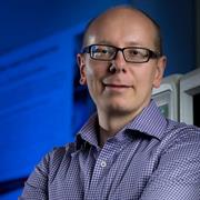
Michael McAlpine is a Professor in the Mechanical Engineering department at the University of Minnesota . He also holds affiliations with the Biomedical Engineering and Electrical and Computer Engineering departments. His research focuses on 3D printing functional materials & devices , Nanoscale inks , Biomedical devices , Bioelectronics , and Flexible Microsystems . Research Interests : 3D Printing, Biomedical Engineering, Nanotechnology, Flexible Electronics, Microfluidics Labs : ME 361/363 Contact : mcalpine@umn.edu , (612) 626-3303, ME 117 Recent Research Trends include 3D Printed Biomedical Devices , Flexible Electronics , and Bioprinting Applications . His work spans from Spinal Organoid Formation to Programmable Drug Release Capsules . Scientific Award : Circulation Research 2020 Best Manuscript Award
Vadim Cherezov, the Ester Dornsife Chair in Biological Sciences and Professor at the University of Southern California (USC), leads groundbreaking research in membrane protein structure and function. Affiliated with the Bridge Institute, Department of Chemistry, and Michelson Center for Convergent Bioscience, his work focuses on GPCRs, ion channels, and transporters—critical targets for drug discovery. His team leverages advanced techniques like Lipidic Cubic Phase (LCP) and Serial Femtosecond Crystallography (SFX) at XFEL facilities to solve high-resolution structures under physiological conditions. Institutional Affiliations: Bridge Institute, USC Michelson Center, Department of Chemistry, Department of Pharmacology and Pharmaceutical Sciences. Key Collaborations: Katritch Lab, Kuhn Lab, NIH, European XFEL. His research explores the role of lipids in modulating GPCR function, addressing diseases like Alzheimer’s, diabetes, and cancer. By solving the structure of the A 2A adenosine receptor via sulfur SAD phasing at XFEL, Cherezov’s lab demonstrated de novo phasing without heavy atoms. This breakthrough enables structural studies of previously intractable membrane proteins. Scientific Awards & Grants: NIH R01 GM108635, U54 GM094618, U54 GM094599, R01 GM095583 Science Signaling Breakthroughs of the Year (2014) Cherezov mentors a dynamic team, including postdocs (e.g., Dong-Gyun Kim), graduate students (e.g., Behnaz Davoudinasab), and alumni (e.g., Benjamin Stauch at Eli Lilly, Nairie Michaelian at Genentech). His lab’s publications span Nature , Science , and Cell , with recent work on Science Advances (2025) addressing ABEL-FRET for GPCR dynamics.
Prof. Waldemar Kolanus leads the Molecular Immunology and Cell Biology department at the University of Bonn's Life & Medical Sciences Institute (LIMES) . His research bridges immunoregulation , stem cell dynamics , and metabolic stress responses in immune cells. Unit 2 member at LIMES Principal investigator in SFB 704 and ImmunoSensation Cluster Leads a multidisciplinary lab with postdocs, PhD students, and technical staff His work focuses on intracellular signaling pathways connecting immune activation to tissue homeostasis, particularly through: Cytohesin proteins in integrin-mediated adhesion and migration TRIM71 in stem cell regulation and congenital hydrocephalus High-salt environments affecting macrophage function Publication trends show expertise in immune cell migration , genetic models , and chemical inhibition , with frequent use of mice and zebrafish for in vivo studies. Key articles explore: TRIM71's dual role in auditory development and germ cell maintenance Cytohesin family's Golgi regulation and insulin signaling Ruxolitinib's off-target migration inhibition of dendritic cells Contact details: Address: LIMES Institute, Carl-Troll-Straße 31, Bonn Email: kolanus.sekretariat@uni-bonn.de Phone: +49 228 73-62788
Christoph F. Schmidt is the Hertha Sponer Distinguished Professor of Physics at Duke University with cross-appointments in the Thomas Lord Department of Mechanical Engineering and Materials Science, Biology, and Biomedical Engineering. He serves as Co-Director of the Duke Materials Initiative and leads an active research program at the intersection of physics and biology. His educational background includes a D.R. from the Technical University of Munich (Germany) in 1988. Schmidt has established himself as a leading researcher in biophysics through decades of innovative work. Professor Schmidt's research spans multiple scales of biological organization, from single molecules to whole organisms. His lab investigates cellular mechanics using advanced techniques including optical trapping, atomic force microscopy, and microrheology. A significant innovation from his group involves single-walled carbon nanotubes for high-bandwidth intracellular tracking. Current research focuses on cardiomyocyte mechanics, Drosophila tissue dynamics, and computational analysis of complex biological systems. His work on motor proteins like Eg5 and ncd has provided fundamental insights into cellular division mechanics. His recent publications (2021-2025) demonstrate increasing integration of computational approaches with experimental biophysics, particularly in analyzing cardiac tissue mechanics and Drosophila sensory systems. The work shows progression from fundamental biophysical measurements toward applications in understanding disease mechanisms and biological function. Professor Schmidt teaches several courses including PHYSICS 995 (Graduate Training Internship), PHYSICS 493 (Research Independent Study), PHYSICS 415 (Biophysics II), PHYSICS 174 (Introduction to Frontiers of Biophysics), and BIOLOGY 425 (Biophysics II). He has successfully mentored numerous graduate students to completion, including recent PhD graduates Dr. Mingru Li and Dr. Xiaoxuan Jian. The Schmidt Lab, part of Duke's Physics Department and the Duke Soft Matter Center, maintains state-of-the-art equipment for optical trapping, atomic force microscopy, and advanced light microscopy. The lab participates in the Triangle Soft Matter Workshop, fostering collaborations with researchers from Duke, UNC Chapel Hill, and NC State University. Current research directions include mechanical responses of suspended cells, tracking non-equilibrium cellular fluctuations, nuclear mechanics, and bacterial membrane mechanics under turgor pressure.
Professor Hala Zreiqat AM is a leading biomedical engineer at The University of Sydney , serving as the Director of the ARC Training Centre for Innovative BioEngineering . A Fellow of all major Australian academies (AAS, ATSE, FAHMS, FRSN), she develops 3D printed bioceramics for bone regeneration while championing diversity through initiatives like the IDEAL Society and BIOTech Futures mentorship program. Her work bridges academia, clinical practice, and industry in musculoskeletal research . Research Focus: Her lab creates synthetic bone scaffolds that mimic natural bone architecture, strength, and porosity, enabling non-rejected bone regeneration via patient-matched implants. Key applications include orthopaedic, dental, and maxillofacial repair , with over $18M in competitive funding and multiple patents. Current projects explore AI-driven scaffold performance prediction and anti-senescence strategies for aging-related bone loss. Scientific Trends: Recent publications highlight 3D printed nanovoxelated ceramics , antisenescence biomaterials , and multifunctional theranostic platforms . Her team integrates machine learning for scaffold design, atom probe tomography for interface analysis, and two-photon imaging for cellular monitoring in 3D environments. 2021-2022 Fulbright Senior Scholar 2018 NSW Premier's Woman of the Year 2019 Eureka Prize for Innovative Use of Technology Fellow of Australian Academy of Science (2021) Over $18M in research funding Teaching & Leadership: She designed core courses like Tissue Engineering and Nanomaterials in Medicine , mentoring 158 students in 2020 alone. As Chair of CAAR (2020-2023), she strengthens Australia-Arab collaborations. Her lab trains early-career researchers , with alumni now in academia and industry.
Jianhua Xing is an Associate Professor in the Department of Physics & Astronomy at the University of Pittsburgh , affiliated with the Dietrich School of Arts and Sciences . His research focuses on applying physics-based approaches to study biological systems, particularly cell phenotypic transitions (CPTs) and their underlying dynamics. He integrates quantitative single-cell measurements with computational and theoretical analyses to understand how cells transition between stable states. Key research areas include: Nonequilibrium systems and rate theories for biological transitions Single-cell trajectory analysis and live-cell imaging Epithelial-mesenchymal transition (EMT) dynamics Gene regulatory networks and stochastic processes Biological applications of dynamical systems theory Recent work highlights the coupling between EMT and cell cycle arrest, leveraging machine learning frameworks (e.g., LivecellX ) for high-resolution imaging analysis. His lab also explores chromosomal dynamics and mechanotransduction in stem cell aging. Publications emphasize data-driven modeling and theoretical insights, with contributions to frameworks like GraphVelo and Graph-Dynamo for inferring cellular state transitions. Collaborative efforts bridge physics, biology, and computational science to address fundamental biological questions. No awards or grants are explicitly listed in the provided texts. His research group focuses on advancing systems biology through interdisciplinary methods, with a lab dedicated to quantitative analysis of cellular processes.
Georg Fantner is an Associate Professor at the Swiss Federal Institute of Technology Lausanne (EPFL) with dual appointments in the School of Engineering (STI) within the Institute of Bioengineering and the School of Life Sciences (SV) for teaching. He directs the Laboratory for Bio- and Nano-Instrumentation (LBNI) and holds leadership roles including President of the Open Science Strategic Committee and the Association des Professeurs de l'EPFL. Research Focus: Bioinstrumentation, Nanotechnology, Scanning Probe Microscopy, and Metrology Teaching: Structural Mechanics for Life Sciences, Metrology, and Metrology Practicals His research pioneers advanced instrumentation for nanoscale characterization, emphasizing data-driven approaches to enhance microscopy techniques. Recent work integrates deep learning with scanning probe microscopy for real-time biological imaging and develops novel MEMS devices for fluid-compatible nanoscale manipulation. Key innovations include hermetically sealed sample chambers for pathogen studies and deterministic nanotopography engineering. Professor Fantner actively mentors 7 current PhD students and has supervised 14 graduates. His laboratory fosters interdisciplinary collaboration across engineering, physics, and life sciences to advance nanoscale measurement technologies and instrumentation development.
Suliana Manley is a Full Professor at the École Polytechnique Fédérale de Lausanne (EPFL) , affiliated with the School of Basic Sciences and the Laboratory of Experimental Biophysics . She also holds teaching and research roles in EPFL's School of Life Sciences and Swiss Plasma Center , focusing on interdisciplinary biophysical studies. Education : PhD in Physics (2004), Harvard University Bachelor's in Physics & Mathematics (1997), Rice University Manley's research centers on super-resolution fluorescence imaging , single-molecule tracking , and quantitative biophysics . Key themes include: Understanding protein assembly dynamics at cellular membranes Elucidating viral assembly mechanisms (e.g., HIV-Gag) Developing 3D imaging algorithms and high-density data reconstruction tools like PALMsiever and FALCON Quantifying nanoscale organization in systems like telomeres and centrioles Her work bridges optical physics , computational image analysis , and cellular biology , with notable Nature and PNAS publications. Collaborations span bioengineering , genetics , and medical research . Scientific Awards : Featured in Nature Methods Research Highlights (3x) Very Important Paper and Cover Article (ChemBioChem, 2012) Postdoctoral Fellow, NIH and MIT Advising & Collaborations : Current PhD students in biophysics, cellular biology, and bioengineering Former students: Anna Archetti, Aleksandr Benke, Andrea Callegari, and others Co-founder of tools for high-density super-resolution microscopy and live-cell imaging Labs & Teams : Leads the Laboratory of Experimental Biophysics at EPFL, integrating physics-based methods into biological questions. The lab focuses on quantitative imaging , computational modeling , and software development for nanoscale analysis.
Adam de la Zerda is an Associate Professor at Stanford University's Department of Structural Biology (School of Medicine) and Electrical Engineering (by courtesy). He develops advanced optical molecular imaging technologies combining nanoparticle contrast agents and adaptive OCT systems for cancer and ophthalmic disease research. Technion-Israel Institute of Technology (BSc, 2005) Stanford University (PhD, 2011) UC Berkeley (Postdoctoral Fellowship) Research Themes : Virtual biopsy using machine learning-enhanced OCT Gold nanorod-based molecular contrast agents Speckle noise reduction for cellular resolution Needle beam optical coherence tomography angiography His 15 most recent publications demonstrate technical innovations in: Metasurface optics for extended depth-of-field Spectral deconvolution of multiple contrast agents Speckle modulation for improved diagnostic clarity Noninvasive lymphatic system mapping Scientific Honors : Pew-Stewart Scholar for Cancer Research AFOSR Young Investigator NIH Early Independence Award Forbes 30 Under 30 (x2) Chan Zuckerberg BioHub Investigator His lab team has developed clinical prototypes including OcuBell Inc. 's ophthalmic imaging systems and Visby Medical 's diagnostic platforms. Current research spans from in vivo glycoprotein imaging to de novo biosensor development for real-time disease monitoring in awake animal models.
Prof. Casper Hoogenraad is a full professor in Molecular Neuroscience at the Department of Cell Biology, Faculty of Science, Utrecht University. His research focuses on understanding how intracellular protein trafficking underlies neuronal development and function, with particular emphasis on the microtubule cytoskeleton, synaptic cargo trafficking, and synaptic plasticity. He leads an active research group within Utrecht University's Cell Biology department and collaborates extensively with other neuroscience research groups. Education: PhD, Erasmus University Rotterdam (1996-2001) Postdoc, Massachusetts Institute of Technology (2002-2005) Hoogenraad's research spans three main themes: cytoskeleton dynamics during neurodevelopment and synaptic plasticity, motor proteins and adaptors as regulators of synaptic transport, and psychiatric and neurologic disease disorders linked to intracellular transport. His work combines genetics, biochemistry, molecular, and cellular biology methods in in vitro (neuron cultures), ex vivo (brain slices), and in vivo (mice) systems, along with advanced microscopy techniques including immunofluorescent confocal microscopy, high-resolution live cell imaging, and photo-activated localization microscopy (PALM). Analysis of Hoogenraad's recent publications reveals a strong focus on microtubule organization, neuronal polarity, and the molecular mechanisms underlying synaptic function and dysfunction. His work frequently explores how disruptions in intracellular transport contribute to neurological disorders including Alzheimer's disease, schizophrenia, and autism spectrum disorders, with particular attention to the relationship between cytoskeletal organization and cargo transport in neuronal compartments. Scientific Awards and Memberships: ZonMW-VIDI (2004) European Young Investigators (EURYI) award (2005) NWO-ALW VICI (2011) ERC Consolidator grants (2013) FENS-Kavli Network of Excellence (2014) European Molecular Biology Organization (EMBO) (2015) Young Academy of Europe (YAE) (2015) IBRO Kemali Prize (2016) Hoogenraad leads a research group studying neuronal development and function, with a particular focus on how intracellular transport mechanisms contribute to both normal brain function and neurological disorders. His laboratory employs a multidisciplinary approach combining molecular, cellular, and systems neuroscience techniques to investigate the molecular basis of neuronal polarity, synaptic plasticity, and the pathogenesis of neurological disorders. He has secured significant research funding through prestigious grants including ERC Consolidator grants. The Hoogenraad lab operates within the Cell Biology department at Utrecht University, collaborating with other research groups focusing on cellular dynamics, biophysics, and neurobiology. The lab utilizes advanced microscopy techniques including immunofluorescent confocal microscopy, high-resolution live cell imaging (spinning disc microscopy and total internal reflection fluorescence microscopy), and quantitative analysis using advanced high-resolution microscopy (photo-activated localization microscopy). Current lab technicians include Phebe Wulf and Bart de Haan.
João F. Mano is a Full Professor at the Department of Chemistry, University of Aveiro, and Director of the Doctoral Program on Biotechnology. He leads the COMPASS Research Group and serves as Vice-Director at CICECO - Aveiro Institute of Materials. His academic appointments include Invited Professor at University of Lorraine (France), Visiting Professor at KAIST (South Korea), and Adjunct Professor at Ajou University (South Korea). Education: PhD in Chemistry (1996, Technical University of Lisbon); D.Sc. in Tissue Engineering, Regenerative Medicine and Stem Cells (2012, University of Minho) Research Interests focus on Biomaterials for Regenerative Medicine , integrating Nanotechnology , Microtechnology , and Biofabrication . His group develops Bioinspired Materials using polymer chemistry, Decellularized Extracellular Matrix , and 3D Bioprinting to engineer Cell Microenvironments for therapeutic applications. Recent Publications highlight advancements in Human-Derived Hydrogels , Photopolymerizable Scaffolds , Magneto-Responsive Biomaterials , and Programmable Bioinks . Trends show emphasis on Organ-on-a-Chip integration, Smart Living Materials , and Green Bioprinting methodologies. Scientific Awards include: European Research Council Advanced Grants (2015, 2020) Fellow at IUPAC, European Academy of Sciences, and American Institute of Medical and Biological Engineering ERC Proof of Concept Grants Doctor Honoris Causa from University of Lorraine and Utrecht UNESCO Chair on Biomaterials George Winter Award (European Society for Biomaterials) Supervisions & Collaborations encompass 74+ MSc, 26+ PhD students, and 40+ postdocs. He co-founded METATISSUE and CELLULARIS Biomodels , and serves as Editor-in-Chief of Materials Today Bio .
Ueli Grossniklaus is an Ordinary Professor at the University of Zurich within the Faculty of Mathematical and Natural Sciences , affiliated with the Department of Plant and Microbiology . His work focuses on plant developmental biology, particularly epigenetic and genetic mechanisms governing reproduction and adaptation. Key Courses: Epigenetics, Plant Biology Workshop, Group Seminars on Current Research Laboratory Techniques: Advanced methods in plant cell mechanics, transcriptomics, and genome editing Research Interests span plant epigenetics, reproductive biology, and the interplay between environmental stress and genetic regulation. He investigates: Mechanistic control of gametogenesis and fertilization Epigenetic contributions to plant adaptation Evolutionary implications of asexual reproduction Biophysical forces in plant cell growth Publication Trends (2025–2018) reveal expertise in: Arabidopsis and fern model systems Epigenetic regulation (DNA methylation, histone dynamics) Apomixis and hybrid seed failure mechanisms Biomechanics of pollen tubes and carnivorous plants Genome editing tools (CRISPR) and long-read sequencing Scientific Collaborations include interdisciplinary projects on: Microfluidic devices for plant cell analysis Gene drive ecology and ethics 3D imaging of plant reproductive structures Advising and Grants focus on mentoring through research internships in developmental biology, genetics, and systems biology. His lab engages in: Epigenetic response to environmental stress Cell wall mechanics in reproduction Computational modeling of plant growth Laboratory Teams integrate plant biologists, bioengineers, and computational scientists to study: Mechanistic gene regulation Evolutionary developmental biology Microrobotics for cellular force measurement
Gilles Hickson is a Full Professor at the Université de Montréal’s Faculty of Medicine, Department of Pathology and Cell Biology. He is affiliated with the Azrieli Research Center at CHU Sainte-Justine and serves as Deputy Director of Academic Affairs, Co-director of the Microscopy Imaging Platform, and responsible for the Molecular Biology Program. His research focuses on understanding molecular mechanisms of cytokinesis, particularly the transition from contractile rings to midbody rings, and the roles of anillin, septins, and actomyosin dynamics in cell division using Drosophila as a model. His work has implications for cancer biology and drug development. Education: BSc (Manchester), PhD (University of Glasgow), Postdoctoral training (UCSF). Research Interests: Cytokinesis mechanisms, cytoskeletal coordination, cell division errors in cancer, and developmental variations in cytokinesis. His awards include the FRQS Senior Researcher Fellowship and multiple grants from CIHR and NSERC. He supervises graduate students and has mentored over a dozen researchers. His lab employs genetic tools and high-resolution microscopy to dissect cytokinesis machinery. He also contributes to teaching, including courses in molecular medicine and microscopy. Awards: Komen Postdoctoral Fellowship, Leukemia & Lymphoma Special Fellow, Cole Foundation Transition Award. Grants: Ongoing CIHR and NSERC funding (2022–2027) for cytokinesis research. Hickson collaborates with institutions like the CHU Sainte-Justine and leads projects on cytokinesis networks, septin assembly, and actomyosin coordination. His lab is part of the Azrieli Center and the Microscopy Imaging Platform.
Anders Sejr Hansen is an Assistant Professor of Biological Engineering at MIT, leading the Hansen Lab focused on understanding 3D genome structure and its functional implications. He holds a PhD from Harvard University and completed postdoctoral training at UC Berkeley. His research integrates advanced imaging, genomics, and computational methods to study chromatin dynamics, enhancer-promoter interactions, and their roles in gene regulation across health and disease. Education: Bachelor's/Master's in Chemistry, University of Oxford (2010) PhD in Chemistry and Chemical Biology, Harvard University (2015) Postdoctoral Research, UC Berkeley (2015–2020) Research Interests: His work spans molecular mechanisms of genome organization, development of novel microscopy techniques (e.g., MINFLUX, expansion microscopy), and computational models for 3D genomics. Key areas include chromatin dynamics, loop extrusion by cohesin/condensin, and the impact of 3D structure on gene expression in cancer and aging. Awards: NIH K99 Pathway to Independence Award (2019) NIH Director’s New Innovator Award (2020) Pew-Stewart Scholar for Cancer Research (2021) NSF CAREER Award (2024) NIH Director’s Transformative Research Award (2024) Advising & Grants: Hansen mentors PhD students and postdocs, including notable advisees Viraat Goel and Domenic Narducci. His lab has secured major grants from NIH, NSF, and private foundations, supporting interdisciplinary projects in imaging, genomics, and synthetic biology. Labs/Teams: The Hansen Lab at MIT collaborates with institutions globally, advancing technologies like Region Capture Micro-C (RCMC) and deep learning models (e.g., Cleopatra) for high-resolution genome mapping. The lab also explores synthetic biology approaches to engineer genome structures.
Professor Donald Wlodkowic is a faculty member at RMIT University's School of Science (Biosciences), leading the Neurotox Laboratory. His research focuses on aquatic ecotoxicology, behavioral ecotoxicology, and eco-neurotoxicology, with expertise in neurotoxins, industrial pollutants, and neuroactive drugs' effects on central nervous systems. He innovates biomicrofluidic technologies and digital video-based analysis for high-throughput behavioral studies in toxicology. Research Interests: Aquatic toxicology, neurotoxicology, water quality, and eco-neurotoxicology. His lab develops real-time biomonitoring systems and early-warning tools for water quality assessment. Current projects include neurotoxicity studies of pollutants, high-throughput behavioral assays, and embryo-based risk assessment. Teaching: Coordinates courses in cell biology, biochemistry (BIOL2146/2333/2420, PROC2048) and supervises honors projects (ONPS2313). He integrates digital tools into bioscience curricula for innovative teaching. Advising & Leadership: Supervises Masters and PhD students in projects such as 'Emerging Pollutants on Aquatic Animal Behavior' and 'Nanotoxicology.' He has led international academic-industry collaborations in Australia, New Zealand, and Europe, fostering motivated research teams. Labs & Teams: NeuroTox Lab pioneers interdisciplinary research in ecotoxicology and neurotoxicology, combining microfluidics, digital analytics, and behavioral assays to address environmental and health challenges.