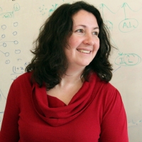
Polina Golland is a Professor in the Department of Electrical Engineering and Computer Science (EECS) at MIT and a Principal Investigator in the Computer Science and Artificial Intelligence Laboratory (CSAIL). Her research focuses on developing novel techniques for biomedical image analysis and understanding, particularly in medical vision, AI/ML, and health care applications. She leads the Medical Vision Group and collaborates with the Vision Group at CSAIL. Her work emphasizes statistical modeling of medical images, shape modeling, and predictive analytics for biological processes. Current projects include fetal MRI analysis, cardiac MRI segmentation, and quantitative assessment of pulmonary edema in chest X-rays. She has secured grants from NIH, MIT-IBM Watson AI Lab, and other institutions to support her research. Dr. Golland teaches courses on inference, probability, and probabilistic systems. She advises graduate students in MIT's EECS program and has mentored numerous postdocs and researchers. Her lab focuses on translating advanced imaging techniques into clinical workflows, with applications in neuroimaging, fetal health monitoring, and cardiovascular disease analysis. Notable collaborations include work with Harvard Medical School affiliates, Brigham and Women's Hospital, and the MIT Jameel Clinic. Her research aims to bridge computational methods with clinical needs, improving diagnostic tools and treatment planning through machine learning and medical imaging innovation.
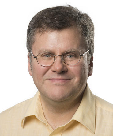
Professor Daniel Rueckert is a leading academic in Artificial Intelligence and Medical Imaging, holding dual positions at Imperial College London (as Professor of Visual Information Processing) and Technical University of Munich (Alexander von Humboldt Professor for AI in Medicine and Healthcare). He obtained his MSc from Technical University Berlin (1993) and PhD from Imperial College London (1997), followed by postdoctoral work at King’s College London. At Imperial, he led the Department of Computing (2016–2020) and founded the Biomedical Image Analysis group. His research focuses on AI-driven medical image analysis, including algorithms for image reconstruction, registration, and clinical decision support. His research interests span AI applications in healthcare, machine learning for medical imaging, and computational methods for clinical diagnostics. Notable contributions include over 500 publications and 60+ PhD graduates, with key works in federated learning, cardiac motion analysis, and biomarker development. Awards include the Leibniz Prize (2025), Royal Academy of Engineering Fellowship (2015), and multiple ERC grants. He leads the BioMedIA research group and is an editorial board member of Medical Image Analysis . Recent publications highlight advancements in AI-driven medical imaging, such as secure federated learning frameworks and deep learning models for disease prediction. His work bridges academic and industrial sectors through initiatives like IXICO, an Imperial spin-out. Current affiliations include roles at both Imperial and TUM, emphasizing interdisciplinary collaboration in healthcare technology. Advising and grants: Supervised over 60 PhD students and 40 post-docs. Secured grants including ERC Synergy (2013) and ERC Advanced (2020). Active in labs focused on biomedical image computing and AI in healthcare systems. Collaborative efforts include the BioMedIA group and TUM’s AI initiatives. Labs/teams: Leads the Biomedical Image Analysis group at Imperial and the TUM AI in Medicine team. Collaborates extensively on projects like cardiac imaging analysis and federated learning for healthcare.
Professor Guy Williams is a leading academic at the University of Cambridge with a focus on imaging science and clinical neurosciences, affiliated with Downing College and the Wolfson Brain Imaging Centre . Holding a PhD in Physics from his initial Natural Sciences degree, he specializes in nuclear magnetic resonance (NMR) and MRI techniques for brain imaging. Education: BA, PhD in Physics His research centers on non-invasive imaging of brain structure and function, particularly in traumatic brain injury (TBI) and dementia. His work involves developing novel MRI pulse sequences and advanced data analysis algorithms, including AI-based diagnostic tools. He leads studies on white matter integrity post-trauma, longitudinal dementia assessment, and applications of MRI in disorders of consciousness and addiction. Recent publications highlight collaborations in traumatic brain injury outcomes, AI-guided dementia prediction, and neuroimaging of post-COVID cognitive deficits. His team's work on ultra-high field laminar fMRI and distortion correction methods has advanced clinical neuroscience applications. Key techniques include diffusion tensor imaging (DTI), 7 Tesla MRI, and positron emission tomography (PET/MR). His research spans from basic NMR physics to clinical translation, with a strong emphasis on multi-site studies and real-world diagnostic implementation.
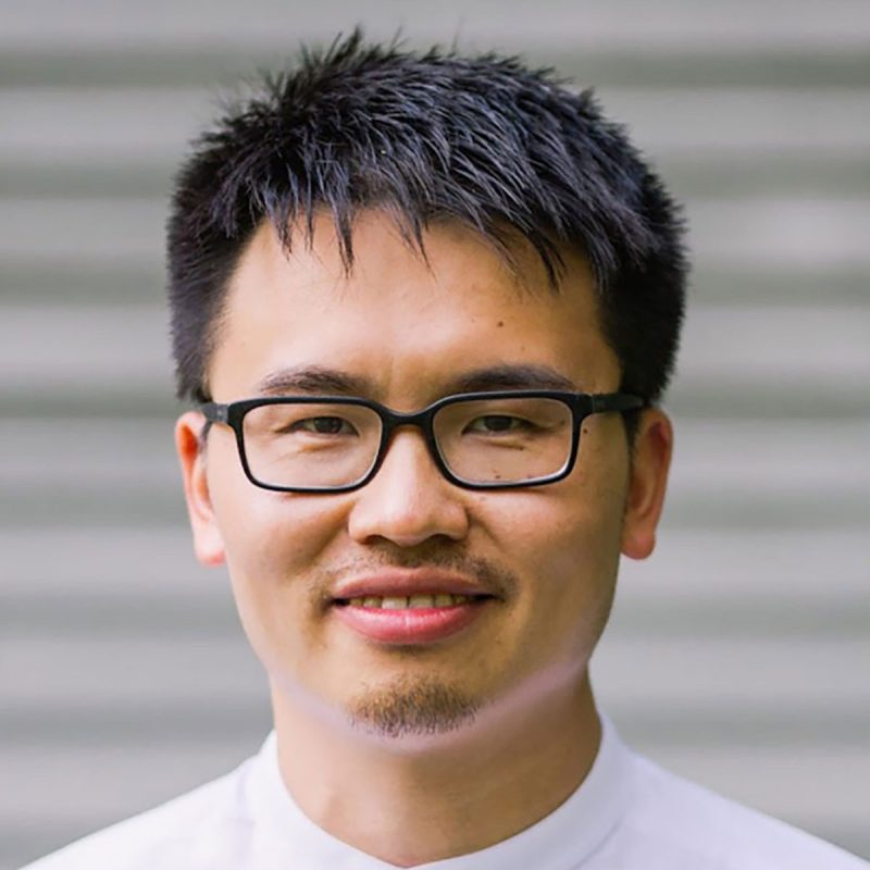
Dr. Hongye Zhang serves as a Lecturer in Superconducting and Cryogenic Electric Machines at the School of Engineering, University of Edinburgh, while maintaining a Visiting Research Fellow position at the University of Manchester. He actively contributes to the European Society for Applied Superconductivity (ESAS) as a Board Member and chairs the international HTS 2026 workshop. His educational foundation includes: BSc and MSc in Electrical Engineering from Xi’an Jiaotong University (2015, 2018) Diplôme d’ingénieur (MEng) from École Centrale de Lyon (2018) PhD in Applied Superconductivity from the University of Edinburgh (2021) Dr. Zhang’s research centers on decarbonizing transport through superconducting/cryogenic electric machines for hydrogen-powered aircraft, integrating artificial intelligence with superconductor technology and cryogenic techniques. His work targets net zero emissions by developing high-power-density propulsion systems that leverage hydrogen energy and advanced numerical modeling of superconductors. Analysis of his 2022-2025 publications reveals dominant themes in superconducting machine design for wind energy and electric aviation, with significant contributions to loss mitigation, flux pump technology, and trapped field magnet applications. His research bridges fundamental superconductor characterization with practical system integration for renewable energy. Recognized with the 2021 IEEE Council on Superconductivity Graduate Study Fellowship, his professional engagements include: Early Career Editorial Board Member for Elsevier’s Superconductivity journal Technical Editor for IEEE Transactions on Applied Superconductivity Program Committee Member for SMT 2023 He leads critical research within the £54-million H2GEAR project developing hydrogen-electric aircraft propulsion, while teaching Power Engineering 2 and Electrical Machines courses. His advisory roles span doctoral supervision and industry collaboration through Energy Systems research institute. Based at the University of Edinburgh’s Faraday Building, Dr. Zhang directs a research group focused on hydrogen energy applications and superconducting machine testing, with strong ties to the H2GEAR consortium and ESAS working groups.
Uwe Himmelreich is a Full Professor at the Faculty of Medicine, KU Leuven , leading the Biomedical MRI unit. He is actively involved in the Medical Imaging Division , KU Leuven Brain Institute , KU Leuven Institute for Integration of Micro- and Nano-scale Technologies , and KU Leuven Cancer Institute . Role: Full Professor and Head of Biomedical MRI Affiliations: Faculty of Medicine, Medical Imaging Division, LBI, LIMNI, LKI His research spans neuroscience , cardiovascular imaging , and nano/micro-scale technologies . Key projects include: MindMAP: Radiotherapy-induced neurotoxicity in juvenile brains Preclinical cancer models for oral tumors Quantitative T2 mapping of lung disease in murine models Neuroinflammation and cognitive decline in cryptococcosis Resistance training effects on cortical thickness in aging cohorts His work integrates MRI , multi-photon microscopy , and novel contrast agents for longitudinal in vivo studies. Methodologies include vascular density mapping , proton therapy verification , and preclinical radiotherapy evaluation . Notable contributions include: 2025: JAK/STAT inhibition in malaria-induced inflammation 2025: Manganese-enhanced MRI for cardiac injury 2025: Quantitative lung imaging at 9.4T 2024: IVIM as vascular density marker in rat brain 2024: Phase-change ultrasound agents for proton therapy
Joachim Weickert is a Professor of Mathematics and Computer Science at Saarland University where he heads the Mathematical Image Analysis Group since 2001. He received his diploma and Ph.D. in mathematics from the University of Kaiserslautern (1991, 1996), and a habilitation degree in computer science from the University of Mannheim (2001). Prior to his current position, he worked as a research assistant at the University of Kaiserslautern, as a post-doctoral researcher at the universities of Utrecht and Copenhagen, and as an assistant professor at the University of Mannheim. His research focuses on image processing, computer vision, and scientific computing, with special emphasis on techniques based on partial differential equations, variational principles, wavelets, morphological and nonlocal methods, as well as neuroexplicit approaches. He has developed mathematical models and efficient numerical algorithms for image restoration, enhancement, segmentation, compression, optic flow computation, stereo reconstruction, shape from shading, and signal processing methods for tensor fields. These ideas have been successfully applied in industry, biomedical image analysis, and other fields. Analysis of his recent publications reveals a strong trend toward combining traditional PDE-based methods with modern deep learning approaches, particularly in the areas of image inpainting and compression. His work increasingly explores the connections between numerical algorithms for partial differential equations and neural network architectures, demonstrating how mathematical foundations can inform cutting-edge AI techniques while maintaining strong theoretical guarantees. Gottfried Wilhelm Leibniz Prize (2010), considered the most important research award in Germany ERC Advanced Grant (2017) for "Inpainting-based Compression of Visual Data" Elected member of Academia Europaea - The Academy of Europe Jan Koenderink Prize for Fundamental Contributions in Computer Vision (2014) Multiple DAGM Prizes and Best Paper Awards throughout his career AAIA Fellow (2021) and Highly Ranked Scholar (2024) distinctions Professor Weickert has supervised over 250 bachelor's and master's theses and initiated the Master Programme in Visual Computing at Saarland University, the first of its kind in Germany taught in English. He has established numerous interdisciplinary collaborations with colleagues from medicine, bioinformatics, pharmacy, physics, mechatronics, and mechanical engineering. As Principal Investigator for Visual Computing within the Multimodal Computing and Interaction Cluster of Excellence, and former dean of the Faculty of Mathematics and Computer Science (2008-2010), he has played a significant leadership role in advancing visual computing research and education. He heads the Mathematical Image Analysis Group, which has been at the forefront of developing mathematical methods for image analysis. The group maintains strong connections with both theoretical mathematics and practical applications, bridging the gap between fundamental research and real-world implementation across various domains including medical imaging, industrial inspection, and multimedia processing.
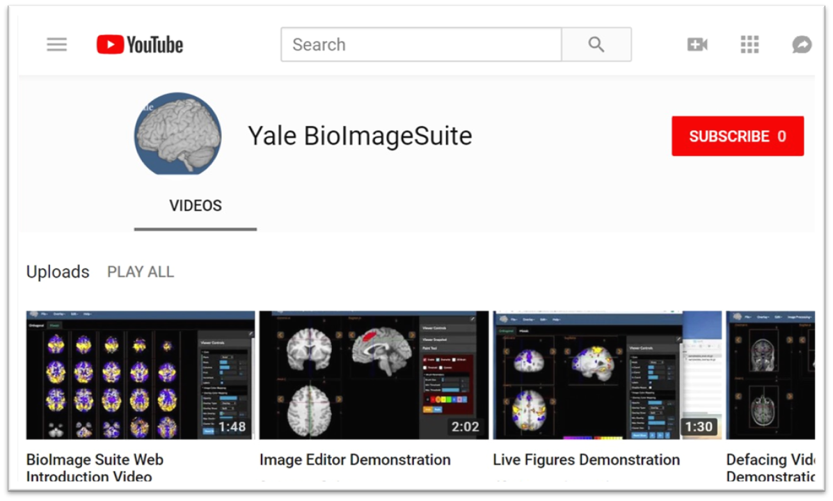
Xenophon Papademetris is a Professor of Biomedical Informatics & Data Science and Radiology & Biomedical Imaging at Yale School of Medicine. He serves as Associate Director of Biomedical Imaging Data Sciences at Yale Biomedical Imaging Institute and directs the Medical Software and Medical Artificial Intelligence Certificate Program. PhD in Electrical and Information Sciences from Yale University (2000) BA from Cambridge University (1994) Postdoctoral Fellowship at Yale University (2002) His research focuses on medical image analysis, machine learning, and biomedical software development. He has developed tools like BioImage Suite Web and contributed to standards committees at the Association for the Advancement of Medical Instrumentation (AAMI). His work spans modalities including MRI, CT, PET, and optical imaging. Recent publications emphasize neuroimaging analysis, explainable AI in healthcare, and multimodal data integration across species. He leads NIH-funded research under the BRAIN Initiative (R24 MH114805) and has authored a textbook on Medical Software published by Cambridge University Press. IEEE Senior Member Yale Brown-Coxe Postdoctoral Fellowship Harding Bliss Prize for Excellence in Engineering He directs the BioImage Suite Project, creating web-based image analysis tools using JavaScript and WebAssembly. His teaching includes both academic courses and a Coursera program on Medical Software with over 14,000 enrollments.
Suyi Li is an Associate Professor in the Department of Mechanical Engineering at Virginia Tech's College of Engineering, where he leads the Dynamic and Architected Robot and structurE (DARE) Lab. Previously, he served as an Assistant Professor at Clemson University from 2016-2022 after completing postdoctoral research at the University of Michigan. Ph.D. in Mechanical Engineering, University of Michigan, Ann Arbor (2014) M.Sc. in Mechanical Engineering, Pennsylvania State University (2008) B.S. Summa Cum Laude in Mechanical Engineering, University of Michigan, Ann Arbor (2006) Dr. Li's research focuses on pioneering new paradigms of intelligent robots and functional structures by exploiting the interplay between geometry, mechanics, actuation, and computation. His work spans origami-inspired morphing structures, physically computing materials that perform machine learning tasks without traditional electronics, and soft/reconfigurable robots that can move like animals or grow like plants. His innovative approach combines mechanical engineering principles with computational thinking to create systems with 'mechano-intelligence'. Analysis of Dr. Li's recent publications reveals a strong trajectory toward embodied intelligence and mechanical computing, where physical structures themselves perform computational tasks. His work increasingly integrates origami/kirigami principles with advanced materials to create systems that can sense, process information, and actuate without conventional electronics. The research shows progression from fundamental mechanics of adaptive structures to sophisticated applications in robotics and computing. Dean's Awards of Excellence – Faculty Fellow, Virginia Tech (2024) C.D. Mote Jr Early Career Award, ASME Design Engineering Division (2022) Gary Anderson Early Achievement Award, ASME Aerospace Division (2021) Junior Researcher of the Year Award, College of Engineering, Clemson University (2020) CECAS Dean's Faculty Fellow, Clemson University (2018) CAREER Award, National Science Foundation (2018) ASME Freudenstein Young Investigator Award Dr. Li has secured nearly two million dollars in research funding, including the prestigious NSF CAREER award and an NSF EFRI project to build mechano-bio hybrid reservoir computers. He advises multiple Ph.D. and Master's students in the DARE Lab, with recent successes including Vishrut Deshpande's Ph.D. defense. His research has generated close to 80 journal and conference papers, demonstrating significant impact in the fields of adaptive structures and materials systems. Dr. Li also serves on editorial boards for several prominent journals including Journal of Intelligent Material Systems and Structures and Philosophical Transactions of the Royal Society A. The DARE Lab at Virginia Tech comprises a multidisciplinary team of researchers working on origami-inspired meta-structures, physically computing materials, and soft robotics. Current projects include developing electronics-free crawling robots with mechanical central pattern generators, creating kirigami-based wearable medical devices, and engineering metamaterials with programmable mechanical properties. The lab actively collaborates with institutions across the country and has received recognition for its innovative approaches to combining mechanical design with computational capabilities.
Frank E. Garcea, Ph.D., is a Research Assistant Professor in the Department of Neurosurgery and Neuroscience at the University of Rochester School of Medicine and Dentistry. His research focuses on the cognitive and neural mechanisms underlying tool use, apraxia, and stroke recovery. He employs neuropsychological testing, fMRI, and lesion-symptom mapping to study brain injury effects on functional connectivity and action knowledge. Education: Bachelor of Science in Psychology, St. John Fisher College (2006–2010) PhD in Brain and Cognitive Sciences, University of Rochester (2012–2017) Research Interests: Dr. Garcea investigates how brain regions like the parietal cortex and dorsal/ventral streams mediate object manipulation and tool use. His work explores stroke-related disconnection syndromes, motor speech coordination networks, and translational brain mapping to preserve neural function during surgery. He collaborates on projects involving epilepsy patients undergoing electrocorticography to study action-related neural pathways. Labs & Affiliations: Principal Investigator of the Garcea Lab at URMC, focusing on tool use deficits in brain tumor/stroke survivors. Affiliated with the Del Monte Institute for Neuroscience and the Neurobiology & Anatomy Program. His lab integrates neuroimaging, lesion analysis, and clinical care to advance personalized brain mapping strategies.
Professor Ananya Choudhury serves as Chair and Honorary Consultant in Clinical Oncology at the University of Manchester, where she is also Co-Group Leader of the Translational Radiobiology Group within the Division of Cancer Sciences. She joined The Christie NHS Foundation Trust in 2008, specializing in urology and sarcoma, and has since focused on radiotherapy-related research in prostate and bladder cancers. Professor Choudhury is clinical lead for advanced radiotherapy, including the groundbreaking MRLinac project, and plays a key role in national radiotherapy research initiatives. Professor Choudhury earned her BA (Hons) in 1993, MB. BChir (Cantab) in 1995, and MA (Cantab) in 1997 from Trinity College, Cambridge. She completed her Clinical Oncology training at the Yorkshire Deanery from 2000-2008, during which she earned her MRCP in 2000 and F.R.C.R in 2004. She completed her PhD in 2008 through the University of Leeds and Princess Margaret Hospital in Toronto, Canada, where she studied the molecular epidemiology of DNA double strand break repair in bladder cancer. Professor Choudhury's research program focuses on optimizing and personalizing radiotherapy using advanced imaging technology to deliver high doses while minimizing side effects. Her work centers on prostate and bladder cancers, with particular interest in predictive biomarkers, hypoxia, and the integration of magnetic resonance imaging to improve treatment precision. She has pioneered research in radiotherapy dose optimization, biomarker development, and the identification of patients who would benefit most from different treatment approaches. Her extensive publication record demonstrates a strong focus on radiation therapy, particularly in genitourinary cancers. Recent work explores MRI-guided radiotherapy, hypoxia biomarkers, and personalized treatment approaches across multiple cancer types. She has made significant contributions to understanding how imaging technology can improve radiotherapy precision and effectiveness while reducing side effects, with several publications appearing in top journals through 2025. Professor Choudhury has received multiple prestigious awards recognizing her contributions to the field: Cancer Research-UK/Royal College of Radiologists Clinical Training Fellowship (2005) Fellowship for the 10th ECCO-AACR-ASCO Workshop on Methods in Clinical Cancer Research (2007) Outstanding Contribution, Greater Manchester Clinical Research Awards (2017) RCR Research Fellowship (2005) Research Fellowship, Princess Margaret Hospital, Toronto (2004) Professor Choudhury has supervised numerous doctoral and master's students across multiple cancer types, with current students expected to complete through 2024. She is Principal Investigator on multiple research grants, including 'Measuring tumour radioresistance to improve radiotherapy outcomes' and the 'MAESTRO Programme' as part of CRUK RadNet. Her research program is supported by significant funding from NIHR Manchester Biomedical Research Centre and other major funding bodies. As Co-Group Leader of the Translational Radiobiology Group, Professor Choudhury collaborates extensively with leading researchers including Peter Hoskin, Catharine West, Corinne Faivre-Finn, and Marcel van Herk. Her team is at the forefront of integrating advanced imaging with radiotherapy to improve cancer treatment outcomes, with active projects spanning from basic radiobiology to clinical implementation of novel radiotherapy techniques.
Essa Yacoub is a Professor in the Department of Radiology at the University of Minnesota, affiliated with the PhD Program in Medical Physics and the Center for Magnetic Resonance Research. His work focuses on advancing MRI and fMRI technologies, particularly at ultrahigh magnetic fields (e.g., 10.5 T), to achieve unprecedented spatial and temporal resolution in brain imaging. He leads projects in RF coil design, noise reduction algorithms, and developmental neuroimaging. Roles: Professor, Medical Physics Program Faculty Affiliations: Center for Magnetic Resonance Research, Department of Radiology Research emphasizes high-resolution fMRI applications, including layer-specific brain mapping, pediatric neurodevelopment studies (e.g., Baby Connectome Project), and translational tools like BIBSNet for infant brain segmentation. His innovations bridge hardware engineering (RF coils) and software (denoising pipelines) to tackle challenges in mesoscopic-scale imaging. Key contributions include optimizing imaging protocols at 7T/10.5T, developing NORDIC denoising for submillimeter data, and advancing understanding of brain networks in aging and neurological disorders. His work is foundational for large-scale initiatives like the Human Connectome Project and non-human primate neuroimaging collaborations. Grants and collaborations focus on translational imaging technologies, while educational contributions include training through the Medical Physics PhD Program. Ongoing efforts aim to refine ultra-high field MRI applications for clinical and basic neuroscience research.
Zhi-Pei Liang is the Franklin W. Woeltge Professor in the Department of Electrical and Computer Engineering at the University of Illinois at Urbana-Champaign, with joint appointments in the Department of Bioengineering, Beckman Institute for Advanced Science and Technology, and Coordinated Science Laboratory. His research spans biomedical engineering, medical imaging, and signal processing with a focus on advancing magnetic resonance imaging and spectroscopy technologies. His educational background includes a Ph.D. in Biomedical Engineering from Case Western Reserve University (1989) and a B.S. in Electrical Engineering from South-China University of Technology (1982), followed by postdoctoral training at UIUC (1989-1991). Professor Liang's research interests center on magnetic resonance imaging and spectroscopy , with particular emphasis on ultrafast imaging techniques , model-based reconstruction methods , and the integration of physics-based modeling with machine learning . His pioneering work on SPICE (SPectroscopic Imaging by exploiting spatiospectral CorrElation) has revolutionized high-resolution metabolic brain imaging by enabling label-free molecular imaging through the marriage of spin physics and machine learning. His research spans pattern recognition, parameter estimation, image formation theory, and algorithms for medical imaging applications. Analysis of his recent publications reveals a strong focus on high-resolution metabolic imaging , particularly using SPICE methodology to map brain metabolism with unprecedented detail. His work bridges fundamental physics of magnetic resonance with advanced computational methods to overcome traditional limitations in imaging speed and resolution. Current research directions include J-resolved spectroscopic imaging, deuterium-based metabolic mapping, and multimodal integration of PET and MRSI for studying neurological disorders. Elected to International Academy of Medical and Biological Engineering (2012) Gold Medal, International Society for Magnetic Resonance in Medicine (2022) Technical Achievement Award, IEEE Engineering in Medicine and Biology Society (2014) Fellow, National Academy of Inventors (2021) Author of influential book 'Principles of Magnetic Resonance Imaging' (1999) President of IEEE Engineering in Medicine and Biology Society (2011-2012) Professor Liang has advised numerous students and postdocs in biomedical imaging research and has received multiple teaching honors including the Ronald W. Pratt Outstanding Teaching Award (2005) and multiple listings among UIUC's Excellent Teachers. His research has been supported by various grants from NIH, NSF, and other funding agencies. He leads the SPICE (Spectroscopic Imaging by exploiting spatiospectral Correlation) research group which focuses on developing novel imaging techniques that combine physics-based modeling with machine learning for ultrafast metabolic imaging. His laboratory, part of the Beckman Institute's Integrative Imaging Theme, collaborates extensively with clinical researchers at Carle Illinois College of Medicine and other institutions to translate advanced imaging techniques into clinical applications for neurological disorders, cancer, and metabolic diseases. Current projects focus on high-resolution mapping of brain metabolism in Alzheimer's disease, stroke, and brain tumors using novel MR spectroscopic imaging techniques.
Allan David serves as the John W. Brown Professor of Chemical Engineering and Associate Dean for Research at Auburn University's Samuel Ginn College of Engineering. His leadership extends across academic administration and cutting-edge nanomedicine research, with a focus on translating laboratory discoveries into clinical applications. His educational foundation includes: Ph.D. in Chemical Engineering, University of Maryland B.S. in Chemical Engineering, University of Maryland Dr. David's research program pioneers nanomedicine applications through the development of smart materials for cancer diagnostics and therapy. His work spans nanoparticle-based MRI contrast agents , ocular drug delivery systems , and vaccine delivery platforms , with particular emphasis on optimizing physicochemical properties for targeted biological interactions. Current projects address critical healthcare challenges including safer contrast agents for patients with kidney impairment and precision cancer targeting mechanisms. Analysis of his 15 most recent publications reveals a cohesive research trajectory centered on magnetic nanoparticles and biomimetic delivery systems . The work demonstrates increasing translational focus, evolving from fundamental nanoparticle characterization (2020-2021) to clinically relevant applications like ocular delivery and cancer theranostics (2022-2024), culminating in commercialization efforts through NanoXort, LLC. Dr. David has secured significant research funding including an $184,773 grant from the Alabama Department of Economic and Community Affairs (ADECA) for developing cardiovascular MRI agents. He leads collaborative efforts that bridge chemical engineering with biomedical innovation, notably co-founding NanoXort, LLC to commercialize safer MRI contrast agents addressing gadolinium toxicity concerns for renal-impaired patients. His laboratory operates at the intersection of chemical engineering and medicine, focusing on nanoparticle-cell interactions and targeted delivery systems. The research group maintains strong industry partnerships through the NanoXort startup, which has secured $1 million NSF funding to advance MRI contrast agent technology toward clinical implementation.
Daniele Loiacono is an Associate Professor at Politecnico di Milano's Department of Electronics, Information, and Bioengineering (DEIB), affiliated with the Artificial Intelligence and Robotics Lab (AIRLab). His research focuses on interdisciplinary applications of Artificial Intelligence, Machine Learning, and Deep Learning in medical imaging, radiation therapy, and procedural content generation for games. He leads projects in synthetic image generation for radiotherapy quality assurance, automated treatment planning, and bias analysis in medical AI systems. Key research areas include medical image synthesis using GANs, radiation therapy optimization, and algorithmic game design. His contributions span clinical applications such as total marrow irradiation (TMI) planning and lymph-node segmentation, alongside innovations in shader generation and interactive evolutionary tools for game development. Loiacono collaborates on multi-center studies to validate AI-driven workflows in healthcare and has pioneered methods combining lean Six Sigma with machine learning for treatment process improvement. His work bridges clinical medicine and computer science, addressing challenges in radiation oncology, anatomical imaging, and procedural content automation. The AIRLab serves as a hub for his research, integrating AI advancements into real-world medical and engineering solutions.
Axel Haase is a Carl von Linde Senior Fellow at the Technical University of Munich (TUM) and Director of the Institute of Medical Engineering (IMETUM). He holds a professorship in Experimental Physics (Biophysics) at the University of Würzburg. His research focuses on magnetic resonance imaging (MRI), including co-inventing the FLASH MRI technique and advancing biomedical applications like cardiac and neurological studies. He previously served as President of the University of Würzburg (2003–2009) and President of the European Society of Magnetic Resonance in Biology and Medicine (ESMRMB). Education: Diploma in Physics (1977), PhD (1980) from University of Giessen, Habilitation in Biophysical Chemistry (University of Frankfurt). Leadership Roles: Max Planck Institute of Biophysical Chemistry (1978–1989), Postdoc at University of Oxford (1982). Research Interests: MRI技术创新,包括快速成像技术、医学成像应用、生物医学工程。His work has led to patents and significant advancements in MRI methodologies. Awards: 包括Bavarian Academy of Sciences Fellow (2001)、ISMRM金质奖章 (1991)、DFG Heisenberg Fellowship (1987)等。 Labs & Teams: Director of IMETUM at TUM, leading interdisciplinary research in medical engineering and imaging technologies.